What is Hepatocellular Adenoma?
Hepatocellular adenoma (HCA), or hepatic adenoma, is an uncommon and non-cancerous liver tumor often associated with the use of birth control pills containing estrogen. Other factors, such as steroid misuse, blood disorders like Fanconi anemia and aplastic anemia, metabolic syndrome, and glycogen storage disease, can also contribute to its development. Although HCA is not cancerous, it may lead to internal bleeding and can potentially turn into cancer.
Usually, there is just one tumor, but there can be multiple in some cases. If there are more than 10 tumors, the condition is referred to as hepatic adenomatosis. With advancements in medical imaging and classifications based on molecular characteristics, doctors can now implement a more organized treatment approach for hepatic adenomas. It is typically advised that men with these tumors and women having tumors larger than 5 cm should consider having surgery to remove the tumors.
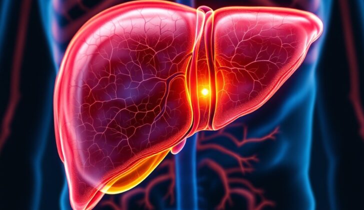
The liver is located below the diaphragm and to the right of the upper abdomen. Its intricate layout supports its various functions. The liver is covered by a protective layer and stretches from the right side of your rib cage to just under your collar bone on the same side. The convergence of the diaphragm and a large vein, the inferior vena cava, forms a bare area on the top side of the liver.
The Couinaud system divides the liver into 8 distinct segments for functional understanding based on their blood supply and drainage. These sectioned segments, shaped like wedges, are pointed towards the center of the liver, and each receives one branch of the bile duct, portal vein, and hepatic artery, respectively.
Blood vessels known as hepatic veins run between these sections, draining into the inferior vena cava. The liver’s two main sections, right and left, are separated by the sizable middle hepatic vein. Smaller sections of the large lobes are separated by the right hepatic vein (for the right lobe) and by a structure called the falciform ligament (for the left lobe). The portal vein divides these sections into upper and lower halves.
The liver’s blood supply primarily comes from the portal vein, which is important for liver imaging for diagnoses. Tumors that receive blood supply from the hepatic artery can be visualized better during the arterial phases of imaging, a principle used in treatments like transarterial chemoembolization. Hepatic veins can be seen as hollow tubes on an ultrasound, emptying into the inferior vena cava.
The fundamental unit, known as the hepatic lobule, is a cluster of liver cells (hepatocytes) that surround the central vein and are arranged in a hexagonal pattern. These liver cells are structured around spaces filled with two types of cells – Kupffer and stellate cells. The arrangement of these cells around structures formed by veins, arteries, and bile ducts, known as portal triads, ensures smooth blood flow. The arrangement also helps differentiate functional areas of the liver, known as zones. Different zones are involved in processes like metabolizing oxygen, breaking down drugs, and other combined functions.
What Causes Hepatocellular Adenoma?
In a 2002 study, Chen and his team furthered our knowledge of how liver adenomas (benign, or non-cancerous, liver tumors) develop. They discovered the important role of a protein called β-catenin in a biological process known as the Wnt signaling pathway. scientists then developed a system classifying these liver adenomas into four main types based on their genetic and physical characteristics:
1. Type 1: Liver cell mutations linked to the Hepatocyte Nuclear Factor -1α (HNF-1α) gene. This represents around 35-40% of all cases. This condition involves a double mutation in the T-cell factor-1 gene, which produces the HNF-1α protein. This protein plays a role in forming liver cells and also affects sugar and fat digestion. People with this type of adenoma often have more fat in their liver when biopsied and are more likely to be women. Moreover, this condition is often associated with a form of diabetes that starts in adulthood, known as MODY3.
2. Type 2: β-Catenin activated mutations. This group accounts for 15-20% of all liver adenoma cases. This condition is often linked with androgen (a type of hormone) exposure, producing glucose, and a genetic disorder causing benign tumors in the intestine called familial adenomatous polyposis. Adenomas for this group bring a higher risk of turning malignant.
3. Type 3: Inflammatory liver adenomas. They make up 40-50% of all liver adenomas. Risk factors include being female, high body mass index (BMI), consuming a lot of alcohol, and systemic inflammatory syndrome (an exaggerated immune response). This group of adenomas doesn’t have mutations associated with the other two types, but instead, inflammation of IL-6 pathway is involved in their development.
4. Type 4: Unclassified types, constituting 10% of all cases. These adenomas don’t have the characteristics seen in the other three types, but when tested, they show typical liver fatty acid-binding protein staining (a marker for healthy liver function).
Thus, the type of adenoma can significantly impact how it behaves and affects the patient’s body, and as such, identifying the type can help in tailoring an optimal treatment plan.
Risk Factors and Frequency for Hepatocellular Adenoma
Hepatocellular adenoma (HCA) happens more frequently in people taking oral contraceptive pills, with 30 to 40 cases per million people, compared to just 1 case per million in those who don’t take these medications. The risk is even higher for people who have been on these pills for more than two years. In fact, a study showed a 25-fold risk increase in women who used these pills for over 109 months, in comparison to women who used them for less than 12 months. Once the use of these pills is stopped, the tumor often shrinks, showing a connection to sex hormones. Women are more likely to get HCA than men, with a ratio of 4 to 1, although the use of sports-related anabolic drugs is changing this ratio.
- Other groups more likely to develop HCA include people with certain glycogen storage diseases (GSD types I and III), iron overload conditions like β-thalassemia and hemochromatosis, and hormonal imbalances like Klinefelter and polycystic ovarian syndromes.
- With the exception of polycystic ovarian syndrome, these conditions mostly affect men and are usually diagnosed in childhood.
Signs and Symptoms of Hepatocellular Adenoma
About half of people with hepatocellular adenomas (HCAs) don’t show symptoms and are often discovered accidentally during an unrelated checkup. For those who experience symptoms, they can vary between mild upper abdominal pain and bloating to severe pain in the right upper quadrant of the abdomen. If the adenoma ruptures, they feel dizzy, may vomit uncontrollably, and become very tired. Sometimes, the pain can spread to the right side, mimicking symptoms of gallbladder inflammation or urinary tract problems. In severe cases, up to 27% of HCAs can rupture, leading to a death rate of 5% to 10%.
It’s crucial to investigate any symptoms, particularly abdominal pain, in people suspected of having HCA. Their medical history could reveal prolonged use of sex hormones, genetic storage disorders, obesity, and any family history of liver disease or cancers, which might make it more likely for HCAs to occur.
A thorough physical checkup can help determine a diagnosis and how stable the patient’s condition is. Unlike patients with kidney stones, individuals with abdominal pain due to digestive system problems tend to stay still. A rapid heart rate and low blood pressure might indicate a state of shock, while a fever could point to inflammation, a symptom also seen in many other conditions, including hepatitis.
Some patients may show signs of dehydration like dry mouth and poor skin elasticity. Paleness and weak pulses could indicate bleeding. Clear findings from listening to the heart and lungs may eliminate heart or lung problems as the cause of referred abdominal pain, unless such illnesses are also present. On examination of the abdomen, an enlarged liver with tenderness might be found. Stiffness of the abdomen could indicate irritation from bleeding into the abdominal cavity due to the rupture of the tumor.
Testing for Hepatocellular Adenoma
When assessing potential liver tumors like hepatocellular adenomas, lab tests can come in handy. These tests won’t necessarily confirm the presence of the tumor, but they can help eliminate other possible conditions and check your overall blood and metabolic health. Some things doctors might look into include α-fetoprotein (AFP) levels, which usually stay normal but can increase if the tumor becomes malignant, turning into liver cancer. Doctors will also likely test for hepatitis to make sure that’s not causing your symptoms.
In some cases, levels of alkaline phosphatase and γ-glutamyl transferase might go up, especially with a specific type of adenoma called inflammatory hepatocellular adenoma. Other measures like white blood cell count, fibrinogen, and C-reactive protein can also be raised.
As for biopsy, where a small piece of tissue is removed for examination, this isn’t always very helpful in diagnosing this type of tumor and is usually saved for cases where images aren’t clear and confirming the diagnosis might change the treatment plan.
Now, let’s talk imaging. Ultrasound is often used in medical settings, but the downside is, it doesn’t always accurately tell benign (non-cancerous) and malignant (cancerous) tumors apart. A specific type of ultrasound, called Doppler ultrasound, might show a basket-shaped pattern of blood vessels at the edge of the tumor. The tumor might also look hyper-echoic, or brighter on the ultrasound, because of fat content within the liver cells.
The preferred imaging technique for diagnosing hepatocellular adenomas, however, is dynamic magnetic resonance imaging (MRI) with a specific contrast agent. Essentially, this is a type of MRI that uses a chemical to make certain structures pop on scans. This is particularly good at differentiating between different types of benign and malignant liver tumors. Sometimes, a tumor will show a central region with lots of parallel vessels coming in from the sides, a bit like a spoked wheel. In some cases, the dynamic MRI might show tortuosity, or a lot of twisting and winding, of the vessels around the tumor with central areas of dead tissue.
A dynamic computed tomography (CT) scan can also be helpful at times. This type of scan might show the tumor enhancing from the outside in during the early phase, leading to a flow towards the center during the later portal venous phase.
Treatment Options for Hepatocellular Adenoma
Small liver tumors known as hepatic adenomas (HCAs) that are less than 5 cm in size and possibly caused by oral contraceptive pills are initially treated in a conservative way. This involves stopping the pill and closely monitoring the tumor with regular imaging. Many times, this approach results in the tumor shrinking. If a woman decides to use the pill again, it’s important that the tumor is regularly monitored with scans. The best length of time for this kind of monitoring is still not certain, with some suggesting it should continue until menopause. Tumors that don’t shrink often have ties to obesity.
Mostly, HCAs remain unchanged during pregnancy. Hospitals with more specialized services will often track small HCAs every three months over the course of a pregnancy and also during the post-pregnancy period. Women with small HCAs are generally not advised against becoming pregnant.
Surgery is recommended for all men, regardless of the size of the HCAs, and for women with HCAs larger than 5 cm. The procedure doesn’t require removal of a large amount of tissue or nearby lymph nodes. In emergency situations of a ruptured HCA with internal bleeding, the surgery has a risk of death between 5% to 10%. The risk of death for non-emergency surgery is less than 1%.
If a liver tumor is causing bleeding, a process known as Transarterial Embolization (TAE) is recommended. This treatment stops the flow of blood to the tumor. If an individual with a bleeding tumor isn’t showing signs of shock, TAE may be used first, followed by planned surgical removal of the tumor. TAE is typically done within 2 to 3 days of the tumor bleeding.
Radiofrequency ablation is a procedure that uses heat to destroy the tumor. It’s only suitable for selected cases, and isn’t appropriate for those that are good candidates for surgery, individuals with hormone-sensitive tumors, people with underlying liver disease, or those wishing to become pregnant. Radiofrequency ablation is typically used for tumors smaller than 4 cm.
Health conditions like obesity, a certain liver condition called nonalcoholic steatohepatitis, and metabolic syndromes increase the risk of developing multiple liver tumors (hepatic adenomatosis). These tumors are less correlated with bleeding or becoming cancerous, but are more associated with their molecular characteristics.
About 4% to 5% of HCAs do have the potential to become cancerous, and those tumors are typically recommended for surgical removal. Genetic counseling is advised for patients with multiple liver tumors, especially if they’re associated with certain genetic conditions like familial adenomatous polyposis or MODY3. Liver transplants aren’t generally recommended for those with non-resectable (unable to be operated on) liver tumors, except for men with specific liver conditions combined with inoperable liver tumors.
What else can Hepatocellular Adenoma be?
When trying to figure out if liver lesions are due to hepatocellular adenoma (HCA), doctors will consider other conditions such as:
- Hemangioma (a benign blood vessel tumor)
- Focal nodular hyperplasia (FNH, a benign liver condition)
- Hepatocellular carcinoma (HCC, a type of liver cancer)
- Metastatic tumors (cancer that has spread from another part of the body)
Medical imaging such as dynamic contrast-enhanced MRI can be very useful. It can distinguish between HCA and hemangiomas, which generally show a specific type of pattern on the MRI. Gadolinium-based contrast MRI usually reveals that FNH absorbs more contrast than HCA during the hepatobiliary phase (a phase during liver imaging), and FNH often has a central scar and a lobulated appearance.
The patient’s clinical scenario is important in differentiating between HCA, HCC, or metastatic disease. For instance, individuals with HCC typically have an underlying chronic liver condition, with or without cirrhosis (scarring of the liver). People with cancer that has spread to the liver from another part of the body typically have a known primary cancer elsewhere and particular risk factors associated with it.
What to expect with Hepatocellular Adenoma
The future health outcomes for patients with a type of liver tumor known as hepatocellular adenoma are not very clear. Stopping the use of oral contraceptives might help in reducing or even completely getting rid of some of these tumors. It’s important to note, though, that there’s a roughly 27% chance of these tumors bursting, which can carry a risk of death ranging from 5% to 10%. Also, there’s a 4.2% chance of this type of tumor turning malignant, or cancerous, and this risk remains even if a patient stops taking oral contraceptives.
In most cases, it’s unlikely that the hepatocellular adenoma will completely disappear. About a quarter of women will still experience pain in the right upper quadrant (RUQ) of their abdomen. Plus, there’s a possibility that between 30% to 45% of women could experience a hemorrhage, or bleeding. This bleeding could occur within the tumor itself or within the abdomen. Tumors that are larger present a higher risk of this kind of bleeding. Pregnancy has also been associated with these tumors growing in size and a higher chance of them bursting.
Possible Complications When Diagnosed with Hepatocellular Adenoma
The complications associated with HCA, a type of liver tumour, include the tumour breaking open (rupturing) and bleeding heavily (hemorrhage). These complications can cause a severe drop in blood pressure (hemorrhagic shock) and can sometimes be fatal. These risks increase for tumours that are larger than 5 cm. Also, there is a higher risk of these tumours turning cancerous (malignant transformation) in men compared to women if the tumour is larger than 5 cm.
Common Complications of HCA:
- Tumour rupture
- Severe bleeding (hemorrhage)
- Severe drop in blood pressure (hemorrhagic shock)
- Potential death
- Increased complications for tumours larger than 5 cm
- Higher risk of turning cancerous in men with tumours larger than 5 cm
Preventing Hepatocellular Adenoma
To prevent the development of Hepatocellular Adenomas (HCAs), which are benign liver tumors, it’s vital to reduce contact with certain risk factors. These include medications like birth control pills and muscle-building steroids. Providing thorough education about the risks associated with these drugs and their ability to trigger HCA growth is crucial. Additionally, maintaining a healthy lifestyle can possibly lower the risk of developing HCAs. This includes managing your weight and limiting the amount of alcohol you consume.
Patients who either have HCAs or genetic predispositions to develop them should be informed about the potential complications like rupture or liver cancer, especially if no treatment is taken. Women with such conditions should know that becoming pregnant is not discouraged, particularly if the tumor size is 5 cm or smaller. But, they should receive consistent professional care and vigilant monitoring of the tumor during their pregnancy.
If surgery is needed during pregnancy, ideally, it should take place during the second trimester, as this is the time when the risks to both the mother and the unborn child are at their lowest. Patients should be given all the necessary information about their condition, the available treatment options, and their associated risks and benefits. This way, they can make informed decisions about their health.


