What is Obturator Hernia?
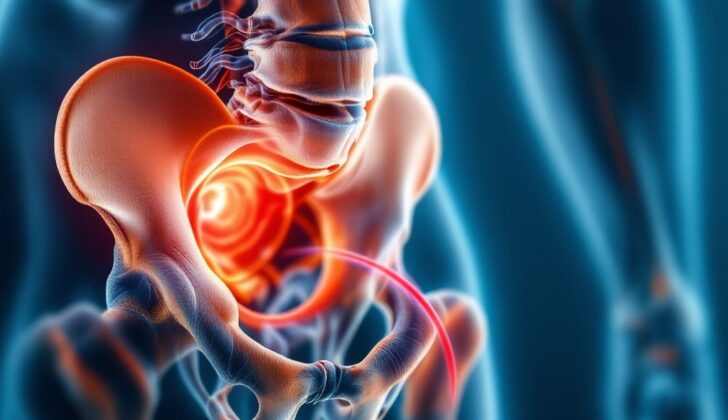
Obturator hernias are a rare cause of stomach pain, making up less than 0.04% of all hernias. An obturator hernia happens when parts of the abdomen move into a small hole in the pelvis, called the obturator foramen. Because this hole is quite small, there’s a higher chance for these hernias to become trapped and eventually strangled, which is a serious condition. See image: Obturator Hernia.
Diagnosing an obturator hernia can be tricky, as patients often only seek help when the condition has advanced, and their overall health may be poor. Diagnosing this condition often requires a high level of alertness in older adults, and even then, a firm diagnosis might not be possible without the help of imaging tests or surgery findings.
The treatment methods for obturator hernias are varied. Today, there are many reports of minimally invasive procedures being used to treat these cases. The choice of treatment depends largely on the patient’s overall health. Quick resolution of the patient’s condition is important in all cases.
What Causes Obturator Hernia?
The obturator foramen is a hole in your hip bone, specifically where the bones called ischium and pubis meet. This hole is covered by a type of muscle and connective tissue membrane called the obturator membrane. A bundle of a nerve, artery, and vein, known as a neurovascular bundle, goes through this membrane from top to front, and then through a small channel known as the obturator canal. This canal starts as a groove and becomes a canal because of a band-like ligament attaching to the bone’s tubercles or small rounded projections. This canal links your hip to the inner part of your thigh. It’s a tiny space, about 0.2 to 0.5 cm wide and 2 to 3 cm long. The obturator nerve is the highest part of the nerve, artery, and vein bundle passing through the canal. It then splits into two branches when it leaves the canal.
The obturator membrane is covered on both sides by obturator muscles. If a hernia, which is when part of an organ pushes through a weak spot in your body, forms, it will pass through three structures in the following order: obturator internus muscle, obturator membrane, and obturator externus muscle. After passing through these structures, the natural position for the hernia is deep and below the pectineus muscle, a muscle located in the thigh close to the hip, and above the obturator externus muscle. But the hernia can push through the obturator externus muscle, carrying with it the obturator nerve. In this position, the hernia sits behind a muscle called the adductor brevis. Very rarely, the hernia may be found between the obturator internus and externus muscles.
The shape of the hole in your hip bone can vary based on whether you’re a woman or a man. Women’s obturator foramens are usually triangular and wider, while men’s tend to be more oval-shaped.
Risk Factors and Frequency for Obturator Hernia
Obturator hernias are often referred to as ‘the skinny old woman hernia’. This name comes from the fact that these types of hernias are usually found in thin, older women, specifically those in their 70s or 80s. It’s very rare to find this type of hernia in men.
The reason for this is thought to be due to the unique structure of the obturator canal in women. Because it is wider and more slanted, it might allow hernia contents to pass more easily. In addition, women who have had multiple children may experience changes in the tissue that make the canal more likely to expand and allow a hernia to develop.
It’s also worth mentioning that obturator hernias usually only happen on one side. The reason for this could be that the sigmoid colon (part of the large intestine) is on the left side, which might prevent hernias from developing on that side.
Signs and Symptoms of Obturator Hernia
An obturator hernia is a rare condition, often discovered in emergency cases when the patient has symptoms of an intestinal obstruction. It’s very rare for this type of hernia to become strangulated, accounting for less than 0.04% of all hernias. Nevertheless, it’s essential to keep this type of hernia in mind as a potential diagnosis when you have an elderly, underweight woman who has symptoms of an intestinal obstruction and no previous history of abdominal surgery, which could otherwise explain the obstruction.
The hernia’s closeness to the obturator nerve can cause certain symptoms, such as the Howship-Romberg sign, where the patient experiences pain along the inner thigh. This pain typically improves with hip flexion and worsens with hip extension, adduction (moving the leg inward), and medial rotation of the hip. Another sign is the Hannington-Kiff sign, where the adductor reflex in the thigh is absent. This reflex is tested by tapping the inner thigh muscle with a tendon hammer. If the muscle contracts, the reflex is present. The Hannington-Kiff sign is a more specific indicator of an obturator hernia compared to the Howship-Romberg sign, but it can be difficult to evoke and depends greatly on the skill of the person performing the test.
Testing for Obturator Hernia
In the case of an obturator hernia, the physical symptoms might not be immediately apparent and can vary depending on who’s examining the patient. So, if symptoms aren’t clear, a Computed Tomography (CT) scan might be done. A CT scan is a type of X-ray that takes detailed pictures of the inside of your body. It’s been reported that a CT scan can correctly diagnose an obturator hernia up to 90% of the time.
In preparation for a potential surgical procedure, the patient will also need to have general lab tests done. These tests are standard procedures that vary based on local hospital or clinic protocols, but they help to ensure that the patient is healthy enough for surgery.
Treatment Options for Obturator Hernia
Hernias typically need treatment to prevent complications like the hernia becoming trapped and not receiving sufficient blood supply, a condition known as strangulation. This is also true for obturator hernias, which are usually problematic and often need prompt attention. They’re unlikely to get better or reduce on their own, so medical intervention is necessary.
The choice of treatment usually boils down to two options: open surgery or laparoscopic surgery. Laparoscopic surgery is a minimally invasive technique that’s becoming more commonly used. For obturator hernias, which are located deep within the body, this approach can be beneficial because it allows for a clear view and treatment of the hernia’s contents. Most of the case studies available have mentioned using a specific laparoscopic technique – the transabdominal preperitoneal (TAPP) approach. This technique allows the surgeon to thoroughly inspect the contents of the hernia. If bowel tissue is identified within the hernia, this can be evaluated for healthy function. Depending on the surgeon’s expertise and the condition of the bowel tissue, the tissue may even be removed laparoscopically or through conversion to open surgery if necessary.
The steps for open surgery to treat a strangulated hernia are standard. It involves an incision made in the midline of the lower abdomen, identifying the hernia, checking the contents, possibly removing and reconnecting (anastomosis) a part of the bowel, and then repairing the area where the hernia formed through either stitching or placing mesh. Other types of open approaches exist but are often performed by surgeons who specialize in these methods. The tried and true lower midline incision is a more common technique and is generally more familiar to most surgeons, thus, it’s less likely to result in problems.
In the case of laparoscopic surgery, a TAPP repair is usually carried out. This procedure involves creating space within the abdomen using a method that fills the abdomen with gas (pneumoperitoneum), often executed using an open technique known as the Hassen technique. Surgical ports, which are small tubes through which surgical instruments are passed, are placed a few inches to either side of the midline below the navel. Notably, even if only one-sided (unilateral) hernia diagnosis was made initially, it could still be valuable to check the opposite side for the presence of a hernia that may have been missed. Once the hernial sac is identified, it’s carefully pulled away (retracted), sometimes involving careful dissection. If bowel tissue is found and is not viable, it may need to be removed, a process that follows certain steps. Any remaining excess hernial sac can be left in place to avoid unnecessary further dissection. Lastly, a prosthetic mesh is placed within the space created earlier to help reinforce the area. This is followed by suturing the protective covering of the abdomen (peritoneum) closed.
What else can Obturator Hernia be?
If an elderly person suddenly can’t go to the bathroom at all, it’s important to check if this could be due to blockage of a small or large intestine. In older people, a small intestine can get blocked because of hernias, adhesions (scar tissue), and tumors, while a large intestine block is most likely caused by tumors, hernia, and twisting of the intestine (volvulus).
Identifying what’s causing the blockage usually involves image scans like a CT scan, which are done after making sure the patient is stable and properly prepared for the procedure.
What to expect with Obturator Hernia
Obturator hernias can be difficult to study because of their infrequency. Past research has reported a mortality rate for these hernias ranging from 13% to 40%, and this continues to be confirmed in more recent studies. The morbidity, or the occurrence of disease related to these hernias, often relates to late diagnosis or identification of the issue. This delay can lead to infectious complications like the accumulation of infected material or ‘collections’. It can also lead to ‘anastomotic leaks’, which occur when there are issues with therapeutic connections made between parts of the bowel due to bowel resections.
Possible Complications When Diagnosed with Obturator Hernia
Like any other hernia, an obturator hernia can lead to various complications. These include strangulation, which may require bowel resection – a surgical procedure where a part of the bowel is removed – and its related complications. Damage to the obturator nerve due to pressure symptoms is also possible, as well as complications that are associated with surgery.
Common Complications:
- Strangulation of hernia
- Bowel resection
- Damage to the obturator nerve
- Complications associated with surgery
Recovery from Obturator Hernia
After surgery for these patients, their aftercare should follow the local hospital’s routine for any bowel surgery or emergency hernia repairs. This includes sufficient pain management, resting the gut (in line with your local hospital’s recovery plan), and taking care of the surgical wound. This is then followed by gently starting to eat and drink and moving around as soon as they can.
There are no standard rules on how often these patients should be checked up on. However, normally, doctors may want to see patients 2 and 6 weeks after the surgery to make sure they are healing properly and are satisfied with their care.
Preventing Obturator Hernia
Obturator hernias, a type of condition where tissue pushes through a weak spot in your pelvic muscles, can’t be prevented, and because they are rare, long-term studies on this condition are difficult. Meanwhile, it’s crucial for doctors to be aware of this condition, particularly when treating patients in the age groups commonly affected.


