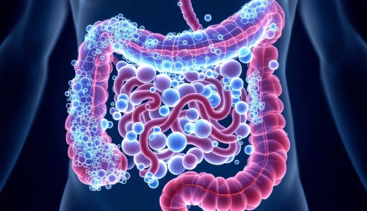
What is Pneumatosis Intestinalis?
Pneumatosis intestinalis (PI) is a medical condition where gas or air is found in places it shouldn’t be in your digestive system. Usually, air or gas in parts of your GI tract, like the lining of your intestines or the blood vessels that supply them, is a sign of an underlying health problem. Please note though, having gas inside your intestines is perfectly normal and is usually released when you pass gas.
Interestingly, PI can go by several other names in medical texts, such as pneumatosis cystoides intestinalis, intraluminal bowel gas, and pneumatosis coli. These names often reflect where and how the air is wrongly present in your digestive system. Specifically, pneumatosis cystoides intestinalis refers to a situation where multiple gas-filled cysts form in the innermost and outermost layers of your intestines. If left unattended, the number and size of these cysts can increase.
The condition was first identified way back in 1730 by DuVernoi while he was studying a body post-mortem. In 1946, Lerner and Gazin were the first to image it using radiographic techniques, but at the time they weren’t sure what caused it. Since then, thanks to advances in radiology, more and more cases of PI have been identified.
What Causes Pneumatosis Intestinalis?
Pneumatosis intestinalis, or PI, is not a condition on its own but a symptom that suggests an underlying disease. Think of PI like an alarm bell signaling that something is going wrong in your body. Its presence means that gas is forming in unusual places, such as within the walls of your intestines.
In 1998, a researcher named BL Pear suggested that this abnormal gas formation could be due to several reasons – like tissue death in the bowel, lung diseases, or a change in the intestines that lets too much stuff pass through its walls. In fact, the reasons behind PI can be a combination of lung-related, mechanical, or bacterial causes. But regardless of the cause, one thing is common in all PI cases: the surface of your intestines is disrupted in some way.
PI can be split into two types: primary and secondary. In primary PI, the gas forms harmless collections and creates a pattern that looks like bubbles in the walls of your intestines. Meanwhile, secondary PI happens when there’s a disease causing it, and the gas appears more like a line.
Risk Factors and Frequency for Pneumatosis Intestinalis
Pneumatosis intestinalis is a condition that affects around 0.03% of people. However, because it doesn’t always cause symptoms, the exact number of people it affects is unclear, and the true figure could be higher. It can happen to anyone, regardless of age, but it’s more common in older people than in young adults or babies. In infants, the condition can be serious and is often connected with a severe gut condition called acute necrotizing enterocolitis. On the other hand, adults usually have a harmless form of the condition, which is often discovered by accident during medical checks. In adults, about 15% of cases are primary, meaning they occur on their own, while 85% are secondary, meaning they happen as a result of another condition.
Signs and Symptoms of Pneumatosis Intestinalis
Pneumatosis intestinalis is a health condition with two types: primary and secondary. Primary pneumatosis intestinalis commonly leads to mild abdominal discomfort and some cases may not show any symptoms at all. On the other hand, secondary pneumatosis intestinalis can produce severe symptoms similar to intestinal ischemia and peritonitis, which can be life-threatening. Additional symptoms might include blood in the stool, anal discomfort, constipation, and abdominal pain. In some cases, complications like pneumoperitoneum, intestinal ischemia, and obstruction can occur. People who have a history of abdominal surgeries or endoscopy are more likely to develop pneumatosis intestinalis because these procedures can disrupt the intestinal lining and increase pressure inside the intestine.
There are also many diseases and conditions that are associated with pneumatosis intestinalis, some of which include:
- Pulmonary diseases like asthma, chronic obstructive pulmonary disease (COPD), and emphysema
- Autoimmune conditions such as Lupus variants, polymyositis, dermatomyositis, polyarteritis nodosa, and celiac sprue
- Drug-induced causes, like glucosidase inhibitors and corticosteroids
- Gastrointestinal issues like Inflammatory bowel disease (IBD), diverticulitis, colitis, Clostridium difficile infection, appendicitis, carcinoma, peptic ulcer, and necrotizing enterocolitis (in children)
- Infectious diseases such as Human immunodeficiency virus (HIV), acquired immunodeficiency syndrome (AIDS), mycobacterium tuberculosis, 2019 novel coronavirus (COVID-19)
- Iatrogenic causes like blunt abdominal trauma, endoscopy, post-surgical intestinal anastomosis, barium enema, and positive end-expiratory pressure (PEEP) ventilation
Testing for Pneumatosis Intestinalis
To diagnose an illness called pneumatosis intestinalis, doctors often use different types of imaging techniques such as X-rays of the abdomen, CT scans, MRI scans, and ultrasound. However, these tests might not always show signs of the disease like bowel walls that have become thicker and contain gas, in about one-third of the patients.
The CT scan is particularly helpful as it can more precisely distinguish between air inside the bowel and fat beneath the mucus layer lining the bowel. This scan can also help identify other causes of pneumatosis intestinalis that might not be otherwise detected, such as air in the portal vein, strands of tissue in the colon, and an enlarged bowel.
To check for the presence and increase of air inside the portal vein and the abdomen, further imaging tests might be needed.
Pneumatosis intestinalis can cause gas to accumulate in different patterns. It can accumulate in a cyst-like pattern as we see in a condition called pneumatosis cystoides intestinalis, or in a more straight line. This linear pattern is often linked with bowel ischemia (an inadequate blood flow to the bowel which leads to tissue damage) and with gas in the portal vein. A study reported that these gas cysts can be found in various parts of the digestive tract: the colon (46% of the time), small intestines (27%), large intestines (7%), and stomach (5%).
Doctors can perform an endoscopy (a test where a long, thin tube is inserted into the body to visualize the digestive tract) to understand how the gas is distributed (in a linear or cyst-like manner) and to rule out other possible problems in the colon.
Lab tests are also very important in figuring out the best treatment plan by distinguishing between relatively harmless cases and life-threatening ones. Some of these lab tests include counting the number of white blood cells, measuring the level of lactic acid, aspartate aminotransferase, alanine aminotransferase, amylase, bicarbonate, and alkaline phosphatase in your body.
Treatment Options for Pneumatosis Intestinalis
Pneumatosis intestinalis (PI) is not a disease, but rather a clinical sign – that is, something that a doctor observes during an examination. Treatment options for PI therefore aim to address the underlying cause that’s creating this sign in the first place. The chosen treatment also depends on specifics related to the patient, such as their overall health and the severity of their symptoms.
For patients without critical conditions like peritonitis (inflammation of the inner lining of the abdomen) or sepsis (a severe response to infection that can lead to organ failure), a non-surgical treatment may be preferred. In these cases, it’s important to keep monitoring the patient closely to ensure that their PI doesn’t develop into a life-threatening situation.
Non-surgical treatment methods include monitoring the patient, giving them oxygen therapy, using antibiotics to manage any potential infections in the intestines, and carrying out endoscopy (a procedure where a thin tube with a camera is used to look inside your body). Hyperbaric oxygen therapy, which involves breathing in pure oxygen in a pressurized room or tube, can help resolve symptoms by reducing the pressure of gases in the veins and assisting gas to move out of the bowel wall.
Breathing in oxygen through a special mask or nasal tubes (entering 4 to 6 litres of oxygen per minute into the body) can also be beneficial. Oxygen therapy is recommended for at least two days after the disappearance of PI-related cysts (small sacs filled with liquid or air) to decrease the risk of them reappearing. Using the antibiotic metronidazole is also common to help manage PI.
To provide the body with all essential nutrients and reduce gas production in the intestines, a special diet known as an elemental diet may be advised. This diet consists of nutrients in liquid or powder form that the intestines can easily absorb.
In some cases, surgical intervention may be necessary to treat PI – this is typically when certain critical conditions are observed, like elevated white blood cell count (signifying an active infection), explicit imaging results showing the gas is affecting the veins supplying the liver (known as portal venous gas), and signs of serious illnesses such as sepsis or acidosis (high levels of acid in the body fluids).
An endoscopy procedure may be used to relieve intestinal obstruction caused by PI. The doctor uses a fine needle to remove gas from the many cysts, helping ease the blockage. This technique can also be used to confirm the presence of gas-filled cysts. If the obstruction persists, the wall of the cyst may be treated to prevent the cyst from refilling with gas or expanding again.
What else can Pneumatosis Intestinalis be?
There can be other medical conditions that resemble the symptoms of the disease being diagnosed, these need to be eliminated from the possible diagnoses. One critical condition to be aware of is bowel ischemia. This condition should be considered, particularly among patients with a history of blood vessel disease, heart failure, or irregular heartbeats. If a patient is suffering from widespread stomach pain along with unusual lab results, bowel ischemia could be a possible cause. Moreover, related underlying conditions such as trapped intestines caused by an internal hernia, abdominal infection or inflamed obstruction in the gut could also be the root cause.
What to expect with Pneumatosis Intestinalis
A poor prognosis for a condition called pneumatosis intestinalis depends on several factors:
- a pH level under 7.3
- the presence of gas in portal veins
- lactate level above 2mmol/L
- amylase (an enzyme that helps digest carbohydrates) level above 200 U/L
- bicarbonate (a substance that helps balance acidity in the body) level under 20 ml/L
There is a potential need for surgery if patients, particularly those over 60, show symptoms of blockage in the intestine, signs of sepsis (a severe response to an infection), shock, or images that suggest the presence of gas in the portal veins. These symptoms are associated with a high risk of death.
According to a study by Greenstein and others in 2007, factors that can lead to a need for surgery include vomiting, a high white blood cell count (above 12 c/mm3, which indicates an infection in the body), and being 60 or older. Additionally, lactic acidosis (a build-up of lactic acid in the body), shock, and sepsis are linked to unfavorable outcomes, including death.
Possible Complications When Diagnosed with Pneumatosis Intestinalis
Pneumatosis intestinalis, a condition involving gas-filled cavities within the wall of the intestine, can lead to a variety of complications. These issues can be divided into those affecting the intestine directly, and those impacting surrounding body parts. The intestinal complications mostly involve obstructions caused by these gas cysts. If not treated, these obstructions can lead to fecal impactions, essentially a blockage of feces. If unnoticed, these can cause perforations that arise from ulcers in the large intestine. This pressure build-up can result in a ruptured intestine, leading to serious conditions like an acute abdomen, septic shock, peritonitis, and more.
If Pneumatosis intestinalis also involves gas in the portal vein (the blood vessel leading to the liver), then this correlates with approximately 70% of bowel ischemia cases, a serious condition where the intestines don’t get enough blood. Complications outside the intestines can include adhesions and compressions of nearby structures due to the cysts. These cysts can push against other organs, and if they rupture, it can lead to pneumoperitoneum, a condition that requires urgent surgery. These complications occur in roughly 3% of secondary pneumatosis intestinalis, with death rates from these complications ranging from 50% to 75%, particularly in those with bowel ischemia.
Key issues include:
- Intestinal obstructions from gas cysts
- Blockage of feces causing perforations
- Ulcers in the large intestine leading to ruptures
- Acute abdomen, septic shock, peritonitis, etc., from ruptures
- Bowel ischemia correlated with portal venous gas
- Adhesions and compressions of nearby structures
- Organs compressed by cysts
- Ruptured cysts leading to pneumoperitoneum
- Death rates from complications up to 75%
Preventing Pneumatosis Intestinalis
Understanding one’s condition and following the doctor’s instructions are key to effectively treating pneumatosis intestinalis, a condition where pockets of gas form in the wall of the intestines. Neglecting to follow the recommended treatments and lifestyle changes can lead to serious consequences, such as the condition coming back, infection spreading throughout the body (septic shock), or even death.
Adopting long-term lifestyle changes is a significant part of the treatment. This includes following an ‘elemental diet’, which is a diet based on a type of meal replacement drink. This diet is designed to be easy on the body and encourages full absorption of the nutrients provided, while also reducing the production of gas in the intestines. Therefore, making these dietary changes can help prevent pneumatosis intestinalis from recurring.


