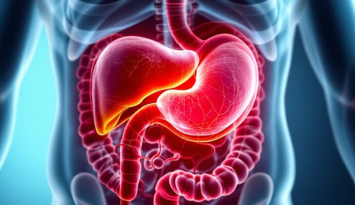
What is Portal Vein Thrombosis?
Portal vein thrombosis (PVT) is a condition where the portal vein, a large vein that carries blood from the digestive organs to the liver, gets narrowed or blocked by a blood clot. This clotting can happen in the main part of the portal vein or its branches within the liver, and it can even spread to veins leading to the spleen or another major vein linked with the small intestine. PVT often happens alongside liver cirrhosis, which is scarring of the liver, but it can also occur without a related liver disease – for example, because of cancer, infection in the abdomen, or inflammation of the pancreas (pancreatitis). When PVT leads to chronic blood vessel formation (known as portal cavernoma) or alternate pathways for the blood to flow around the clot (known as collaterals), it is seen as a separate issue.
The portal venous system begins developing very early in a baby’s life when still in the womb (from the fourth to the twelfth weeks of pregnancy), and it carries blood from most of our digestive tract, including the spleen, pancreas, and gallbladder to the liver.
The veins that take the blood away from our gastrointestinal organs sit near the major arteries, the large blood vessels that bring the blood from the heart to these organs. These veins carry the blood back to the portal vein, which guides the blood into the liver. The liver receives about 75% of its blood supply from this system, with the rest coming from the hepatic artery. The blood then exits the liver through the hepatic veins and enters the general circulation.
The portal vein is formed where the splenic and superior mesenteric veins come together, carrying blood away from the spleen and small intestine. PVT typically affects two groups of people: those with liver cirrhosis and those with abnormalities in the blood clotting system. Acute PVT takes place when the portal vein gets suddenly blocked by a clot. This blockage can be complete or partial and may spread into the veins leading to the small intestine and spleen. In acute PVT, there are no signs of chronic PVT like alternate pathways for the blood (collateral circulation) or high blood pressure in the portal venous system (portal hypertension). However, if it’s not clear when the clot formed and there are no signs of chronic PVT, the condition can be called “recent”. The treatment for recent PVT is the same as for acute PVT.
What Causes Portal Vein Thrombosis?
Portal Vein Thrombosis (PVT) is a condition where a blood clot forms in the portal vein, which carries blood from your intestines to your liver.
The most common reason for PVT is a liver disease called cirrhosis. In a healthy liver, PVT mostly happens due to situations that increase the tendency of your blood to clot, which could be inherited or acquired. Primary myeloproliferative disorders, which cause your body to make too many red blood cells, white blood cells, or platelets, are the most common situation that cause your blood to clot.
Other conditions that could increase your blood clotting and lead to PVT are paroxysmal nocturnal hemoglobinuria (a rare disease that destroys red blood cells), antiphospholipid syndrome (an autoimmune disorder that causes abnormal blood clots), and hyperhomocysteinemia (high levels of an amino acid called homocysteine in the blood). There are also inherited disorders such as deficiencies in proteins C and S, and antithrombin III that increase blood clotting risk. Also, certain uncommon genetic mutations present in some people can cause a tendency for blood clotting.
PVT is also associated with rare conditions like pregnancy, chronic inflammatory diseases, oral contraceptive use, and cancers, regardless of previous clotting conditions. About 25% of all PVT cases are caused by cancer.
Inflammation in the abdomen that injures the vascular endothelial (the inner layer of your blood vessels) can also lead to PVT. Conditions like pancreatitis (inflammation of the pancreas), cholangitis (inflammation of the bile duct), appendicitis, and liver abscesses (pus-filled pockets in the liver) can all cause this. Local injury to the portal venous axis (the veins that bring blood to liver from the digestive organs) following surgery or abdominal trauma in people with clotting disorders can also cause PVT.
In children, a condition called extrahepatic portal venous obstruction (EHPVO) often happens due to a hardened vein (phlebosclerosis) with clotting occurring afterward. Conditions or events like inflammation of the umbilical cord at birth (omphalitis), infection of the umbilical cord in newborns, repeated abdominal infections, sepsis (a severe infection that spreads in the bloodstream), abdominal surgery, and trauma can result in EHPVO.
Other factors that could lead to PVT are complications from abdominal surgeries, especially weight loss surgeries, complications of a rare autoimmune disease called Behçet’s syndrome, collagen vascular diseases (where your immune system attacks your own skin, joints, blood vessels, and other organs), and cancers like liver and pancreatic cancer. PVTs can also be caused by certain treatments like endoscopic sclerotherapy (injecting a chemical to treat swollen blood vessels), oral contraceptive drugs, or even a total removal of the pancreas with islet autotransplantation (transplanting some insulin-producing parts of pancreas back in the body). Acute pancreatitis, paroxysmal nocturnal hemoglubinuria, pregnancy, retroperitoneal fibrosis (a rare disease that results in a fibrous mass in the back of your abdomen), transjugular intrahepatic portosystemic shunt (a treatment to reduce high blood pressure in your liver), and trauma to the portal vein can also lead to PVT.
Risk Factors and Frequency for Portal Vein Thrombosis
Portal vein thrombosis (PVT) is a condition found in 0.6% to 16% of patients with cirrhosis, especially in those awaiting a liver transplant. The occurrence of PVT is even higher – up to 35% – in patients with both cirrhosis and hepatocellular carcinoma. The risk of experiencing PVT in one’s lifetime for the general population is around 1%.
A large-scale study reviewing 8549 research articles investigated PVT occurrence in cirrhosis patients. This included the analysis of information from three major databases: PubMed, Excerpta Medica Database, and the Cochrane Library. The findings of 54 studies showed that the prevalence of PVT in patients with cirrhosis was about 13.92% with a confidence interval between 11.18% to 16.91%.
Several factors increase the risk of PVT in patients with cirrhosis. These include:
- Having a Child-Pugh class B or C rating (a clinical grading system for the severity of cirrhosis)
- High levels of D-dimer (a small protein fragment related to blood clotting)
- A history of using non-selective beta-blockers
- Being at risk for moderate to severe esophageal varices (abnormally enlarged veins in the esophagus)
- Presence of ascites (a buildup of fluid in the abdomen).
Signs and Symptoms of Portal Vein Thrombosis
Portal vein thrombosis is a condition where blood clots form in the portal vein, which carries blood from the digestive organs to the liver. For many patients, they don’t have any symptoms and don’t even know they have it. However, when symptoms do show up, they can be either sudden (acute) or happen over a longer period of time (chronic).
Portal vein thrombosis can lead to a condition called portal hypertension, which is high blood pressure in the vein mentioned earlier. This can cause feelings of fullness in the upper left part of your stomach due to an enlarged spleen (splenomegaly), or bleeding from dilated veins in your esophagus or stomach (varices).
If the cause of portal vein thrombosis is not liver cirrhosis or cancer, it often manifests as:
- Stomach pain (91% of cases)
- Fever (53% of cases)
- Ascites, which is a buildup of fluid in the stomach (38% of cases)
In severe cases, if the blood clot expands into a vein that provides blood to the intestines, it may lead to conditions such as intestinal ischemia (lack of blood supply to the intestines), bowel death, or ileus (lack of movement in the intestines) presenting as bloody stools, fever, and sepsis. This particular scenario results in high death rates.
If someone with liver cirrhosis develops new portal vein thrombosis, they may experience conditions such as ascites, jaundice, or bleeding from esophageal or gastric varices. Ascites in these patients usually happen when they are given a lot of intravenous fluids to treat major bleeding. Patients with obstruction in the portal vein due to blood clots generally show symptoms only related to portal hypertension, such as a generally non-threatening upper GI bleed, enlarged spleen, anemia, low platelet counts. Some of them may not show any symptoms and are diagnosed incidentally through imaging procedures.
Testing for Portal Vein Thrombosis
If suspected of portal vein thrombosis (PVT), a condition where a blood clot forms in the main vein carrying blood to the liver, doctors conduct various tests. Liver function tests are generally normal unless the patient already has liver cirrhosis. Increased pressure in blood vessels supplying the liver due to chronic PVT may result in low platelet count because of enlarged spleen. If PVT affects the bile ducts, an increase in enzymes within the liver (alkaline phosphatase) and a yellow pigment found in bile (bilirubin) may be seen.
Doppler ultrasound is a commonly used diagnostic tool to confirm PVT, with an accuracy between 80 to 100%. Doctors can see isoechoic or hypoechoic material (solid regions of equal or lower-than-normal echoes) within the portal vein during an ultrasound, suggesting a clot. Another sign of chronic PVT may be a network of small, twisted blood vessels replacing the portal vein-known as portal cavernoma. Splenomegaly, or enlarged spleen, is another associated condition that can be detected with ultrasound. Advanced ultrasound techniques, like contrast-enhanced ultrasound and endoscopic ultrasound, can give more precise information about blood flow in the portal vein when it is tiny.
Computed Tomography (CT) scans and Magnetic Resonance Imaging (MRI) provide additional information about the clot, like its complete extension, signs of bowel tissue dying due to insufficient blood supply, and status of nearby organs. CT scans using contrast (dyed fluid) can help differentiate a “bland” non-cancerous blood clot from a cancerous one. An MRI is extremely sensitive and specific for detecting the main PVT and is helpful in determining the possibility of surgically removing a tumor involving the portal venous system and monitoring the condition after treatment. Doctors may also use a technology called Positron Emission Tomography CT to distinguish between benign (non-cancerous) and malignant (cancerous) blockages in the portal vein.
In the past, a test called Splenoportovenography was used, which involved injecting dye into the spleen and visualizing the blood network involved in liver blood supply. This procedure was mainly done to determine the openness of the blood network for potential surgery and provided portal pressure measurements. However, this technique is now obsolete.
Doctors may also perform endoscopy in patients with PVT because they often have portal hypertensive gastropathy- a condition where the lining of the stomach is damaged due to high blood pressure in the liver. Large abnormal veins in the esophagus or stomach are present more often in patients with chronic PVT.
Once PVT is diagnosed, all patients should have a thorough investigation of potential blood clotting disorders and local factors. The evaluation may include testing for a disease where the blood is more prone to clotting (antiphospholipid syndrome), measuring various proteins and factors involved in blood clotting, and checking for genetic mutations that may increase clotting risk, among others.
Treatment Options for Portal Vein Thrombosis
The goal of treatment for portal vein thrombosis (PVT), which is a blood clot in the vein that transports blood from the stomach, intestines, spleen, and pancreas to the liver, is either stopping the clot from enlarging or preventing further clots from forming. Anticoagulation therapy, such as heparin or novel oral anticoagulants (new types of blood thinners), are often prescribed. While the role and effectiveness of these medications is well-known in people without liver disease, their effects are less clear in those who do have liver disease.
What we do know is that anticoagulation therapy is often used in patients who have PVT alongside other symptoms and conditions, such as the risk of intestines getting insufficient blood supply, liver disease that could lead to a liver transplant, or liver disease with a new PVT diagnosis. On the other hand, anticoagulation therapy may not benefit patients with advanced liver disease who are not candidates for a liver transplant.
Meanwhile, thrombolytic therapy, a treatment that dissolves blood clots, can be used for PVT in patients without liver disease. But because it may carry the risk of creating emboli, small clots that can block blood vessels, it is often delivered directly into the vein with the clot to reduce such risks.
In some cases, thrombectomy, a surgical procedure that removes clots, might be considered. However, given the risks of clot recurrence, venous ruptures, and potential trauma to the portal vein, this approach requires careful consideration.
Other treatments include placing a transvenous intrahepatic portosystemic shunt (a type of stent that creates a pathway within the liver for blood to flow) and management with anticoagulation therapy.
Overall, the aim is to prevent the clot from extending and create the best situation for the clot to dissolve naturally. While anticoagulation therapy plays a significant role in managing acute PVT, its role in chronic PVT patients is still subject to further research.
Before embarking on anticoagulation therapy, certain patients, especially those with liver disease, should be screened for esophageal varices, which are enlarged veins in the esophagus that can possibly rupture.
Lastly, recent research suggests that more studies are needed to understand how factor Xa inhibitors, a type of blood thinner, work in treating acute PVT, and to weigh their potential benefits against the risks.
The duration of treatment can range from 3 to 6 months, although longer treatment might be considered depending on the patient’s individual circumstances such as inherited blood clotting disorders and extended portal vein thrombosis. The best plan of care should be personalized, taking into consideration the patient’s unique needs and conditions.
What else can Portal Vein Thrombosis be?
When it comes to diagnosing this condition, there are other possible diseases that could be causing the symptoms. These options need to be considered and may include:
- Arsenic poisoning
- Budd-Chiari syndrome (a rare liver condition)
- Cirrhosis (scarring of the liver)
- Sarcoidosis (inflammation that produces small lumps of cells)
- Schistosomiasis (a type of infection caused by parasites)
What to expect with Portal Vein Thrombosis
In simpler terms, when it comes to acute non-cirhhotic portal vein thrombosis – a condition involving a blood clot in the vein that supplies blood to the liver in people without liver disease – an early diagnosis and rapid treatment could lead to a survival rate as high as 85% over 5 years. The issue of death primarily arises due to the underlying cause or a consequence of increased pressure in the portal vein, which is the vein that conveys blood to the liver.
Acute non-cirrhotic portal vein thrombosis generally has a good outcome if it doesn’t progress to intestinal infarction – a serious condition where blood supply to the intestines is disrupted.
In cases of chronic extrahepatic portal vein thrombosis – chronic blood clot in the vein outside of the liver – the risk of death from
bleeding is much lower thanks to preserved liver function, compared to cirrhosis – scarring of the liver due to liver diseases. On the other hand, for those who have portal vein thrombosis with cirrhosis, the survival rate over 2 years is reduced by 55%, primarily because of poor liver functioning.
Possible Complications When Diagnosed with Portal Vein Thrombosis
Portal hypertension, or high blood pressure in the veins connected to the liver, is a common side effect of chronic PVT (Portal Vein Thrombosis). It can lead to an enlarged spleen, varices (swelling) in veins other than those in food pipe and stomach, and accumulation of fluid (ascites) in the belly.
- Portal Hypertension
- Enlarged spleen
- Varices (swelling in veins)
- Ascites (fluid accumulation in the belly)
When acute PVT obstructs the venous outflow of the small intestine, it constricts and blocks the artery leading to intestinal ischemia.
- Intestinal Ischemia (obstruction in small intestine)
Septic portal vein thrombosis is the situation where PVT strikes someone with an infection in the abdomen, such as appendicitis or diverticulitis.
- Septic Portal Vein Thrombosis (acute pylephlebitis)
Portal cholangiopathy is another complication that can occur with long-standing PVT. It happens due to the pressure on the large bile ducts from swollen veins around the portal vein. This condition may worsen, leading to narrowing of bile ducts, presenting obstructive yellowing of skin and eyes (jaundice) and inflammation of bile duct (cholangitis).
- Portal Cholangiopathy
- Obstructive Jaundice (yellowing of skin and eyes)
- Cholangitis (inflammation of the bile duct)


