What is Pseudomyxoma Peritonei?
Pseudomyxoma peritonei (PMP), often referred to as “jelly belly,” is a rare health condition. It results in a jelly-like substance filling the abdomen and creating small growths on the inner lining of the belly, known as the peritoneum. This term was first introduced by Werth in 1884 and was initially associated with a burst growth in the appendix.
However, nowadays this condition is now more commonly used to describe the spread of a kind of tumor that produces mucus within the abdomen. The tumor can originate from various organs such as the appendix, small and large intestines, stomach, pancreas, lungs, breasts, gallbladder, fallopian tubes, and ovaries.
Because PMP develops slowly, it’s often found by chance with advanced stages during abdominal surgeries or imaging scans done for other health issues. This condition can be categorized as a ‘borderline malignancy’, meaning it sits between being a non-cancerous and cancerous condition. The prognosis or forecast of the condition may change depending on where the tumor initially starts.
What Causes Pseudomyxoma Peritonei?
People who have a condition called familial adenomatous polyposis (FAP) are at greater risk for developing a type of cancer in the appendix known as mucinous adenocarcinoma. Also, a mutation in a gene known as KRAS has been found in about 70% of a type of growth in the appendix called adenomas.
Risk Factors and Frequency for Pseudomyxoma Peritonei
Pseudomyxoma peritonei is a rare condition, with about 1 to 4 out of a million people diagnosed per year. Typically, this condition is linked to a type of cancer known as mucinous appendiceal adenocarcinoma. On average, people are diagnosed around the age of 53, and it’s found more often in females than in males.
Signs and Symptoms of Pseudomyxoma Peritonei
Pseudomyxoma peritonei is a condition that is often symptom-less at first, or it might cause non-specific symptoms that are mistaken for irritable bowel syndrome. As it advances, the disease may start to cause symptoms similar to appendicitis, along with increased belly size, detectable pelvic masses or new hernias. Further progression of the disease may lead to a distended abdomen, a build-up of fluid in the abdominal cavity (ascites), blockage of the intestine, and compromised nutrition. In some cases, an infection can occur, leading to sepsis.
Physically examining the patient may show an expanded abdomen with a tangible omental cake – a mass in the layer of fat covering the abdominal organs. If a rectal examination is performed, deposits may be found in the pouch of Douglas or the rectovesical pouch, both of which are spaces within the pelvic cavity. In female patients, ovarian masses may also be detectable.
- Silent or misunderstood symptoms at the starting stage
- As the disease evolves, symptoms similar to appendicitis can appear
- Enlarged belly size
- Pelvic masses or new hernias
- Belly distension, ascites, intestinal blockage, and nutritional issues in more advanced stages
- Severe cases can result in infection, leading to sepsis
- Physical examination can reveal an expanded abdomen, pelvic deposits and, in women, ovarian masses
Testing for Pseudomyxoma Peritonei
If your doctor suspects that you have pseudomyxoma peritonei, a rare cancer that affects your abdomen, they will likely use a couple of different tools to help diagnose and manage your condition.
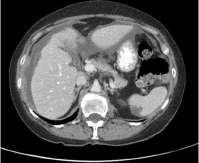
One of these tools is an imaging test called a CT scan with contrast. This is a special type of X-ray that gives a detailed picture of the inside of your abdomen. The CT scan essentially takes multiple pictures of your body from different angles, which are then combined to produce a cross-sectional view. This can help the doctor detect any abnormalities or changes in your body. In pseudomyxoma peritonei, a typical CT appearance includes a ‘scalloping’ or irregular outline of the liver and spleen surface, caused by small, enclosed accumulations of the jelly-like substance, mucin. These mucin pockets can help distinguish pseudomyxoma peritonei from other diseases.
In some cases, the doctor might not be able to identify the original appendiceal tumor using the CT scan, and they might need to use gadolinium-enhanced MRI instead. Compared to a CT scan, this type of MRI is more sensitive and better at locating small tumors, small bowel assessments, and checking the ligament that connects the liver and the duodenum (‘hepatoduodenal ligament’). If the disease appears to be more aggressive, your doctor might also use a PET/CT scan to check if the disease has spread beyond the abdomen.
Additionally, your doctor might test your blood for specific proteins, or ‘tumor markers,’ which can provide useful information about how your disease might progress and help monitor your condition after treatment. These markers, called CEA, CA 19.9 and CA 125, tend to be at high levels in people with pseudomyxoma peritonei of appendiceal origin. CA 125 may also be high if the ovaries are involved.
Last but not least, to obtain a conclusive diagnosis, the doctor may need to examine a sample of your tissues under the microscope – this is called a histopathological examination. This is especially necessary when the clinical signs and imaging tests don’t provide a clear answer. A small tissue sample can be obtained by performing minor procedures such as laparoscopy or laparotomy. A needle biopsy procedure guided by an imaging technique typically doesn’t provide much valuable information for this condition, as the punctured material may not contain cellular elements and can be simply acellular mucin.
Treatment Options for Pseudomyxoma Peritonei
In the past, periodic surgical debulking, a process of removing as much of the tumor as possible, was commonly used for treating Pseudomyxoma Peritonei (PMP), a rare form of cancer that begins in the appendix. However, this method is not preferred anymore due to the high probability of the cancer coming back and the increased risk of multiple surgeries.
Today, the favored treatment for PMP is a combination of two procedures: complete cytoreduction surgery (CRS) and hyperthermic intraperitoneal chemotherapy (HIPEC). The goal of CRS is to remove all visible tumors. The surgeons consider it a success if they don’t leave any tumors larger than about the size of a pea (2.5mm).
HIPEC is conducted to kill any remaining cancer cells. In this therapy, a warmed chemotherapy drug is inserted directly into the abdominal cavity. The drug most commonly used is called mitomycin C. The reason they warm the drug is to help it penetrate the remaining cancer cells better. After this treatment, a scan and certain blood tests for tumor markers are recommended to be done after 3 months and then every 6 months to check if the cancer is coming back. Additional surgery may be considered in some patients, based on their specific situation.
However, it’s important to note that CRS/HIPEC treatment is quite intensive. Therefore, doctors need to thoroughly assess a patient’s overall health before deciding this treatment. In fact, patients with a more aggressive cancer type or specific cellular features (signet ring cell components) that affect their daily living significantly may not survive for long after the surgery. Instead, these patients may be helped more by palliative care, which focuses on relieving their symptoms and improving their quality of life.
After CRS/HIPEC, additional chemotherapy may be beneficial for patients with aggressive cancer. However, for those with less aggressive cancer, this may not be the case. Despite this, there is currently no well-accepted standard plan. Furthermore, giving chemotherapy before the surgery generally doesn’t help.
What else can Pseudomyxoma Peritonei be?
When diagnosing certain conditions, doctors should also think about a few other possibilities that can cause similar symptoms. These might include:
- Ascites, a buildup of fluid in the abdomen that can be caused by cirrhosis (liver disease) or congestive heart failure (CHF)
- Ruptured cystadenomas (a type of tumor) in the appendix or the ovary, which are filled with a jelly-like substance
- Endometriosis, a condition where tissue similar to the lining of the uterus is found outside of the uterus, that has undergone certain changes
- A ruptured internal organ causing mucus to spill into the abdominal cavity
- Soft tissue tumors that have undergone certain changes
What to expect with Pseudomyxoma Peritonei
The outlook for patients with pseudomyxoma peritonei (a rare cancer that affects the abdomen) depends upon the classification of the tumor when examined under a microscope. A recent study found that patients with low-grade tumors survived for ten years at a rate of 63%. However, for patients with high-grade tumors, the ten-year survival rate dropped to 40.1% and for those with a specific high-grade tumor represented by ‘signet ring cells’, there were no survivors recorded. Please keep in mind, this data can vary from study to study.
The treatment procedure known as CRS/HIPEC (a combination of surgery and heated chemotherapy), tends to result in better long-term outcomes for patients than a procedure known as debulking surgery (surgical removal of as much of the tumor as possible).
Possible Complications When Diagnosed with Pseudomyxoma Peritonei
After undergoing the surgery known as CRS/HIPEC, patients may experience serious complications. These can include the formation of dangerous blood clots, leaks in the areas where the surgeon connected pieces of the digestive tract, tearing in the bowel, the development of a fistula (an abnormal connection between two parts of the body), abscesses, and the splitting open of the surgical wound. Other potential complications are a high risk for low white blood cell count which can weaken the immune system, serious infections that spread throughout the body (sepsis), a build-up of fluid between the lungs and chest wall (pleural effusion), and difficulty with breathing (respiratory insufficiency).
Possible Complications:
- Formation of blood clots
- Leaks at surgical connection sites of digestive tract
- Tearing of the bowel
- Development of a fistula
- Abscesses
- Splitting open of surgical wound
- High risk of low white blood cell count
- Serious widespread infections
- Fluid build-up between lungs and chest wall
- Difficulty with breathing
Preventing Pseudomyxoma Peritonei
If a patient is recently diagnosed with pseudomyxoma peritonei, a rare condition where a jelly-like substance accumulates in the abdominal cavity, their doctor may recommend a procedure known as CRS/HIPEC if their overall health permits it. The aim of this treatment is to remove as much of the disease as possible.
Many patients with low-grade (less aggressive) types of this condition experience a very positive long-term response to treatment. However, the disease has quite a high chance of coming back. Therefore, it’s really important for patients to maintain regular check-ups with their doctor. This lets the doctor monitor for any signs that the disease might be getting worse, and helps ensure the best possible care.


