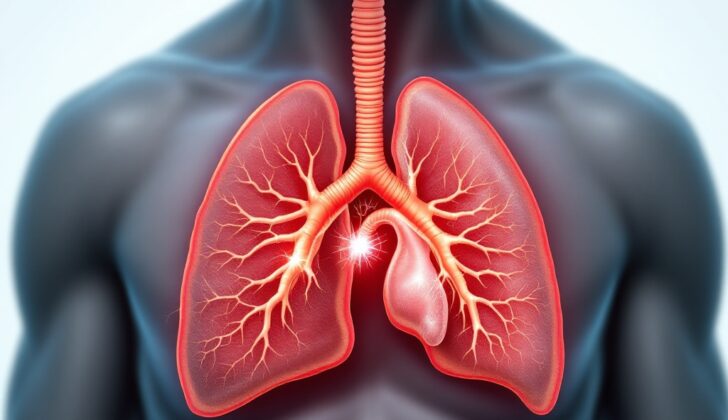
What is Diaphragm Eventration?
Diaphragmatic eventration (DE) occurs when part or all of the diaphragm muscle raises abnormally due to the loss of muscle or nerve function, while still remaining attached to its normal structures. This condition can occur from birth (congenital) or develop later (acquired), so it can affect both children and adults. The diaphragm is a dome-like muscle that is vital for breathing and serves as a boundary between your chest and stomach areas. This muscle gets its signals from the phrenic nerve, which originates from certain areas in your spine.
Every half of the diaphragm is controlled by the respective left and right phrenic nerves. If these nerves are not developed properly, or get injured, it can lead to diaphragm paralysis and reduced lung activity. In both cases of eventration that are present from birth or develop later, part of the diaphragm is weakened, thinner, and functions less. This can produce varying degrees of symptoms, from no symptoms at all to respiratory issues. The confirmation of this condition is done via imaging techniques. The treatment usually involves supporting the patient’s respiratory function and, in some instances, a type of surgery called plication.
What Causes Diaphragm Eventration?
Congenital causes, or those that are present from birth, are because of a defect in the development of the diaphragm muscle. This is due to an issue with the migration of certain cells, which results in them being replaced with a different kind of tissue. This makes part of the diaphragm thin and weak, leading to an upward shift of the affected side. Eventration, or when the diaphragm is positioned too high in the chest, can happen alongside other birth defects and infections. Some of the disorders include several syndromes, chromosomal defects, lung underdevelopment, spinal muscular atrophy, malrotation, and heart disease. In newborns, a type of disorder linked to the cells’ energy production has also been associated with diaphragmatic problems. Certain infections during fetal development, like rubella and cytomegalovirus, have also been linked to this condition.
Acquired cases, which develop after birth, are often caused by injuries that damage the nerve controlling the diaphragm and result in muscle wasting. This nerve injury could occur due to various reasons, like trauma from accidents or during birth, chest surgeries, or as a complication of other illnesses, including multiple sclerosis, Guillain-Barre syndrome, nerve compression, radiation therapy, and diseases affecting connective tissues. Any damage or paralysis of this nerve can increase muscle thinning and upward shift of the diaphragm.
Eventrations of the diaphragm can be complete, partial, or affect both sides of the diaphragm. If we categorize them based on their origin during embryo development, congenital eventration can be at the front, back and side, or middle of the diaphragm.
Risk Factors and Frequency for Diaphragm Eventration
Diaphragmatic eventration is a very rare condition, which means there’s not a lot of data on how often it occurs or how many people it affects. Some case reports suggest that it is extremely rare, perhaps affecting less than 0.05% of people. It appears to be more common in males and more likely to affect the left side of the diaphragm. Some other data suggest it could occur in as many as 1 in 10,000 live births. However, the true occurrence rate might be higher than these numbers suggest. This is because most people with the condition don’t have noticeable symptoms and thus, many cases likely go undiagnosed.
Signs and Symptoms of Diaphragm Eventration
Eventration of the diaphragm, a condition affecting the muscle that helps us breathe, can have different symptoms. Many people with this condition have no symptoms and learn they have it when a chest X-ray is done for other reasons. However, others may experience breathing, chest or stomach problems. In both children and adults, there might be breathing difficulties when active, shallow breathing, fast breathing, and chest pain. For adults, there may be frequent chest infections, difficulty swallowing, heartburn, and/or discomfort in the upper stomach. Infants might throw up, struggle to eat enough, and not gain weight as expected.
Different signs might be observed when a doctor examines a patient with eventration of the diaphragm, but these can vary widely and may not be obviously related to this condition. But, there are some common signs that doctors might look for in both adults and infants:
- Fast breathing
- Blue skin color if the patient is not getting enough oxygen (this is rare if only one side is affected)
- Struggling muscles in the chest
- Abnormal chest movement while inhaling
- Fewer sensations when touched and dull sound during tapping on the chest affected side
- Stomach pain when the doctor feels the area around the stomach button
- Sound of bowel movements in the chest
Some signs are more specific to infants:
- Signs of injury to the chest
- A sunken belly
- Severe upper stomach discomfort because of twisted stomach
- Signs of a nerve injury to the upper arm associated with birth trauma
For adults, the doctor might additionally find:
- Fast heart rate or irregular heartbeat associated with chest pain
- Stomach pain due to things that can drastically increase pressure in the abdomen, such as build-up of fluid in the abdomen, infection, fluid retention or pregnancy
Testing for Diaphragm Eventration
Diaphragmatic eventration (DE) – which means thinning or raising of the muscle that separates chest and abdomen – often goes unnoticed as the majority of patients do not experience symptoms. For many, the condition is discovered accidentally through chest imaging. However, patients who do show symptoms should have chest x-rays performed, a process which usually involves a detailed medical history and physical exam.
X-ray images – both from the back to front (posterior-anterior or PA) and the side (lateral or LAT) – help confirm the diagnosis. These images typically show a raised section in the affected part of the diaphragm along with unchanged heart and midpoint contours. In cases where the diagnosis is not clear or if the abnormalities are suspected inside the chest or abdomen, a computed tomography (CT scan) of the chest may be required. This can differentiate DE from a hernia given the normal attachment points of the diaphragm.
Further evaluation usually involves measuring lung volumes and diaphragm function. These tests include lung function tests, maximum inspiratory pressure (MIP), maximum expiratory pressure (MEP), fluoroscopic sniff testing, and ultrasound. Additional lab tests may be necessary if congenital diseases or infections are suspected.
The testing of lung function indicates a restrictive pattern with a reduced forced vital capacity (FVC) and forced expiratory volume in 1 second (FEV1). This restrictive pattern is more common when DE affects both sides that can improve with diaphragm surgery.
MIP and MEP tests measure the pressure generated during a deep breath or a strong exhale and reflect the strength of the involved respiratory muscles. In patients with DE, MIP usually lowers reflecting the diaphragm’s role in inhalation while MEP generally remains normal.
Fluoroscopic sniff testing is used to check diaphragm function. The patient is asked to take hard, quick breaths (sniffs) while each diaphragm half’s movement is observed under continuous fluoroscopy. In DE, abnormal movement may be seen in part of the diaphragm. If non-surgical measures fail, both DE and diaphragm paralysis are treated with surgery. In infants, who might find the sniff test difficult, ultrasound is preferred to evaluate diaphragm function due to its ability to avoid exposure to radiation.
Even before birth, DE can potentially be detected through high-resolution fetal ultrasound, CT, or MRI scans. However, it can be hard to distinguish from congenital diaphragmatic hernia (CDH) as the fetal stomach or liver is visualized in the same plane as the heart and the lung appears underdeveloped. After birth, DE is suspected with diaphragm elevation on chest x-rays when checking for breathing difficulties. Fluoroscopy, dynamic MRI, or ultrasound can confirm DE by evaluating diaphragm function. Depending on the patient’s history and physical findings, further examination may be needed to figure out the underlying cause like infections, cancer, and developmental or degenerative diseases.
Treatment Options for Diaphragm Eventration
The treatment approach for diaphragm eventration, or a weak and elevated hemidiaphragm, usually depends on the cause and severity of the condition. If symptoms are mild or aren’t present, the main treatment recommendation is to provide supportive care.
For people who are not getting enough oxygen due to this condition, oxygen therapy can be given to help maintain an appropriate level of oxygen in the bloodstream. If oxygen supplementation through the use of a nasal cannula isn’t successful, continuous positive airway pressure (CPAP) may be used instead.
Infants with this condition often experience symptoms related to digestion and growth, so getting proper nutrition is a key focus. Infants who are in severe respiratory distress might need to be fed through a tube, or parenterally. This involves intravenously providing them with essential nutrients and fluids. Physical therapy and lung rehabilitation exercises are other approaches that can be part of conservative treatment.
In more severe cases, when mechanical ventilation is required or when other attempts at treatment are unsuccessful, surgical intervention could be needed. Plication, which involves stitching pleats into the weak part of the diaphragm and anchoring it down, is a well-established surgical treatment. This procedure helps increase the space inside the chest and allows for better lung expansion. It is important to note, however, that plication surgery doesn’t improve the function of the weakened part of the diaphragm.
The surgery can be performed through different methods like open thoracotomy, video-assisted thoracoscopic surgery, laparoscopic surgery, or robot-assisted surgery. After surgery, a small tube is inserted to drain fluid from the chest for 1-2 days until the output becomes less than 200 mL per day.
Surgical intervention is usually considered when:
– Respiratory distress doesn’t improve with conservative treatment
– Difficulty breathing that is not caused by other conditions like heart failure or lung diseases
– Infants who are not getting enough nutrition or are not gaining weight properly
– Recurrent or life-threatening pneumonia
– Patients can’t be taken off mechanical ventilation
Following the operation, patients are monitored, and they return for follow-ups based on their symptoms or potential complications. The follow-ups may include chest X-rays and lung function tests. To assess the patient’s quality of life, level of activity, and the impact on their social and mental well-being, the St. George’s Respiratory Questionnaire might be used.
What else can Diaphragm Eventration be?
The following conditions can often be mistaken for one another due to their similar symptoms:
- Paralysis of the diaphragm (an entire half of the diaphragm might be affected, making it difficult to distinguish from a condition called “eventration,” which may impact either a smaller part or the entirety of the diaphragm)
- Damage to the phrenic nerve, which controls the diaphragm
- Diaphragmatic hernia (a condition where the stomach or other abdominal organs push through a hole in the diaphragm)
- Lung consolidation (a condition where a part of the lung is filled with liquid instead of air)
- Accumulation of fluid under the lung (subpulmonic pleural effusion)
- Pleural mass (a growth in the pleura, which is the thin tissue that lines the lungs and the chest cavity)
- Damage due to pulling or stretching (traction injury)
- Injury caused by medical procedures in the chest area
- Ascites (a condition where fluid builds up in the abdomen)
- Enlargement of the liver (hepatic enlargement) or spleen (splenic enlargement)
What to expect with Diaphragm Eventration
The outlook for patients with eventration, a condition where the diaphragm is abnormally stretched, is generally positive and often depends on the severity and cause. Many patients show no symptoms and may not need treatment. However, those experiencing intense eventration can have serious breathing troubles, necessitating the use of a machine to assist in breathing.
In these cases, a surgical procedure known as diaphragm plication has significantly improved symptoms and patient’s quality of life. This surgery has also been demonstrated to improve two key measures of lung function, FEV1 and FVC, by up to 25% and 30% respectively as shown by post-surgery lung function tests.
Possible Complications When Diagnosed with Diaphragm Eventration
Some possible issues that can occur due to certain medical conditions or treatments include:
- Acute or chronic failure of the respiratory system
- Pneumonitis, or inflammation of the lungs
- Nutritional deficiency, or not getting enough essential nutrients
- Failure to thrive, or not growing or developing as expected
- Cardiac arrhythmias, or irregular heartbeats, due to rapid changes in the space inside the chest
- Complications that may arise from surgical correction, such as:
- Pneumonia, or a lung infection
- Deep venous thrombosis, or a blood clot in a deep vein
- Pleural effusions, or excess fluid around the lungs
- Cardiac events, or severe heart-related issues
Recovery from Diaphragm Eventration
After surgery, especially for those who need assistance with breathing, care in the intensive care unit is crucial. The process of weaning them off the assisted breathing is critical. This is because the surgery could cause changes in the compliance of the chest and abdomen, and there could also be a change due to the adjustment of contents in the abdomen.
Keeping a check on the pressure inside the abdomen is also an important part of post-surgery care. To help prevent lung problems, patients are encouraged to use an incentive spirometer and maintain good lung hygiene.
Adequate pain management during recovery is important too. When pain makes coughing difficult, it can lead to the build-up of secretions in the lungs and a condition called atelectasis where the lungs cannot expand fully.
Preventing Diaphragm Eventration
Diaphragm eventration, or the abnormal elevation of the diaphragm, is often discovered by chance and most people don’t experience any symptoms or require extensive treatment. It’s important for patients and parents of children who have been diagnosed with diaphragm eventration to be educated about the symptoms and know when to seek medical help. Pulmonary rehabilitation, a program that teaches about diaphragm eventration and includes breathing exercises, can also be beneficial.


