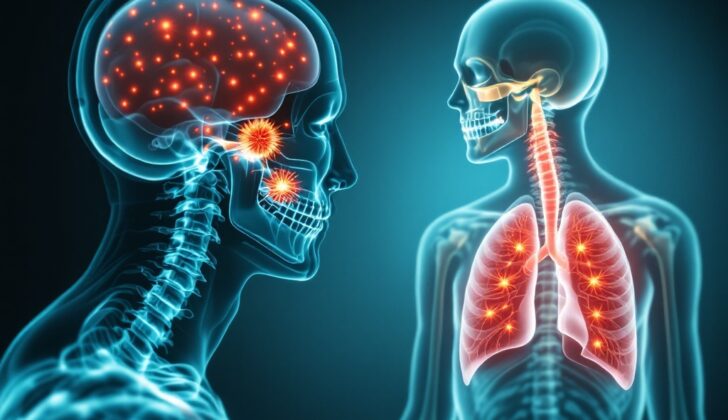
What is Eosinophilic Granuloma (Langerhans Cell Histiocytosis)?
Eosinophilic granuloma (EG) is the mildest type of a disease called Langerhans cell histiocytosis. EG was first identified in 1940 and was classified under the umbrella of a disorder known as histiocytosis X, along with two other diseases: Hand-Schuller-Christian disease and Letterer-Siwe disease. Histiocytosis X was named as such because it involved an abnormal growth of certain cells (called Langerhans cells) of unknown cause. These conditions are now known as Langerhans cell histiocytosis (LCH).
EG is like a benign tumor (a tumor that won’t spread) caused by an abnormal growth of Langerhans cells which come from a type of cell found in the bone marrow, not from the skin. EG is the most common type of LCH.
EG mostly affects the body’s central framework of bones, including the skull, jaw bone, spine, pelvis, ribs, and long bones. Lesions or abnormal areas are generally found in the main part of long bones, and can often involve the tissues surrounding the bones. The affected bone sites can differ between children and adults. In kids, the skull, especially the forehead bone, is most commonly affected, whereas in adults it’s the jaw. The upper part of the spine is typically affected in kids, while the neck part of the spine is more commonly affected in adults. EG can also occasionally impact the skin, important glands in the brain, lungs, liver, spleen, and parts of the digestive system.
EG makes up less than 1% of all bone tumors and can present in two ways: as a single lesion, which rarely needs treatment, or as multiple lesions across the body, requiring aggressive treatment.
EG is classified into three types:
1. Eosinophilic Granuloma:
– Single bone lesion (Monostotic): This is more common and found in nearly 90% of patients.
– Multiple bone lesions (Polyostotic): This is less common and found in roughly 10% of patients.
2. Hand-Schuller-Christian Disease: Identified by a significant three symptoms: bulging eyes, a particular form of diabetes, and breakdown of skull bone.
3. Letterer-Siwe Disease: Marked by enlarged lymph nodes, skin rash, swelling of the liver and spleen, and a decrease in all types of blood cells.
What Causes Eosinophilic Granuloma (Langerhans Cell Histiocytosis)?
There’s a continuing discussion about whether eosinophilic granuloma (EG), a condition marked by the unusual increase of a type of cell called Langerhans cells, is a response to an issue in the body or due to uncontrolled cell growth. These Langerhans cells originate from particular cells and are usually found in the bone marrow. They have a unique ability to move into the body’s tissues and display antigens – substances that cause an immune response – to T lymphocytes, which are a type of white blood cell.
The increase in the number of Langerhans cells could be triggered by viral infections like Epstein-Barr virus and Human Herpes virus-6, bacteria, or dysfunction in the immune system that results in a rise in substances known as cytokines, specifically interleukin-1 and interleukin-10. An interesting fact is that EG in the lungs is strongly associated with the habit of cigarette smoking. As per the revised classification by the Histiocyte Society in 2016, Langerhans cell histiocytosis is now considered as an inflammatory condition resulting from an issue in certain white blood cells.
The gathering of Langerhans cells in the lungs is believed to occur in response to exposure to cigarette smoke, which is supported by initial testing results that highlight changes around the bronchial tubes. It’s suspected that the stimulation from one or more components in cigarette smoke might be the cause for this condition.
Risk Factors and Frequency for Eosinophilic Granuloma (Langerhans Cell Histiocytosis)
Eosinophilic granuloma (EG) is a rare disease that is more common in kids, adolescents, and young adults, especially those of northern European descent and Hispanics. Most reported cases are found in individuals aged 5 to 10. An interesting fact is that males are more likely to get it than females. However, it’s still not clear if this disease can be inherited. As far as EG affecting the lungs, it’s even more uncommon, and we don’t have a clear number of how many people have it. Thanks to a study done in Japan, we can guess that for every 100,000 people, about 0.27 males and 0.07 females will have lung EG.
- Eosinophilic granuloma is a rare disease.
- It generally affects children, teenagers, and young adults, most commonly those aged 5 to 10.
- Every year, there are 4 to 5 cases per million kids and 1 to 2 cases per million adults.
- Males are slightly more likely to get it than females, with a ratio of 1.2:1.
- We still aren’t sure if it can be passed down through families.
- In the case of lung eosinophilic granuloma, it’s even rarer.
- A study in Japan suggests about 0.27 males and 0.07 females per 100,000 people might have it.
Signs and Symptoms of Eosinophilic Granuloma (Langerhans Cell Histiocytosis)
Eosinophilic granuloma, or EG, is a complex disease that can affect a person in many different ways. EG can occur in one or many bones and can affect various other body parts such as the skin, pituitary gland, liver, spleen, and lungs. Symptoms vary based on what part of the body is affected. They can include:
- Swelling and pain in the area of the affected bone
- Unexpected bone fractures
- Back pain, stiffness, and a hunched posture, associated with spinal bone involvement
- Symptoms like an ear infection, including swellings near the ear, ear discharge, and hearing loss – these could be associated with involvement of the temporal bone
- Jaw pain, loose teeth, and a mass in the jaw area, associated with involvement of jaw bones
- Headaches, excessive thirst, and frequent urination, associated with skull involvement
- Coughing, weight loss, shortness of breath, chest pain, fatigue, and a sudden lung collapse, associated with lung involvement. Note that these lung symptoms might not always be present.
Physical examination may present several findings:
- Patients may present with tenderness in the affected area and restricted movement, especially in the spine if it is the affected area
- Patients could be limping if the pelvis or the long bones of the lower extremities are involved
- Hearing loss, both from sound conduction and nerve-related issues, may be noticed
- If the spine is extensively affected, neurological examinations may reveal associated deficits
- With multi-organ involvement, the liver and spleen may be enlarged and lymph nodes swollen, as detected by an abdominal exam
- If the lungs are affected, abnormalities may be detected during a chest examination using a stethoscope
Testing for Eosinophilic Granuloma (Langerhans Cell Histiocytosis)
Eosinophilic granuloma (EG) is a condition that requires a variety of tests for a proper diagnosis. This could involve lab tests, imaging such as x-rays and finally, a tissue sample or biopsy for further detailed examination.
Starting with lab tests, the first one undertaken is often a complete blood count. This test is usually normal in people with EG, or it might show a small increase in white blood cells. It’s important to run this test to rule out other diseases that might appear similar to EG, such as infections in the bone (osteomyelitis) or cancer. If the patient has unexplained low blood cell counts, a bone marrow biopsy might be needed. Another test, Erythrocyte Sedimentation Rate (ESR), may show increased rates in people with EG. However, this is not always the case.
Next, x-rays of the bone affected by the disease may reveal a hole or a ‘lytic lesion’. The bone surrounding the hole might have an abnormal reaction, it can also be thinned, expanded, or destroyed. If the disease is in the skull, x-rays will show a similar lytic lesion. Both outer and inner table involvement gives the beveled-edge appearance. X-rays of the spine can show other lytic lesions, bone collapse or increased curvature of the spine. Additionally, a full body x-ray can be done to check for health issues in other regions together with a chest x-ray to look for problems in the lungs.
If the disease has affected the lungs, a chest x-ray may reveal ill-defined round shapes and infiltration of tiny nodules and threads. Over time, cyst-like structures might show up. It is common for the upper part of the lungs to be predominantly affected, with the angles near the ribs typically uninvolved. CT scanners and techniques like magnetic resonance imaging or positron emission tomography may be necessary if the disease has affected the skull, jawbone, or spine. Some structures might be difficult to see with a standard x-ray.
Finally, a biopsy might be needed to confirm the diagnosis of EG. This is done by obtaining a sample of the suspected bone tissue via a needle or guided by a CT scan. Stains for CD1a and CD207 are then applied to this sample to check for EG. An additional test (BRAF V600E mutation) can be beneficial in challenging cases or in scenarios where the disease has affected the central nervous system.
Extra tests, like a lung function test, may be recommended if lung involvement is suspected. A procedure called bronchoalveolar lavage can be diagnostic in pulmonary EG – it involves washing out a small portion of lungs and examining the fluid under a microscope. An increase of 5% or more in a certain type of white blood cells called Langerhans cells in the fluid is indicative of EG. If the lesions involve the skull and orbit or temporal bone, further neurological, visual and auditory tests might be needed.
Treatment Options for Eosinophilic Granuloma (Langerhans Cell Histiocytosis)
The treatment for this condition can be split into two categories: non-operative, which doesn’t involve surgery, and operative, which does involve surgery.
Non-operative treatments include:
- Observation: Single lesions often get better on their own. So, people with these lesions, particularly younger patients and patients without any symptoms, are closely monitored over time to see if the lesions disappear by themselves.
- Immobilization: If the lesion is in the spine and causing pain but not major neurological issues, patients may need to be immobilized. Bracing can be used for patients with curvature of the spine (kyphosis).
- Low Dose Irradiation: This involves exposure to a controlled amount of radiation energy. It’s used for lesions that continue to cause problems or come back even after other conservative treatments. It’s also given to patients with mild neurological issues.
- Injections and Chemotherapy: Injections of a drug called methylprednisolone are used for lesions of the spine and extremities that are causing symptoms. This drug, as well as chemotherapy, may be offered if the condition has spread to the lungs (pulmonary eosinophilic granuloma) and is causing significant symptoms. In more serious cases, chemotherapy (with drugs like vinblastine, prednisone or cytarabine) may be used for a period of 12 months. It’s also important for patients who smoke to quit, as this can help stabilize lung conditions, slow their progression, and prevent lung cancer.
Operative treatments include:
- Curettage and Bone Grafting: Single lesions on the skull are treated by removing tissue from the lesion area (curettage), and then filling the area with new bone or bone substitute (bone grafting). Lesions at risk of causing a bone to break need similar treatment.
- Surgical Fixation: Surgery is performed to stabilize and reduce pain in parts of the body with severe pain, limited movement, worsening neurological problems, or an unstable spine.
- Chest tube insertion and Pleurodesis: In cases where the condition causes a lung to collapse (pneumothorax), a tube may be inserted into the chest to remove air and allow the lung to expand. Medical substances are then instilled inside the pleural space (pleurodesis) to prevent a recurrence of lung collapse. If these problems persist, further surgical procedures (pleurectomy) may be necessary.
- Lung Transplantation: In very severe cases, replacing the diseased lung with a healthy one from a donor may be the most effective treatment.
What else can Eosinophilic Granuloma (Langerhans Cell Histiocytosis) be?
When a doctor is trying to diagnose eosinophilic granuloma (EG) that’s affecting the spine, they may consider a list of other conditions that could cause similar symptoms. These conditions include:
- Neurofibroma
- Schwannoma
- Leukemia
- Lymphomas
- Multiple myeloma
- Osteomyelitis
- Tuberculosis
- Plasmacytoma
- Metastasis
These conditions might show up similarly on a scan, but a lack of additional symptoms and minimal lab results can point towards EG. To be sure, a biopsy of the suspicious area would be needed.
When EG is suspected in the skull, there are different diseases to consider such as:
- Otitis media
- Mastoiditis
- Seborrheic dermatitis
- Periorbital cellulitis
If EG is thought to be in the long bones of the arms or legs, diagnosis would involve ruling out conditions like:
- Ewing’s sarcoma
- Osteochondroma
- Osteoblastoma
- Osteosarcoma
- Paget’s disease
Lastly, in cases where EG is suspected in the lungs, a doctor will want to rule out diseases such as:
- Tuberous sclerosis
- Lymphangioleiomyomatosis
- Pulmonary histiocytic sarcoma
- Cystic fibrosis
- Emphysema
- Hypersensitivity pneumonitis
- Sarcoidosis
To differentiate EG from these conditions, a biopsy and detailed microscopic analysis are usually done, including a special staining for CD1a.
What to expect with Eosinophilic Granuloma (Langerhans Cell Histiocytosis)
Patients with a single bone lesion often get better without treatment and usually have a good outcome. However, patients at high risk, such as those with multiple system involvement, adults or patients with more than one bone affected, and particularly if the bones at the base of the skull (the sphenoid, ethmoid, orbital, and temporal bones) are involved, run a greater risk of disease coming back, complications and generally poor outcome. Surgery generally provides faster relief from pain compared to non-surgical treatment. Sadly, more than 10% of patients pass away from this disease, including some cases that reactivate and continue causing health problems over time.
The outcome for patients with lung condition known as eosinophilic granuloma (EG), varies and is closely tied to whether the patient quits smoking or not. Those who continue to smoke tend to see the disease get worse, while those who quit see their disease stabilize or even get better. Some factors worsen the prognosis, including extreme age, large cysts, a certain pattern seen on X-rays known as honeycombing, involvement of multiple organs, and long-term use of steroids.
Possible Complications When Diagnosed with Eosinophilic Granuloma (Langerhans Cell Histiocytosis)
Eosinophilic granuloma (EG) can lead to a variety of complications. These issues can range from physical difficulties to mental health problems, and could also increase the risk of certain diseases:
- Limited movements or disability due to musculoskeletal problems
- Disruption in normal growth
- Broken bones from weakened or diseased bone
- Problems with hearing and sight
- Mental health issues such as depression and anxiety
- The development of tumors and inflammation of the spinal cord following radiation therapy
- Bone infection linked to the use of a specific steroid injection (methylprednisolone)
- Collapsed lung that occurs spontaneously
- Increased risk of specific types of cancer and blood disorders, namely Hodgkin and non-Hodgkin lymphoma, myeloproliferative disorders, and bronchogenic carcinoma
- High blood pressure in the arteries of the lungs and related heart complications
Preventing Eosinophilic Granuloma (Langerhans Cell Histiocytosis)
Eosinophilic granuloma (EG) is a non-dangerous condition that affects the bones. Sometimes, if a singular symptom-free lesion, or abnormal area, appears, it might disappear on its own and doesn’t need treatment. However, it’s important to know that there is a chance of more bone lesions developing within a period of six months to two years. This makes regular check-ups and complete full-body bone scans necessary.
If a child has multiple bone lesions, it’s crucial to check if the disease has spread to other parts of the body. Furthermore, it’s worth noting that relatives of a patient with EG may have a higher chance of thyroid disease, a condition that affects the small gland at the base of the neck responsible for regulating the body’s metabolism.


