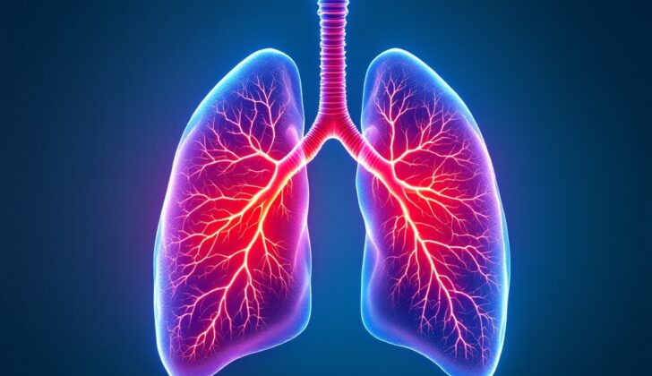
What is Hamartoma?
A hamartoma is a non-cancerous lump made up of an abnormal mix of cells and tissues found in a specific area of your body. Even though most hamartomas aren’t harmful, there’s still a small chance they could turn into cancer. Hamartomas can be found anywhere in the body, but they’re most often discovered in the lungs, hypothalamus, breasts, and colon.
Most of the time, hamartomas don’t cause any symptoms and are found unexpectedly when doctors are investigating other health issues. However, they can still cause health problems. These can include infections, tissue death due to lack of blood supply, pressure or blockage on other structures, bleeding or anemia, fractures, and transformation into cancer.
What Causes Hamartoma?
Hamartomas are abnormal growths that occur because of an malfunction during body tissue formation. Their growth rate matches that of the original tissue they come from. Hamartomas can arise for no apparent reason, but sometimes, they are part of certain syndromes. These growths can appear anywhere in the body.
Certain genes, including SMAD4, PTEN, STK11, and BMPR1A, play a part in the formation of hamartomas.
There are various inherited conditions that can result in hamartomas. Some of these syndromes include:
* Tuberous Sclerosis
* Cowden syndrome
* PTEN hamartoma tumor syndrome
* Peutz-Jeghers syndrome, and others.
Risk Factors and Frequency for Hamartoma
Hamartomas, a type of benign growth, are generally found more often in males than females. There’s no evidence that one race is more likely to develop them than another. The prevalence of most types of hamartomas isn’t well-known, except for those found in the lungs. Pulmonary hamartomas make up about 0.25% of all cases and represent 8% of all lung tumors. They’re most often discovered by accident rather than through symptoms. People aged between 40 and 70 are the most likely to be diagnosed with them. Meanwhile, benign breast lesions are associated with age, hormone levels, obesity, and family history, while an increased age and body mass index (BMI) can raise the risk of developing colonic polyps.
- Hamartomas are more commonly found in males than females.
- There’s no evidence that any particular race is more likely to develop hamartomas.
- The prevalence of most hamartomas is unknown except for pulmonary hamartomas, which make up about 0.25% of all cases.
- Pulmonary hamartomas comprise 8% of all lung tumors and are often discovered incidentally.
- The common patient age range for hamartomas is 40 to 70.
- Benign breast lesions are linked to age, hormonal factors, obesity, and family history.
- Increased age and BMI increase the risk of colonic polyps.
Signs and Symptoms of Hamartoma
Hamartoma is a type of benign tumor, and its diagnosis requires a detailed understanding of a patient’s history, including their family and medical background. While hamartomas often don’t cause symptoms, understanding a patient’s history can help predict the course of the condition. It’s also necessary to know about any related health conditions the patient has. There are several common symptoms and physical signs that can occur depending on the hamartoma’s location:
- Hypothalamus: Seizures, mental state changes, vision changes, early onset puberty, behavioral changes
- Lung: Chronic cough, coughing up blood, fever, abnormal breath sounds, symptoms of blocked airways
- Heart: Chest pain, heart palpitations, swelling, difficulty breathing, bluish skin color, cold and sweaty skin, heart murmur, irregular heartbeat
- PTEN/Cowden Syndrome: These syndromes affect multiple tissues, such as the breast, thyroid, gastrointestinal, genitourinary, and the skin and lining of body cavities. Symptoms depend on which tissues are affected.
Less common symptoms related to kidneys, spleen, and other organs can also occur. These could include: pain in the side or stomach, frequent infections, fever, night sweats, a noticeable lump in the abdomen, enlarged testicles, or enlarged breasts.
Testing for Hamartoma
When it comes to testing for hamartomas, common laboratory tests used include a complete blood count, tests for liver function, and levels of various minerals like calcium, phosphate, potassium, and urea. A CD8 test is also commonly conducted.
A chest X-ray can help to detect lung hamartomas. Typically, these show up as clearly defined lung nodules and a type of calcification that looks like popcorn. However, this method isn’t helpful in detecting other types of hamartomas.
A CT scan is the preferred test for diagnosing a hamartoma. Here, the hamartoma shows up as localized patches of fat alternated with spots of calcification.
MRI scans are another important diagnostic tool. On these scans, a hamartoma presents as a varied signal in the T1 sequence and a high signal due to its fat and cartilage components in the T2 sequence. MRI is particularly useful for hamartomas in the hypothalamus and in certain organs in the abdomen like the kidney, spleen, and pancreas.
In addition, an ultrasound might be useful in diagnosing hamartomas in the spleen. Furthermore, bronchoscopy – a technique to view the airways of the lungs – can be beneficial in diagnosing hamartomas inside the bronchial tubes.
Treatment Options for Hamartoma
Hamartoma is a benign tumour-like growth that is often found unintentionally, as most cases don’t show any symptoms. If there are any symptoms like pain, people are typically given pain relief medication. If a patient doesn’t respond to the treatment, then additional tests are considered. In such cases or for cosmetic reasons, surgery is usually the preferred treatment option.
In the case of a hamartoma found in the lung, it’s often discovered by chance as a nodule, which is a small growth that can be seen in an imaging test like an x-ray. The way it’s handled depends on its features and the chance it could be cancerous. A growth below 30mm is less likely to be cancerous, but if it’s larger, there’s a higher risk. For lesions that carry an intermediate risk of being cancerous, or with patients who can’t have surgery, a type of biopsy can be performed with a bronchoscope, which is a thin tube passed down the throat into the lunges. However, the best way to diagnose a pulmonary nodule is by surgically removing the nodule for testing. This surgical removal can also help cure some types of cancerous growths. Scans like CT or PET scans can help doctors choose the best place to take a biopsy from or to check the extent of disease.
Hamartomatous polyps, benign growths that typically don’t cause symptoms and are found in the digestive tract, usually don’t require treatment. But there are certain cases where these polyps can grow bigger and become symptomatic or even transform into cancer, especially in patients with Peutz-Jeghers syndrome. In such cases, these polyps need to be removed surgically. In more complicated situations, additional treatments like nutritional support, steroids, immunosuppressants, stomach acid reducers and antibiotics might be used.
Breast hamartomas are generally also asymptomatic and are often found during routine mammograms, or they might appear as painless, contained lumps in the breast. These growths are typically non-cancerous, and simple observation or monitoring is usually all that’s needed. If the hamartoma cause symptoms or are suspicious, surgical removal is recommended.
In a condition known as Cowden Syndrome, which affects multiple organs and can cause complications, a team approach is usually needed. In this disease, benign and symptom-free lesions are managed conservatively, while symptomatic or suspicious growths are surgically removed. Other suggested interventions for these patients include regular cancer screenings and genetic testing.
What else can Hamartoma be?
Here are some conditions that can sometimes be mistaken for a health issue called a hamartoma because their symptoms are often similar:
- Hypothalamic-chiasmatic glioma
- Craniopharyngioma: This is a type of tumor just above an area called the sella turcica, coming from something called Rathke’s pouch. It mainly occurs in 5-14 year olds, or people aged between 50 and 75. It can lead to vision problems because it puts pressure on the optic pathway.
- Rathke’s Cleft Cyst: This condition usually doesn’t cause any symptoms, but when it does, they might include headaches, vision problems, and hormone imbalances.
- Pituitary macroadenoma: Symptoms may include nausea, headaches, vomiting, abnormal growth and vision problems.
- Myelolipoma: This condition usually doesn’t cause any symptoms and is often found as a lung nodule in check-ups.
- Pulmonary Chondroma: This type of lung tumor is often diagnosed in teenagers or young adults and is seen in the chest CT scan as a small, rounded area containing fat. It is usually a part of a condition called Carney’s triad.
- Other conditions that can present with similar signs include benign fat-based tumors (lipomas), cancer that has spread (metastasis), muscle-based tumors (rhabdomyomas), fibromas, paragangliomas, blood-vessel based growths in the spleen (splenic hemangiomas), fat-based tumors in the back of the abdomen (retroperitoneal liposarcomas), and tumors in the adrenal gland combined with bone marrow and fat tissue (adrenal myelolipomas).
Surgical Treatment of Hamartoma
Here is a simplified explanation of various treatment options for different anatomic locations:
Lung Hamartoma
* A surgery known as a ‘wedge resection’ is usually the best treatment for patients with a lung hamartoma.
* In some situations, more radical surgeries like removing an entire lobe of the lung or the entire lung may be considered.
* In any case, a small sample is taken from the tumor during the operation and is immediately studied under a microscope, to confirm whether it’s cancerous or not.
Hypothalamic Hamartoma – affecting the hypothalamus in the brain
* A process called MRI-guided stereotactic laser ablation is often quite effective – this uses MRI imaging to guide a laser for accurate removal of the hamartoma.
* Other options include making a small incision in the sphenoid sinus right behind the nose and advancing instruments to the skull base (transsphenoidal microsurgery), using radiation therapy (gamma knife radiosurgery), or using heat to remove the growth (thermocoagulation).
* There are also other surgical methods available, with varying results.
Breast Hamartoma
* To both verify the diagnosis and treat a breast hamartoma, it is often removed through surgery.
* Even though a breast hamartoma typically has the same risk of becoming cancerous as normal breast tissue, sometimes a portion or all of the breast is removed if the lump is large or if the appearance of the breast and patient’s mental outlook warrants it.
What to expect with Hamartoma
Hamartomas are typically harmless, but in some instances, they can become cancerous. For example, people with Cowden syndrome face a higher risk of breast, thyroid, and womb cancer. The outcome for someone with a hamartoma often depends on where the growth is located in their body, how big it is, and if they have any other health conditions.
Larger growths in the kidneys, hypothalamus (part of the brain), or spleen can pose more serious health problems.
Possible Complications When Diagnosed with Hamartoma
Hamartomas, which are usually harmless and symptom-free, can sometimes become unusually large. These large growths can result in internal disfigurement and place pressure on nearby organs, potentially causing life-threatening problems. Depending on where these growths are located, they can result in a variety of health issues including:
- Seizures
- Heart failure
- Breathing difficulties
- Breast changes
- Abdominal pain
Preventing Hamartoma
There are a number of non-profit groups that dedicate themselves to educating and supporting patients with hamartoma and their healthcare providers. These groups also promote research to improve early detection, develop better treatments, enhance quality of life, and find a cure. Health awareness campaigns, discussions, and public education programs across different platforms can significantly help in preventing the condition from progressing or getting worse.


