What is Innominate Artery Injury?
Innominate artery injury, also known as brachiocephalic artery injury, is a rare yet highly dangerous condition. Most injuries of this kind result from blunt, rather than sharp or piercing impacts. The injury can also be iatrogenic, meaning it is unexpectedly caused by a medical treatment or procedure.
If the innominate artery is injured, it needs to be diagnosed quickly, and a skilled surgeon is required to repair the injury, either through a traditional surgery or an endovascular repair which is less invasive. When this artery is injured, especially in the chest area, the surgeon must move quickly to repair it because incorrect choices or procedures could result in the patient’s death.
The primary objective is to control any immediate bleeding to prevent issues like insufficient blood supply to distant organs, the development of pseudoaneurysms (a false aneurysm, or a leakage of blood between the layers of an artery wall), or the rupture of the artery itself.
Over time, advancements in technology have helped us greatly enhance the imaging and techniques used to diagnose and manage innominate artery injuries. The surge in diagnostic tools now allow patients to undergo traditional, endovascular or even non-operative management for traumatic injuries to the innominate artery.
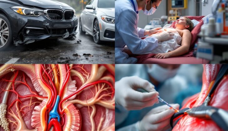
The innominate artery sprouts from the early section of the aortic arch (the top part of the main artery carrying blood from the heart) and splits into the right common carotid and subclavian arteries. On the other hand, the left common carotid artery and the left subclavian artery branch off from the later portion of the aortic arch.
However, in some people the configuration of the aortic arch might be different. The most common unusual configuration is known as a bovine aortic arch. These types of configurations must also be considered during the diagnosis and treatment process. The different arch types of bovine aortic arch exist in 9% and between 13% and 20% of the population, respectively. Other less common abnormalities include variations in the left vertebral artery, the right or left subclavian artery, or the right aortic arch.
What Causes Innominate Artery Injury?
Injury to the innominate artery, an important blood vessel in the body, is typically due to severe physical trauma. This trauma can, most often, be caused by a direct hit or sharp object like a bullet or knife.
Sometimes, this injury can be an unforeseen consequence (known as an “iatrogenic” injury) of a medical procedure or treatment. One of the most common ways this injury happens is in motor vehicle accidents, especially head-on collisions which occur at high speeds or involve sudden changes in speed.
Some signs that this injury might have occurred during a car crash include distinct marks from a seat belt or the imprint of a steering wheel on the body. The part of the innominate artery that’s most often injured is where it attaches to the aorta, another major artery.
Interestingly, studies have shown that pedestrians hit by cars have the highest rate of injuries to major blood vessels. Due to the extreme nature of this injury, patients who arrive at the hospital with it are often in a critical condition.
For injuries caused by sharp objects, like gunshots or stabbings to the chest, the innominate artery is often affected. Medical procedures like inserting a central venous catheter (a large, thin tube put into a large vein) or creating a tracheostomy (an opening in the neck for breathing) can also accidentally injure it. In rare cases, a tracheostomy can lead to a dangerous condition called the “tracheoinnominate artery fistula” where a hole forms between the trachea and the innominate artery.
Risk Factors and Frequency for Innominate Artery Injury
Injuries to the innominate artery, a major blood vessel in the chest, are not very common and can be difficult to evaluate. These injuries are usually a result of blunt or penetrating trauma. Annually, between 7,500 to 8,000 cases of blunt chest injuries occur, and the fatality rate is above 80%. From this data, we know that 4% of patients lose their life on the way to the hospital and 19% pass away during the initial medical examination. Notably, injury to the innominate artery is the second most common type of chest blood vessel injury due to blunt chest trauma.
- The collapse of the inner layers of the artery and the development of a fake aneurysm is a typical finding in this condition.
- In-hospital death rate for patients under non-surgical management reached 46% based on a 2011 study. Yet, for those treated via endovascular methods and open artery repair, the death rates were 9% and 19% respectively.
- Patients with penetrating injuries tend to be more critical than those with blunt injuries as they need larger amounts of fluid and blood products.
Innominate artery injuries can also occur during medical procedures, but these instances are rare. Fistula, an abnormal connection between the trachea and innominate artery, happens in less than 0.7% of patients who’ve had a tracheostomy and usually appears two to three weeks after the procedure in 72% of cases. Innominate artery injury due to the insertion of a central line happens in less than 1% of cases.
Regardless of the injury type, whether blunt or penetrating, patients with multiple injuries have a higher risk of dying.
Signs and Symptoms of Innominate Artery Injury
When patients come to the hospital with serious chest wounds, they should immediately receive advanced trauma care. This entails a complete medical examination, imaging tests, and stabilization before focusing on individual injuries. A detailed history of the incident causing injury should be taken, along with a physical check-up for possible aortic injuries. Symptoms can include chest pain, difficulty breathing, problems with swallowing, a raspy voice, or noisy breathing.
A physical exam may reveal active bleeding from the chest wound or strong signs of vascular damage. This could be a growing lump caused by internal bleeding, loss of pulse in the affected limb, symptoms of reduced blood flow to distant tissues, or a vibrating sensation in the injured area. Some patients with blunt chest trauma might not show any signs of injury initially, or could display signs such as skin discoloration, voice changes, abnormal heart sounds, difficulty breathing, paralysis in the lower body, or uneven blood pressure in the arms. Patients’ vital signs may show instability and signs of shock due to blood loss, while others may have normal vital signs. The most common injuries are broken ribs (46%) and collapsed lungs (36%).
Testing for Innominate Artery Injury
If a patient’s heart rate and blood pressure are not stable, they need immediate surgery regardless of whether their injury comes from a blunt or sharp impact.
However, if a patient’s body is stable, doctors will take an x-ray while the patient lies flat on their back. If the x-ray finds anything unusual like a heart or breathing tube shift to the right side, the patient may have to undergo a computed tomography (CT) scan of their chest. This is a more detailed type of imaging that uses x-rays and computers to produce images. Also, doctors must check for potential injuries to the brain and other body parts when the injury comes from a sharp impact to the chest. CT scans are usually very reliable at detecting these types of injuries.
Transesophageal echocardiography is a newer imaging technique where a small device passed down your throat sends sound waves to your heart, which create video images of your heart’s chambers and vessels.
An expert anesthesiologist uses this technique during surgery to show what the heart and its arteries look like on the inside. This method, however, has its limitations. It requires a skilled operator and might not give clear pictures in cases where there’s bleeding in or around the chest.
The best way for doctors to picture a patient’s aortic arteries is via an angiogram, a procedure during which a special dye is inserted into the bloodstream showing the insides of the arteries. Unstable patients who’ve suffered a heavy blunt or penetrating chest injury should not undergo this procedure. But, if the patient meets the criteria, they should have an angiogram immediately. If upon the procedure doctors find injuries that are well suited to endovascular repair, they may be able to place a stent graft to cover the damaged area during the same procedure. A stent graft is a tube composed of fabric supported by a metal mesh used to repair the artery.
Treatment Options for Innominate Artery Injury
In simplified terms, open repair is recommended for unstable patients, or patients who have experienced a sudden and large expansion of a mass inside their chest or neck, suffered a penetrating chest injury, or if a less invasive treatment (known as endovascular repair) has failed.
In the case of such patients, an operation involving an incision down the middle of the sternum and potentially extended up the right side of the neck may be required. This would be to handle injuries to a particular artery called the innominate (or brachiocephalic) artery, which is often located at the beginning of this artery network. For a more clear view of this artery, a nearby vein can be tied off (ligated). The main goal of any treatment involving blood vessels is to safely control the blood flow, both upstream and downstream of the injury. It’s important to tailor the treatment for each and every patient since everyone’s body and circumstances can be different. Depending on the location of the injury, you may need a method to help maintain blood flow to the brain (cardiopulmonary bypass).
Fixing the innominate artery involves fully exposing the artery, up to where it connects with the aortic arch (main pipeline leaving the heart). Control of the artery is achieved using a drug, heparin, that prevents clotting, and a device known as a vascular clamp. Once under control, doctors can then clean out the wounded area (debridement), and decide whether a graft is needed. A graft is a piece of tissue taken from elsewhere in the body or created from synthetic materials and used to bridge the gap between the two cut ends of the artery. The graft can be connected end-to-end or end-to-side. Once it’s identified what sort of graft (for example, a straight graft or a y graft) and what length is needed for the injury, the graft can be sewn in place. Sometimes, the area where the graft is sewn in may need to be reinforced with additional stitches for a better seal and strength.
Depending on the extent of the injury and the patient’s age, understanding the blood supply to the brain and the risk of inadequate supply is very important. There are several techniques for monitoring and maintaining blood supply to the brain. In some cases, a partial or full-stop of the circulation, cooling the body (hypothermia), and redirecting blood to the brain (carotid shunting) are potential options for ensuring the brain is protected during the procedure.
If patients show bleeding after having a tube placed in their windpipe (tracheostomy), doctors must consider a potential complication known as a ‘sentinel bleed.’ This could be a sign of a fatal event where the windpipe and innominate artery are abnormally connected (a tracheoinnominate fistula). If damage to the innominate artery is due to a tracheostomy, urgent medical care is required. The treatment may involve a less invasive method (endovascular graft placement), which may need to be combined with open surgery if necessary.
On the other hand, a less invasive method (endovascular repair) could be suitable for patients who are stable and have specific conditions, such as a false aneurysm (pseudoaneurysm) or a tear in the arterial wall (dissection) of the innominate artery.
With advancements in medical imaging and techniques, this less invasive approach is becoming more popular. It involves placing an artificial graft inside the vessel to strengthen and support it. Depending on the patient’s condition, access for this kind of procedure could be gained either through the groin (femoral artery) or arm (brachial artery). The main limitation of this method lies in securing the graft before the artery branches out. In some cases, one of the branches could be closed off, and a surgical bypass from the carotid to the subclavian artery could be done if there’s inadequate blood supply.
Typically, this less invasive method involves an X-ray of the blood vessels (angiogram) to measure the injured area and the surrounding regions. After the area is measured, a special filter (embolic-protective device) is placed in a nearby artery. Then the artificial graft (stent-graft) is deployed in the blood vessel. If required, the physician may dilate the blood vessel using a balloon (balloon angioplasty) that matches the blood vessel diameter. Afterward, a final x-ray (completion angiogram) may be done to check the vessel for any further leaks or blockages at the injured segment.
What else can Innominate Artery Injury be?
When a person experiences a blunt or sharp force to the chest, there could be many possible injuries that may have occurred. These include but are not limited to:
- Injury to the carotid artery, a major blood vessel in the neck
- Injury to the subclavian artery or vein, located in the collarbone area
- Injury to the ascending or descending aorta, the main artery in the body
- Aortic dissection, a severe condition where the inner layer of the aorta tears
- Cardiac injury, an injury to the heart
- Lung contusions or lacerations, bruises or cuts on the lung
- Pneumothorax, also known as a collapsed lung
- Hemothorax, which is blood collecting in the space between the chest wall and the lung
- Tracheal injury, an injury to the windpipe
- Esophageal injury, an injury to the food pipe
- Fractures of the sternum (breastbone) or the first or second ribs
What to expect with Innominate Artery Injury
Injuries to the chest area, whether caused by blunt force or piercing objects, can prove fatal, particularly before a patient reaches a hospital. Even for those who get to a hospital, such injuries carry a significantly high risk of severe complications or even death, particularly if the patient has other injuries.
The exact chances of complications or death can vary depending on the specific nature of the injuries and any accompanying medical conditions. According to studies, even for patients who arrive at a hospital, the mortality rate can be over 30%.
Possible Complications When Diagnosed with Innominate Artery Injury
The complications associated with the endovascular approach can consist of:
- Stroke
- Blood accumulated in the groin area
- Adverse body reactions to contrast dye
- Kidney damage due to contrast dye
- Formation of a false blood vessel in the femoral artery
- Sealing the healthy artery unintentionally
- Bursting of the artery
On the other hand, complications related to open techniques could be:
- Harm caused to important nerves in the neck like the recurrent laryngeal nerve or vagus nerve
- Stroke
- Problems with surgical wound healing
Preventing Innominate Artery Injury
Remember to always use safe driving methods and to always wear your seat belts. This simple measure can prevent many injuries.
Medical professionals should always keep you informed about potential risks and complications that come with procedures or treatments. It’s crucial that you are aware of these aspects, so you can make informed decisions about your health and treatment options.


