What is Lymphangioma?
Lymphangiomas are rare, harmless abnormalities in the lymphatic system that can appear anywhere on the skin and inside the mouth. Lymphangiomas are grouped as either deep or surface-level, depending on how deep and big the unusual lymphatic vessels are. They can also be categorized as either existing from birth or developing later in life.
Two specific types of deep lymphangiomas are quite unique and are present at birth: cavernous lymphangiomas and cystic hygromas. On the other hand, surface-level lymphangiomas include lymphangioma circumscriptum and acquired lymphangioma. Acquired lymphangioma is also known as lymphangiectasia in medical terms. Both types have similar visual and microscopic characteristics.
However, the term lymphangioma circumscriptum implies the widening of the lymphatic pathways due to a birth defect in the lymphatic system. Meanwhile, the term lymphangiectasia, or acquired lymphangioma, indicates the enlargement of previously normal lymphatic pathways that have been blocked by external causes.
What Causes Lymphangioma?
Congenital lymphangiomas, also known as benign tumors that occur in the body’s lymph system, occur due to blockages in the lymphatic system when a baby is developing in the womb. However, doctors are unsure what exactly causes these blockages. Certain genetic disorders have been found to be linked with cystic lymphangiomas, which are a type of this condition. These disorders include several types of trisomies (conditions where a person has an extra chromosome), Noonan syndrome, Turner syndrome, and Down syndrome.
In contrast, lymphangioma circumscriptum, another type of this condition, can develop later in life. This can occur when chronic lymphedema, a condition that causes swelling in the body’s tissues, disrupts normal channels in the lymphatic system. However, the reason why this disruption leads to lymphangioma circumscriptum is not fully understood.
Risk Factors and Frequency for Lymphangioma
Lymphangiomas are not a common health issue in the United States, making up 4% of all tumors related to the blood vessels, and about 25% of all non-cancerous child-related vascular tumors. They don’t affect any specific race or gender more than others. Usually, lymphangiomas show up at birth or during early childhood. However, there is a specific type of lymphangioma, known as cutaneous lymphangioma circumscriptum, that often comes up in adults.
Signs and Symptoms of Lymphangioma
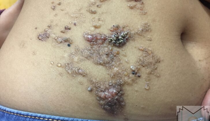
Lymphangioma circumscriptum is a skin condition that is visible as many small, clustered or spread-out, clear or bloody looking bumps that remind one of frog spawn. Purple spots may be seen within these small bump-like growths, as they contain a mix of blood and lymph fluids. These growths can sometimes look like warts, especially when they appear in the genital area. They can also show up in the armpit region and in the inguinal (groin) areas. Sometimes, they can be accompanied by swelling due to fluid buildup (lymphedema). People might experience itching, pain, burning sensation, fluid drainage from the lymphatic system, infections, and concerns about their appearance.
Cavernous lymphangioma generally shows up during infancy as a soft, undefined swelling just under the skin. It is usually painless, although it can cause tenderness when pressed deeply. Sometimes, it can involve an entire limb. This type of growth can be a few centimeters in size and doesn’t cause any changes to the skin on top of it. People often confuse these swellings with cysts or fat deposits (lipomas).
Cystic hygromas, another type of lymphatic malformation, are usually more defined than cavernous lymphangioma. They often appear on the neck, armpit, or groin. These growths are soft, and they can vary in size and shape. They will usually continue to grow if they’re not surgically removed. When there’s a cystic hygroma on the back of the neck, it might be linked to Turner syndrome, hydrops fetalis, or other birth defects. Doctors can see these lesions before birth using ultrasound scans through the stomach or vagina. MRI scanning can be helpful in understanding the extent of these types of lymphangiomas.
Testing for Lymphangioma
Diagnosing most illnesses includes asking about your medical background and conducting a physical examination. If needed, your doctor may use two additional methods – dermoscopy and biopsy – to confirm the diagnosis. Imaging, like an x-ray or ultrasound, might also be used to understand the size and reach of the illness.
Dermoscopy is a skin examination technique that helps differentiate a superficial lymphangioma (a type of skin lesion) from other skin problems. Dermoscopy can reveal two unique patterns for lymphangioma: first, yellow-looking spaces surrounded by light-colored borders without any blood involvement; second, an alternation of yellow to pink spaces with dark-red or bluish ones, which shows that there is blood involved.
A recent dermoscopic observation is a ‘hypopyon-like’ pattern. It shows a change in color from dark to light in some spaces, caused by the settling of blood which separates its components, with cells accumulating at the bottom and serum (liquid part of the blood) at the top part of the spaces.
Treatment Options for Lymphangioma
Lymphangiomas, which are abnormal clusters of lymph vessels, can be difficult to treat, whether they are close to the skin’s surface (superficial) or deep within the body. However, if they can be safely reached, the best treatment is often to surgically remove them. This involves surgically removing the pockets of abnormal lymph vessels. It’s important to note that these lymphangiomas may come back, with studies reporting up to a 23% chance of recurrence.
For smaller lymphangiomas located near the skin’s surface, surgical removal has higher success rates. However, there are other treatments available for lymphangiomas that can’t be surgically removed or are not suitable for surgery. These treatments aim to reduce the symptoms and prevent potential complications.
These treatments can involve using a carbon dioxide (CO2) laser or a particular type of long-pulsed laser (Nd-YAG) to destroy the abnormal vessels. Electrosurgery, a procedure using an electric current to cut or remove tissue, has also been reported to help alleviate symptoms. Other less commonly used treatments include cryotherapy (use of extremely cold temperatures for treatment), superficial radiotherapy (using radiation to kill abnormal cells), and sclerotherapy (a procedure in which a solution is injected into the vessels to shrink them).
In some cases, a swelling-reducing technique known as compression can be used. It’s also paramount to prevent infections as they could worsen the condition.
What else can Lymphangioma be?
Here are different skin conditions that a doctor might consider when examining a patient with dermatological symptoms:
- Skin melanoma (a type of skin cancer)
- Dabska tumor (rare skin cancer)
- Dermatitis herpetiformis (a chronic skin condition)
- Herpes simplex skin manifestations
- Skin lipomas (fatty tumors under the skin)
- Skin manifestations of metastatic carcinomas (cancers that spread to other parts of the body)
- Skin manifestations of neurofibromatosis type 1 (a genetic disorder that causes skin changes and growth of tumors on nerves)
- Herpes zoster (shingles)
- Lymphangiectasia (swelling of lymph vessels)
- Stewart-Treves syndrome (a rare form of skin lymphoma)


