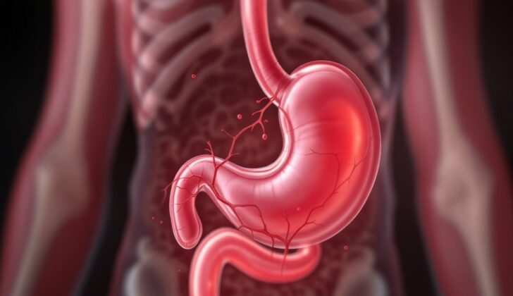
What is Pancreatic Pseudoaneurysm?
A pancreatic pseudoaneurysm is a rare but serious condition that can have life-threatening complications if not identified early. Essentially, a pancreatic pseudoaneurysm happens when a blood vessel near or in the pancreas erodes into a cavity filled with fluid known as a pseudocyst. Usually, this happens because of pancreatitis, which is inflammation of the pancreas. However, it can also happen in patients who don’t even have a pseudocyst. What distinguishes a pseudoaneurysm from a regular aneurysm is the wall; in a pseudoaneurysm, the wall is not made up of regular blood vessel components but is instead fibrous and connective tissue.
About 10% of people with chronic pancreatitis, or ongoing inflammation of the pancreas, will show some signs of a pseudoaneurysm when they have imaging tests done. These pseudoaneurysms can bleed into the pseudocyst, or form a channel (called a fistula) into nearby hollow organs. Most of these are nowadays managed with procedures involving tiny tubes inserted into blood vessels (endovascular procedures).
What Causes Pancreatic Pseudoaneurysm?
A pancreatic pseudoaneurysm is a condition that usually develops after conditions like pancreatitis, which is inflammation of the pancreas, or after having surgery on the pancreas or bile duct area. Sometimes, it can also occur after a pancreas transplant. Other common causes include physical trauma or car accidents. Some people can develop a pancreatic pseudoaneurysm after they’ve had surgery on the pancreas due to cancer or received a pancreas transplant.
This condition tends to happen when there is a leak from a surgical connection in the pancreas, which can then cause an abscess, or a pocket of infection, in the abdomen. Pancreatic pseudoaneurysms are often seen when there are large amounts of digestive enzymes from the pancreas. These enzymes can cause self-digestion and weakening of the walls of nearby blood vessels.
Risk Factors and Frequency for Pancreatic Pseudoaneurysm
In people with chronic pancreatitis who have an angiography, a medical imaging technique, up to 10% may experience it. It’s reported that 5% to 10% of such people may experience bleeding due to issues in their arteries. When pancreatitis is combined with the formation of a pseudocyst, a type of fluid-filled sac, the possibility of bleeding caused by artery problems increases to between 15% and 20%.
Signs and Symptoms of Pancreatic Pseudoaneurysm
A pancreatic pseudoaneurysm is a condition that often shows no signs until it ruptures. However, knowing what to look for can help in early detection. The symptoms can sometimes be vague and may occur in patients with a history of pancreatitis, alcoholism, or pancreaticobiliary surgery. In the case of repeated vomiting of blood (hematemesis) or passing blood in stools (hematochezia), a pancreatic pseudoaneurysm should be ruled out. Even a sudden increase in abdominal pain, coupled with falling blood counts or becoming unstable after pancreaticobiliary surgery, could indicate a bleeding pseudoaneurysm. It’s important to keep in mind that there can be some intermittent bleeding from an abdominal drain placed during surgery before a major bleed – this initial bleed is known as a sentinel bleed. Besides, a rapid increase in the size of a previously identified pseudocyst along with the appearance of a pulsating mass can strongly suggest a bleeding pseudoaneurysm. Finally, notable signs of considerable blood loss such as pallor (pale skin), tachycardia (fast heart rate), and hypotension (low blood pressure) may also be apparent. A bruit (an abnormal sound heard with a stethoscope) is very rare but can occur in the case of a large pseudoaneurysm.
Key signs to look out for include:
- Repeated vomiting of blood or passing blood in stools
- Sudden increase in abdominal pain
- Falling blood counts or becoming unstable after pancreaticobiliary surgery
- Intermittent bleeding from an abdominal drain placed during surgery
- Rapid increase in the size of a pseudocyst along with the appearance of a pulsating mass
- Notable signs of considerable blood loss such as pallor, tachycardia, and hypotension
- A bruit in the case of a large pseudoaneurysm
Testing for Pancreatic Pseudoaneurysm
Angiography is a type of medical test that doctors use to diagnose and sometimes treat diseases in your blood vessels. It is considered to be one of the best ways to both diagnose and treat a specific issue. CT angiography, which uses X-rays and a special dye to see inside the arteries, is highly accurate but cannot provide immediate treatment for the problem.
With angiography, doctors can understand more about the problem’s nature and location. Another benefit is that it can help to control bleeding by inserting a small tube (transcatheter) to block blood flow or possibly place a stent, a small mesh tube that’s used to treat narrow or weak arteries.
Pancreatic pseudoaneurysms, or abnormal dilations in the wall of a blood vessel in the pancreas, can be classified based on several characteristics:
- The type of blood vessel from which the pseudoaneurysm arises,
- Whether there is a connection with the gastrointestinal tract, and
- If there is highly concentrated pancreatic fluid at the bleeding site.
It’s necessary to mention that CT angiography and angiography should only be performed on patients who are in stable condition.
Treatment Options for Pancreatic Pseudoaneurysm
The standard approach to treat a bleeding pseudoaneurysm, a damaged blood vessel, typically includes procedures like endovascular transarterial catheter embolization or the placement of a covered stent. These methods aim to control the bleeding. If this approach fails or the patient experiences bleeding after embolization, surgery becomes the necessary option. The surgical procedures used can either be directly tying off the bleeding vessel or removing the pancreas alongside the pseudoaneurysm.
The success rates lie between 70% to 90% for the percutaneous intervention, a procedure that is of less risk and is minimally invasive. Embolization, which is the blockage of a blood vessel to prevent more bleeding, is often the preferred treatment for patients who are critically ill and unstable or for those where the bleeding is widespread. This procedure can also be an alternative solution if bleeding occurs following unsuccessful surgery or a complication arises from the surgery. However, even with the widespread use of percutaneous methods to stop the bleeding, we don’t fully understand the long-term progress of the condition of the pseudoaneurysm. Data are limited on whether these patients may have repeated complications in the future. Sometimes, thrombin, a blood-clotting substance, can also be injected into the body, but this procedure calls for a pseudoaneurysm with a narrow opening.
When a bleeding pseudoaneurysm connects to a pseudocyst, an abnormal pocket of fluid, located at the tail end of the pancreas, it might imply the need for surgical intervention. Compared to the angiographic vascular treatment, referring to the medical imaging method used to visualize blood vessels, surgery involves a higher risk.
Surgical intervention may also be chosen when it’s necessary to clean up (debride) an abscess and manage the bleeding effectively. If patients bleed from pseudocysts, draining them endoscopically, that is, with a thin, flexible tube with a light and camera attached to it, is generally not advised. If surgical intervention is selected, it’s important to stabilize the patient first. After opening the patient’s abdomen, the surgeon must pack all four areas to control the bleeding and then remove the packs starting from the less suspicious area of bleeding to the most suspicious one. It’s common to initially stop the bleeding by applying pressure, compressing digitally, or cross-clamping the aorta above the abdomen. To access the bleeding site, openings have to be made in the duodenum (the first part of the small intestine), stomach, or they may have to remove the stomach. It’s often not advised to tie off the bleeding vessel because the tissues are weak. The tying off should be done away from the area without risking the blood supply to nearby organs.
What else can Pancreatic Pseudoaneurysm be?
When diagnosing a condition called pancreatic pseudoaneurysm, doctors need to rule out other conditions that might cause similar symptoms. These include:
- Pancreatic abscess (a collection of pus in the pancreas)
- Cystic adenocarcinoma (a type of pancreatic cancer that develops from a cyst)
- Mesenteric cyst (a rare abdominal tumor)
- Hydatid cyst (a type of parasite infection that can form cysts)
- Aortic aneurysm (an abnormal bulge in the aorta, the main artery of the body)
What to expect with Pancreatic Pseudoaneurysm
Patients who only receive basic care for pancreatic pseudoaneurysms, or abnormal dilation of blood vessels in the pancreas, can face death rates exceeding 90%. If patients undergo surgery for this condition, the death rates can range between 20% and 30%. The location of the swollen blood vessels in the pancreas heavily influences this death rate, not the particular type of treatment.
When the pseudoaneurysm is found in the head of the pancreas, death rates tend to be the highest. On the other hand, if a pseudoaneurysm is located at the tail end of the pancreas and requires surgical intervention, the risk of dying is notably lower.
A treatment technique known as embolization therapy, a process aimed at blocking the blood vessels supplying the pseudoaneurysm, has overall improved the success rates. However, the problem can often reoccur, and the total death rate still hovers around 16%.
Possible Complications When Diagnosed with Pancreatic Pseudoaneurysm
A pseudoaneurysm in the pancreas can burst, with the most common area for this to occur being within the cyst itself. This event often causes sudden abdominal pain or bleeding. Other less common yet serious complications involve blockage of the bile ducts outside the liver and the formation of an abnormal connection between an artery and vein, known as an AV fistula. However, the more frequently seen complications include:
- Bleeding
- Shock
- Failure of multiple organs
- Death
Recovery from Pancreatic Pseudoaneurysm
After surgery, it’s crucial to keep a close eye on the patient for any signs of hemorrhage, or serious bleeding. This happens in around 20-70% of cases. The cause of this bleeding could be damage to blood vessels, improper control over the vessels, or loose stitches.
Preventing Pancreatic Pseudoaneurysm
A pancreatic pseudoaneurysm is a condition that can happen because of pancreatitis (an inflammation of the pancreas) or injuries like those from car accidents. It’s crucial for patients to avoid things that might cause pancreatitis. The most crucial thing patients can do is to limit or avoid excessive alcohol consumption.


