What is Thrombosed Popliteal Aneurysm?
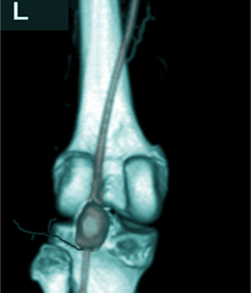
Popliteal artery aneurysms are a type of aneurysms that affect all layers of the artery wall. An aneurysm is when a part of an artery wall weakens, causing it to widen or balloon out. Specifically, for the popliteal artery, which is found in the back of the knee, an aneurysm is defined as the artery enlarging to 1.5 times its average size. This means if the size of this artery (normally between 0.5cm and 1.1cm in diameter) increases to 1.5cm or more, it is considered as an aneurysm.
These aneurysms can be problematic as they can cause blockages or blood clots that interrupt blood flow, leading to acute limb ischemia, a serious condition where the limb doesn’t get enough blood flow. They’re also prone to being filled with thrombus, a technical term for a blood clot, which can block the artery completely.
What Causes Thrombosed Popliteal Aneurysm?
When the popliteal artery, an artery behind the knee, starts to expand unusually in a spindle-shaped or balloon-like manner, it’s because the vessel wall is losing its strength. This causes the vessel to enlarge, which can eventually lead to an aneurysm, a dangerous ballooning of the artery. This usually happens due to unbalanced production and breakdown of materials that make up the artery wall. These materials include elastin and collagen, which provide elasticity and strength, glycosaminoglycans, which help to bind cells together, and smooth muscle in the artery wall.
However, it’s important to note that scientists still don’t know the specific cause of why the popliteal artery might form an aneurysm. The process is complex and likely involves many factors.
Risk Factors and Frequency for Thrombosed Popliteal Aneurysm
Popliteal artery aneurysms, which are the most common type of peripheral arterial aneurysms, make up about 70% of these types of aneurysms. Tracking them is tough since there isn’t a lot of widespread screening. Even so, they’re still quite rare, with just about 0.1 to 2.8% of the population suffering from them. These types of aneurysms mostly occur in males, with around 95% of cases being men. On top of that, about half of the patients have aneurysms in both popliteal arteries, and over a third also have abdominal aortic aneurysms.
- Popliteal artery aneurysms are the most common type of peripheral arterial aneurysms, accounting for 70% of these cases.
- Lasting rates are hard to determine due to a lack of screening.
- They are relatively rare, with an estimated occurrence of 0.1 to 2.8% in the population.
- About 95% of popliteal artery aneurysms occur in males.
- Between 50 to 54% of patients have aneurysms in both popliteal arteries.
- Abdominal aortic aneurysms are also found in 36 to 51% of patients with these kinds of aneurysms.
Signs and Symptoms of Thrombosed Popliteal Aneurysm
Acute or chronic ischemia, a condition where blood flow to your limbs is reduced, could affect up to 50% of patients. They might report worsening leg pain when walking, skin sores, sharp pain at rest, or a blue discoloration in the toe. A physical check usually reveals a lump in the back of their knee, which may or may not have a pulse. Anyone who shows these symptoms should be evaluated for a bulging blood vessel in the knee.
How doctors manage this condition depends on how severe it is. They assess the risk by checking for loss of touch or muscle function and seeing whether there’s a pulse or blood flow sound in the affected limb. The Rutherford classification for acute limb ischemia helps determine how urgently treatment is needed and whether the limb can be saved. This is its breakdown:
- Grade I: The limb is healthy and not in immediate danger. No loss of touch or muscle function. The blood flow sound and pulse are normal.
- Grade II: The limb is at risk, and there are two kinds of risk:
- IIa: Low risk but must be treated urgently. No or very little loss of touch. No loss of muscle function. Pulse not present but blood flow sound is present.
- IIb: High risk that needs instant widening of the affected vessel. Mild loss of touch and muscle function, associated with pain at rest. Pulse not present but blood flow sound is present.
- Grade III: The limb can’t be saved, tissue death or permanent damage to nerves is unavoidable. Deep loss of touch and muscle function. Both pulse and blood flow sound are absent.
Testing for Thrombosed Popliteal Aneurysm
If you have a sudden blockage of blood flow to a limb due to a ballooned and blocked blood vessel known as a ‘popliteal artery aneurysm’, your doctor will likely recommend an arteriogram. An arteriogram is a special test that uses X-rays to visualize your blood vessels. This test is crucial to identify the appropriate outflow vessels, basically the vessels where blood should normally flow, for future treatments which might include reopening the blocked vessel (revascularization) or creating a new pathway for blood flow (bypass).
This important test could be performed using two different methods: a CT arteriogram or a formal arteriogram. The decision on which one to use depends on how severe the blockage is and how urgently intervention is needed. Both tests can provide your doctor with the necessary information to plan the best treatment path forward.
Treatment Options for Thrombosed Popliteal Aneurysm
The treatment of blood clots in the knee artery (known as popliteal artery aneurysms) depends on several factors. These include how severe the blocked blood flow (ALI) is, other health conditions the patient has, and the patient’s ability to withstand surgical procedures.
Sometimes, when the blocked blood flow is not severe (grade I or IIa), patients are treated with blood thinners immediately (for example, a heparin drip), and then assessed for surgical procedures like bypass or exclusion using arteriography. Arteriography is a test that visualizes the inside of your blood vessels. If suitable inflow and outflow vessels, which are responsible for controlling the blood flow to and from the heart, are identified during this test, surgeons should perform the surgical repair during the same hospital stay.
When no visible blood vessels are identified, surgeons may consider other possibilities, such as utilizing drugs to dissolve blood clots (intra-arterial thrombolytics) and removing the clot (thrombectomy), in an effort to prepare for bypass or exclusion.
When ALI is more severe (grade IIb or III), patients will need quick re-establishment of blood flow (revascularization) to try and save their limb. Research has shown that using clot-dissolving drugs can improve outcomes for patients who have poor blood flow on detailing arteriography. However, the use of these drugs in severe ALI cases depends on each individual’s situation and the doctor’s assessment. If during surgery it’s found that there’s not enough blood flow (inadequate outflow), clot-dissolving drugs or physical clot removal may be used to establish an outflow target vessel for bypass.
In extreme cases, where the patient’s limb is no longer viable, they should be given blood thinners, and the surgeons should wait and evaluate the extent of dead tissue (demarcate) to decide the level of necessary amputation.
To enhance surgical results, the use of a saphenous vein graft is preferred when available. This involves using one of the patient’s own veins (the saphenous vein) as a bypass graft. This method is typically associated with better long-term outcomes. In addition, in any patient with significant blocked blood flow (ischemic time exceeding 4 to 6 hours), or signs of compartment syndrome (a painful and potentially serious condition caused by bleeding or swelling within an enclosed bundle of muscles), the surgeons may consider doing a four-compartment fasciotomy. This is a surgical procedure to relieve pressure and treat the loss of blood flow to an area of tissue or muscle.
What else can Thrombosed Popliteal Aneurysm be?
Acute lower extremity arterial occlusion, or the sudden blockage of blood flow to your lower limbs, can be caused by several things. According to medical sources, these include:
- Cardioembolic events, which occur when a clot forms in the heart and is pumped down to the leg, blocking an artery
- Thromboembolic events, which happen when a blood clot forms in a blood vessel in the leg, blocking blood flow
- Hypercoagulability, a condition which makes your blood clot more easily than normal
What to expect with Thrombosed Popliteal Aneurysm
The outcome of a certain condition called acute limb ischemia (which means sudden lack of blood flow to a limb), usually caused by the clotting of an artery in the knee area (popliteal artery), can depend on various factors. These include a patient’s co-occurring health conditions, the level of severity of the lack of blood flow when a patient first seeks medical help, how long the lack of blood flow has been happening, the use of drugs that break up blood clots (thrombolytics), and the availability of a vein from the leg (saphenous vein) for grafting.
When this condition occurs, the chances of losing the limb vary greatly among various reports, but some say it could be as high as 20 to 60%. That being said, most recent studies suggest an incidence of about 14 to 17%. However, there’s some positive news: due to improvements in medical techniques and the use of blood clot-dissolving drugs, several recent studies show encouraging long-term results. Approximately 68 to 80% of patients experience effective blood flow through their bypass grafts five years after the procedure, and more than 95% successfully save their limbs after urgent revascularization (a procedure to restore blood flow).
Possible Complications When Diagnosed with Thrombosed Popliteal Aneurysm
If a person has a popliteal artery clot—known medically as Acute Limb Ischemia (ALI)—there is a roughly 14 to 17% chance of losing the affected limb. This situation may also lead to a condition known as compartment syndrome that causes pain and swelling in an enclosed area, like a limb. The limb could suffer from lack of blood flow, making the symptoms of the compartment syndrome less noticeable but no less dangerous.
Medical professionals should be highly alert to the risks of this condition and take steps to prevent further damage. Medicines to dissolve the blood clot, referred to as thrombolytics, are available, but they carry their own risks, including bleeding, bleeding into the skull, and potentially stroke.
Precautions to Remember:
- Chance of losing the limb (14-17%)
- Possibility of Compartment syndrome
- The need for high medical suspicion
- Risks of using thrombolytics: bleeding, intracranial bleeding, stroke
Recovery from Thrombosed Popliteal Aneurysm
Today, many doctors recommend using blood-thinning or anti-clotting medications after bypass surgery. Also, patients who needed a limb removed during any part of the surgery will require rehabilitation. This is necessary to help them regain their independence after the operation.
Preventing Thrombosed Popliteal Aneurysm
Doctors should explain to patients the usual course and outcome of their condition. It is also advised for patients to consult with a vascular surgeon, a doctor who treats blood vessel issues, for assessment and regular monitoring. The doctor should educate the patients about the dangers of their limbs receiving inadequate blood supply if their aneurysms, or abnormal bulges in their blood vessels, are not treated. Patients should also be informed of risk factors that they can control to improve their condition.


