What is Hypoplastic Left Heart Syndrome?
Hypoplastic left heart syndrome (HLHS) is a birth defect of the heart where the left side is underdeveloped. This includes parts such as the mitral valve, left ventricle, and the aortic valve, which are crucial for the heart to properly function. HLHS is not a common condition – it affects 1 in 5,000 newborns, or 3% of all babies born with a heart defect.
In the past, there were no treatment options for this condition and sadly, all babies with this condition would pass away within their first week of life. For a baby born with HLHS to survive until they can get surgery, they depend on a certain part of their heart to stay open that usually closes shortly after birth, known as the patent ductus arteriosus, and a connection between the two upper chambers of the heart. A continuous supply of prostaglandin E1 (PGE1), a hormone-like substance, is needed to keep the ductus arteriosus open.
Nowadays, we are able to provide several treatment options for babies with HLHS, even before they are born or shortly after birth. These options include different types of surgeries like the Norwood procedure or a heart transplant, providing care to ease their symptoms, and fetal intervention, which is treating the baby while they are still in the womb. Usually, a series of three different surgeries are necessary for the baby to survive past the newborn period and infancy. Even though HLHS is uncommon, it is responsible for about 23% of all heart-related deaths in the first week of living, which shows the serious nature of this condition.
What Causes Hypoplastic Left Heart Syndrome?
HLHS, or hypoplastic left heart syndrome, develops because of multiple factors but mainly due to the underdevelopment of the parts of the heart on the left side. This underdevelopment could be due to a blockage either out from the left ventricle (a condition known as aortic valve stenosis or a problem with the outflow tract of the left ventricle) or a blockage into the left ventricle (like stenosis or atresia, a defect in the mitral valve or restrictive foramen ovale, which is the hole or opening between the upper two chambers of the heart). Most often, it is a narrowing of the left ventricle’s outlet, also known as aortic stenosis, which causes this under-development.
During the development of a baby in the womb, when there’s a blockage to the blood flowing out of the left ventricle, it can lead to enlargement of the left side of the heart, finally causing a weakening of the heart muscle and decrease in its function. This restricted blood flow also affects the development of the ventricle, causing it to be unusually small (a condition known as hypoplasia). As a result, the pressure in the left atrium (the upper chamber on the left side of the heart) increases, leading to abnormal blood flow that could decrease blood flow to the left ventricle. If there’s a blockage of blood flow due to a defective mitral valve, it will also cause the left ventricle to be smaller than usual because of the decreased preload and pressure inside it. Any decrease in blood flow to the heart while it’s developing in the womb can disrupt the normal development process.
Newborn babies with HLHS depend on a normal connection between the two main arteries (the ductus arteriosus) to provide blood to the body and arteries of the heart. As soon as they’re born, they need constant medication to keep this connection open.
Unlike most people, those with HLHS have their systemic (oxygen-carrying) and lung circulations running parallel instead of one after the other. The left ventricle, which is usually the heart’s main pumping chamber, serves no function in an infant with HLHS. When these infants have an open ductus arteriosus, the blood is pumped out of the heart through another valve (the pulmonary valve) into the pulmonary artery which takes blood to the lungs, and from there it flows into the various arteries. From the left lung artery, some blood flows through the ductus arteriosus which then supplies the aorta and heart arteries in a backward fashion and the rest of the body in a forward direction.
The amount of blood going to the body and lungs can vary, and in some cases, can be controlled by medicine or surgery to keep the blood flow equal. At birth, newborns have high pressure in the lung arteries, favoring more blood flow to the body. But shortly after birth, this pressure starts dropping, causing an increased blood flow towards the lungs at the cost of the body. If the ductus arteriosus closes completely, the body’s blood flow reduces severely, leading to an immediate collapse of the cardiovascular system.
In babies with HLHS, there is little or no blood flowing out from the left ventricle, and hence oxygen-rich blood from the lungs exits the heart through a connection either at the foramen ovale or an atrial septal defect. Oxygenated and deoxygenated blood mix in the right atrium and then flow into the right ventricle from where it is pumped as described above. Because of this mixing, typical oxygen saturation is between 75% to 85% which is why some babies may look blue. Babies with a small foramen ovale or with an intact atrial septum might need immediate intervention after birth to create a bigger opening for interatrial communication.
Risk Factors and Frequency for Hypoplastic Left Heart Syndrome
HLHS, short for Hypoplastic Left Heart Syndrome, is a type of congenital heart disease that affects around 1.4% to 3.8% of newborns. It results in about 1000 to 2000 infants every year in the United States being born with this particular heart defect. Sadly, HLHS is accountable for 23% of heart-related deaths in the first week of life.
Signs and Symptoms of Hypoplastic Left Heart Syndrome
HLHS, short for Hypoplastic Left Heart Syndrome, is typically identified in babies before they are born through the use of an ultrasound. Most of the time, babies who have this syndrome are born full-term with a normal weight. They often seem healthy in the initial hours of their lives as their ductus arteriosus, a blood vessel that’s part of typical fetal circulation, remains open during the first day or so following birth. A heart murmur, which is a common sign of heart problems in children, may not be present in these cases.
The American Academy of Pediatrics recommends a universal pulse oximetry screening for all newborns. This screening, performed 24 hours after the baby is born, involves placing a device that measures blood oxygen levels on the right hand and one of the feet. These two measurements can help doctors identify any heart defects that lead to abnormal blood flow.
If a baby fails the pulse oximetry screen, or if they start showing symptoms like poor blood circulation, unusually fast heart rate, disturbed acid balance in the body, low blood pressure, and weak pulses, they should be referred to a cardiologist immediately. These symptoms often start appearing when the ductus arteriosus begins to close, affecting the blood flow in the body.
Testing for Hypoplastic Left Heart Syndrome
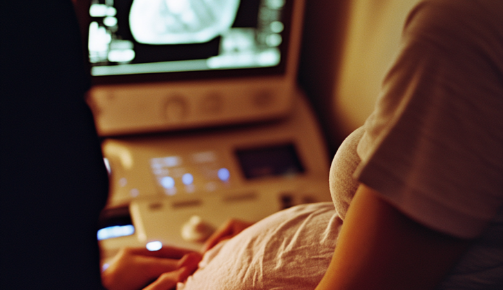
An echocardiogram, which is a type of ultrasound for the heart, can pick up Hypoplastic Left Heart Syndrome (HLHS) either before or after a baby is born. The condition evolves during fetal development, with the left ventricle, a part of the heart, shrinking as the pregnancy progresses. Sometimes, it can be so small that it isn’t detected until the third trimester. The rate at which this condition is diagnosed before birth varies and can be anywhere between 39% and 75%.
With an echocardiogram, the heart’s different parts can be examined. It will reveal a small left ventricle, abnormal heart valves, a small aorta (the main blood vessel), and an aortic arch, which is the curved part of the aorta. It will also show if the right ventricle and right atrium – other parts of the heart- are enlarged due to increased blood volume during the prenatal period. The echocardiogram is also used to see how blood is flowing through the heart and if an emergency heart procedure is required.
A chest x-ray can show if the heart is larger than usual. It can also show if there is high pressure in the veins that carry blood from the lungs to the heart.
Blood tests, including arterial blood gas (which measures oxygen and carbon dioxide levels in your blood), a complete blood count (which measures the number of blood cells), electrolytes (which measure minerals in your blood), and lactate (which measures acid in your blood) can give doctors an idea about the baby’s overall health and function of its organs.
Genetic tests can also highlight if the baby has certain chromosomal conditions, like Turner syndrome, DiGeorge syndrome, or Down syndrome, which are sometimes associated with HLHS. Babies who have a chromosomal abnormality may have higher health risks and longer hospital stays after surgery.
With severe congenital heart disease like HLHS, there’s a higher chance of bleeding within the brain’s ventricles, the cavities that contain cerebrospinal fluid. That’s why a head ultrasound is often needed before heart surgery.
Treatment Options for Hypoplastic Left Heart Syndrome
Newborns with a heart defect known as hypoplastic left heart syndrome (HLHS) typically require immediate care and specialized management in an intensive care unit (ICU). Before they can undergo surgery in their first week of life, they need to be stabilized. The main aims of this initial treatment include keeping an important vessel in the heart (the ductus arteriosus) open, preventing excessive blood flow to the lungs, and making sure blood flows normally from the left upper chamber (atrium) of the heart to the right atrium.
Shortly after birth, babies suspected of having HLHS will have an ultrasound scan of their heart (a transthoracic echocardiogram) to confirm the diagnosis. Once this is established, they’ll be started on a medication called prostaglandin E1 (PGE1) given through a vein (intravenously). This medication keeps the ductus arteriosus open and maintains blood flow in the body. The dose will be gradually lowered once the ductus arteriosus is confirmed to be open.
These babies might suffer from poor blood flow to the body (systemic hypoperfusion) as the resistance in the blood vessels of the lungs (pulmonary vascular resistance or PVR) decreases after birth. It’s important that the right level of oxygen is achieved in the baby’s blood. Surprisingly, a lower oxygen level (around 70-80%) is acceptable. If the level is too high, the baby might need assistance with their breathing to increase the PVR, ensuring balance between the pulmonary and systemic circulations. Any abnormalities in body acidity (metabolic acidosis) can be corrected with sodium bicarbonate, and the baby’s blood’s ability to carry oxygen can be optimized by maintaining a certain measure of red blood cells (hematocrit) in the blood.
An assessment of the baby’s atrial septum (the wall that separates the upper chambers of the heart) will also be made. If there’s a narrow opening (restrictive foramen ovale) or if it’s completely closed (intact atrial septum), the baby will be rushed for a procedure called a septostomy to enlarge this opening and relieve pressure from the left atrium. Failure to do so can cause a persistent rise in PVR, which can lead to higher chances of death or severe health issues in infants with HLHS.
Survival for these babies depends on a series of three palliative surgeries, each designed to separate the body’s blood circulation into two loops – one for oxygen-rich blood and another for oxygen-poor blood. The first of these procedures is performed during the baby’s first week of life, the second is done when the baby is about 4 to 6 months old, and the third is performed around the age of two years.
Following these surgeries, these babies are usually very unwell and may require a kind of life support called extracorporeal membrane oxygenation (ECMO) that takes over the work of the heart and lungs. Despite undergoing the procedures, they still face the risk of death between the stages of surgeries.
In certain cases, heart transplantation may be considered; however, due to the shortage of suitable donor hearts for newborns, it usually is reserved for high-risk cases. Unfortunately, approximately 20% of newborns listed for a heart transplant will not survive while waiting for a donor.
What else can Hypoplastic Left Heart Syndrome be?
When trying to diagnose left heart blockages where the body’s blood circulation depends on a vessel called the ductus arteriosus, doctors usually consider the following conditions:
- Critical aortic stenosis (narrowing of the heart’s aortic valve)
- Hypoplastic left heart syndrome (underdevelopment of the left side of the heart)
- Coarctation of the aorta (narrowing of the major artery leading from the heart)
- Shone’s complex (a rare heart condition with several defects)
- Interrupted aortic arch (a rare heart defect where the aorta is not fully formed)
What to expect with Hypoplastic Left Heart Syndrome
Unfortunately, only two-thirds of children with Hypoplastic Left Heart Syndrome (HLHS) survive past the age of 5. Furthermore, about 1% of patients who undergo the Fontan procedure (a surgery used in treating this condition) pass away each year.
Before any form of surgical intervention can be initiated, about one-third of newborns with HLHS sadly don’t survive. Moreover, newborns with a single heart ventricle face an increased risk of cardiac arrest (12.7%) and death (62.3%).
On a brighter note, some hospitals are reporting that up to 90% of patients survive the initial Norwood procedure. This is the first surgery in the 3-stage treatment series for HLHS.
In 2019, the Society of Thoracic Surgeons Congenital Heart Surgery Database revealed mortality rates for various stages of the surgeries. The mortality rate for the initial palliation stage is around 15%, with subsequent stages Hemi-Fontan/Glenn and Fontan presenting significantly lower mortality rates at 1.8% and 1.0% respectively.
The Aristotle score, ranging from 1.5 to 25, was developed by pediatric cardiac surgeons to more accurately predict the potential for death, surgical complexity, potential complications, and quality of care. This score factors in the complexity of the surgical procedure and the patient’s current condition. Points are often added for different anatomical variants, respiratory failure, shock, prematurity, and low weight. A score higher than 20 indicates a high probability of mortality, and is often associated with the Norwood procedure. This score is calculated before surgery as a guide to provide parents with honest expectations.
Possible Complications When Diagnosed with Hypoplastic Left Heart Syndrome
The Norwood procedure is a heart surgery performed in three stages to treat serious heart birth defects. Just like any other surgical procedure, there are chances of complications that can occur.
In Stage 1, complications could be:
- Thrombosis of the BT shunt, which is a condition where a blood clot forms, reducing blood flow to the lungs. This can result in severe low oxygen levels in the body.
- Inadequate blood supply to the body due to an excessive blood flow to the lungs. This condition, known as “coronary steal”, can lead to sudden death.
- Abnormal heart rhythms, also known as arrhythmias.
- Respiratory failure.
- Bleeding.
- Infections.
- Malfunction of the kidneys, also known as renal dysfunction.
In Stage 2, complications can include:
- Decreased blood flow to the lungs leading to low oxygen levels in the body and decreased pumping ability of the heart.
- Abnormal heart rhythms, or arrhythmias.
- Blood clot-related problems, known as thromboembolic events.
In Stage 3, complications may be:
- Poor functioning Fontan, which is the new circulation route created in the heart.
- Atrial arrhythmias, which are abnormal heart rhythms that occur in the upper chambers of the heart.
- Plastic bronchitis, a rare condition that leads to a blockage of the airways with material that is similar to plastic. This occurs in 3% to 10% of patients.
- Protein-losing enteropathy, a condition where proteins are lost from the body. This occurs in 1% of patients.
- Liver issues such as congestion, cirrhosis, and an increased risk for liver cancer.
- Thromboembolic events in the lungs.
Recovery from Hypoplastic Left Heart Syndrome
Patients who have undergone the first stage of heart surgeries, such as Norwood, Sano, or hybrid procedures, are typically very unwell after their operation and need intensive care in an ICU. They often need assistance with breathing (mechanical ventilation), support for heart function (inotropic support) and other ongoing treatments in a specialized heart ICU. The aim of treating these patients is typically to reach certain health measurements:
- Blood pressure around 40 to 45 units (mmHg)
- Normal body acidity (pH of 7.4)
- Normal levels of carbon dioxide and oxygen in blood
- A percentage (40%) of their blood being red blood cells (Hematocrit or Hct)
- Oxygen saturation between 75 and 85%
- Normal levels of lactic acid
These health measurements are maintained by manipulating the resistance in the blood vessels and adjusting how much the person is inhaling or exhaling, as well as administering blood products as needed.
Transitioning to the second stage of heart surgeries, like Hemi-Fontan or Bidirectional Glenn procedures, the resistance in the blood vessels leading to the lungs needs to be kept low to ensure blood flow and heart performance. In this stage, patients benefit from being taken off the ventilator as soon as possible as this promotes blood flow from the main vein (superior vena cava) to the lungs. The primary aims for this stage of the recovery period include:
- An oxygen saturation between 75 to 85%
- Higher levels of carbon dioxide in the blood, which may help improve blood flow in the brain and lungs, thereby increasing oxygen levels in the body.
- Optimal mixed venous saturation
In the third and final stage of heart surgeries, known as Completion Fontan, the flow of blood from the body to the lungs is passive and ensuring the right volume of blood is maintained is crucial. Lower pressure within the chest encourages blood flow in the arteries leading to the lungs. It is also beneficial for patients to be removed from the ventilator as soon as possible to reduce pressure within the chest and therefore increase the amount of blood the heart is pumping. Typically, oxygen levels in the blood are closer to normal in this stage. However, over time, the right side of the heart, which isn’t usually responsible for pumping blood to the entire body, may start to fail. This leads to long-term health issues such as liver disease, cirrhosis, protein-losing enteropathy, lymphatic dysfunction, and plastic bronchitis. The heart may struggle to cope with these long-term stressors, leading to both systolic and diastolic dysfunction. In severe cases, patients may require a heart transplant.
Preventing Hypoplastic Left Heart Syndrome
If a pregnant woman has an ultrasound scan that indicates her baby might have Hypoplastic Left Heart Syndrome (HLHS), a condition where parts of the left side of the heart do not develop completely, she is usually referred to a children’s heart specialist known as a pediatric cardiologist. This specialist typically sees the expectant mother between the 20th and 24th weeks of her pregnancy. The parents are presented with possible treatment options for their baby. They are also openly informed about the serious risks associated with this heart defect, including severe illness and the possibility of a higher than normal rate of death.


