What is Interventricular Septum Abscess?
An abscess inside the heart can be located within the heart muscle, inner lining of the heart, or heart valves (natural, artificial, or mechanical). This condition has a high risk of causing serious health problems and potentially death. One type of heart infection, known as infective endocarditis, mainly affects the heart valves. If not treated quickly, a heart abscess could develop in 20% to 30% of these cases.
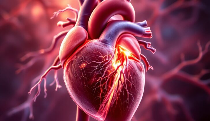
Sometimes an abscess in the heart can present as a rare condition known as an interventricular abscess. The interventricular septum, the thick muscular wall that divides the heart chambers, can become vulnerable and develop an abscess. The abscess usually occurs as a result of infective endocarditis spreading from the heart valves, most commonly from an infection of the aortic valve, the main outflow valve for the heart. The ensuing complications could be a disruption of the heart’s electrical system or heart failure. Despite the best available treatment, aortic valve infections often have high death rates during hospitalization.
There are many risk factors for acquiring infective endocarditis. These range from existing heart conditions such as rheumatic heart disease, heart defects present from birth, or age-related changes to the heart valves. Other risk factors include having a mechanical device inside the heart, the use of intravenous drugs, or having a long-term catheter in place for medical treatment. The bacteria Staphylococcus aureus is the most common cause of infective endocarditis. Studies have shown an increasing rate of this bacterial infection, and in a separate study, it was found to be responsible for over half of all cases. Bacteria can start to grow and cause a whole body infection as a result of intravascular catheters, surgical wounds, artificial devices, or use of kidney dialysis machines.
What Causes Interventricular Septum Abscess?
An interventricular septal abscess is an infection that usually develops from severe heart valve disease, known as infective endocarditis. This infection can spread to the wall separating the lower chambers of the heart, the interventricular septum, and give rise to this abscess. A similar abscess can also form around a heart valve impacted by infective endocarditis and eventually extend into the interventricular septum.
The main cause of an interventricular septal abscess is the spread of bacteria in the blood, also known as bacteremia. This usually happens due to a spreading infection or a localized infection. This abscess can also show up in people with specific heart conditions like degenerative valves or mechanical or prosthetic valves, and among people who misuse intravenous drugs.
Other common causes of an interventricular septal abscess reported are:
- Injury or trauma
- Deep wounds that damage the heart tissue
- Infected coronary stents (small tubes placed in the coronary arteries)
- Infected surgical scars on the sternum (the flat bone in the middle of the chest)
- Infected hearts that have been transplanted
- Severe burns
- Immune-compromised patients (those with a weakened immune system)
- HIV patients
- Parasitic infections
- Pseudoaneurysms (false aneurysms characterized by a blood-filled sac formed by the outer layers of a damaged blood vessel)
- Suppurative pericarditis (a condition characterized by pus around the lining of the heart).
Risk Factors and Frequency for Interventricular Septum Abscess
An interventricular abscess, which is an uncommon consequence of a heart infection known as infective endocarditis, is more usual in underdeveloped nations. In these countries, a lack of antibiotics to treat simple bacterial infections may lead to serious heart infections. In the U.S. and Europe, however, individuals often have certain medical conditions or devices that might increase the chance of an infection. Staphylococci are the main cause of these heart infections, which can further cause a septal abscess. Between 2000 and 2011, the occurrence of heart infections in the United States rose from 11 to 15 per 100,000 people. Pinpointing an exact occurrence rate of interventricular abscess is tough due to ongoing changes in diagnosing criteria of infective endocarditis. Other factors such as variants in risk factors, predisposing medical conditions, microbiological agents, valvular pathologies, the use of intravenous illicit drugs, and socioeconomic factors might also contribute to the rising occurrence of the interventricular abscess with infective endocarditis.
Various infectious organisms can cause an interventricular abscess, including:
- Staphylococcus aureus
- Haemophilus species
- Enterococci
- Escherichia coli
- Beta-hemolytic streptococci
- Streptococcus pneumoniae
- Bacteroides species
- Parasitic organisms
- Hydatid cysts
- Listeria monocytogenes
- HIV
- Immunocompromised states
- Other miscellaneous causes
Signs and Symptoms of Interventricular Septum Abscess
An interventricular abscess is a serious condition that involves an infection in the heart. The presentation and symptoms can vary depending on several factors such as how acute the issue is, the extent of heart muscle involvement, the impact on electrical pathways in the heart, and the condition of the heart valves. This condition should be considered in patients who present with irregular heart rhythms, heart blockages, and spreading of the abscess.
The symptoms of interventricular abscess usually become more apparent during an acute infection. Key areas to focus on would be the area affected by the abscess, risk factors, the level of blockage in the conduction pathway, and any anatomical or contraction anomalies. Signs that an interventicular abscess could be the cause include:
- Dizziness
- Difficulty breathing
- Chest pain similar to possible heart attack or angina symptoms
- Shortness of breath at night
- Blue discoloration of the skin due to lack of oxygen
- Difficulty breathing while lying flat
- Fainting or near fainting
- Joint pain
- Cough
- Skin rashes
- Prior heart surgery or injury
- Travel to areas known to have diseases
- Exposure to an animal
- Exposure to infection risk factor
Physical examination of a patient with this condition might reflect signs of infective endocarditis, which is an infection of the heart lining and valves. However, there’s no specific physical examination that can confirm an interventricular abscess, the diagnosis usually comes from identifying a combination of symptoms such as:
- Slow heart rate
- Signs of heart failure such as breathlessness, swelling in legs, fluid in the lungs, and swollen neck veins
- Apparent issues with the aorta, the body’s main artery, including indications of aortic regurgitation (backflow of blood into the heart) or an enlarged aortic root
- Signs of a hole between the heart chambers, such as cyanosis or heart failure
- Signs of increased pressure in the lung arteries
- mitral regurgitation findings that include a holosystolic murmur at the apex, and pulmonary edema
- Diverse signs across multiple organs due to potential spread of bits of the abscess
- Back pain, potentially a sign of bone infection
- Petechia (tiny red spots), sublingual or splinter hemorrhages
- Osler nodes (painful bumps on the hands or feet), Janeway lesions (small, painless red spots), Roth spots (spots on the retina), Splenomegaly (an enlarged spleen)
Testing for Interventricular Septum Abscess
The Duke’s criteria is a method first introduced in 1994 to help diagnose infective endocarditis, an infection that can cause damage to the heart’s inner lining. This is a crucial step towards diagnosing an interventricular septal abscess, a type of infection within the walls separating the chambers of the heart. The Duke’s criteria involves a mix of major and minor clinical signs, and can confirm a diagnosis when two major and one minor, one major and three minor, or five minor signs are present.
Simplified, diagnosing an interventricular abscess involves identifying infective endocarditis, issues with the heart’s electrical signals, and visible symptoms via imaging. Tests that may be conducted for this diagnosis could include blood tests, infection marker testing, an electrocardiogram (EKG or ECG), and various imaging scans.
In acute, or sudden, cases, routine blood tests may show high white cell count, normocytic anemia (which is a form of blood disorder), and thrombocytopenia (meaning low platelet count). In less sudden, or subacute, cases, the white blood cell count might appear normal.
Tests checking for inflammation or infection, such as the erythrocyte sedimentation rate (ESR), C-reactive protein (CRP), and ferritin tests, can also be helpful. Lack of significant rises in ESR and CRP could indicate there’s no ongoing acute or chronic infection.
Blood cultures are also important. Sampling blood from three different sites can detect up to 98% of infections found in the bloodstream. Valve cultures can also be useful in some cases, though they might lead to false-positive results, which means the test indicates an infection when there isn’t one.
A urine analysis could show proteinuria (excess protein in the urine) and microscopic hematuria (tiny amounts of blood in the urine), which provide further indicators.
An EKG can reveal a variety of conduction blocks, or problems with the heart’s electrical signals. The appearance of a new AV block, a type of heart block, on an EKG is a good indicator of infective endocarditis extending to the heart wall or the interventricular septum.
Imaging techniques such as echocardiography (a type of ultrasound that uses sound waves to create live images of your heart), cardiac CT, MRI, scintigraphy (which uses a special camera and radioactive tracer to examine internal organs), and angiography (which checks your blood vessels for abnormalities) can help to identify signs of infective endocarditis and interventricular abscess. Echocardiography is the primary imaging technique; it might involve transthoracic or transesophageal techniques, depending on the specific requirements of the case.
Scintigraphy is usually used where echocardiography falls short, and is particularly useful for diagnosing prosthetic valve endocarditis. This imaging technique allows doctors to identify and monitor an abscess formation earlier than other imaging modes.
Treatment Options for Interventricular Septum Abscess
The aim of treatment plays two roles – find and address the underlying cause of infection and reduce risk factors. This can be done with both medical treatment and potentially surgery. What treatment your doctor decides on will primarily depend on if there are any complications. This could include an interventricular abscess (a pocket of pus between the heart’s chambers) or infective endocarditis (a heart valve infection), which could trigger unstable blood pressure, an irregular heart rhythm, or heart failure.
Firstly, doctors usually work to stabilize vital signs with IV fluids if needed. They also manage irregular heart rhythms, and treat heart failure, often using a combination of different medications. A specialist in infectious diseases will help choose the right antibiotic and monitor progress. The treatment can last from two to six months and may involve rounds of oral or intravenous antibiotics. Initially, doctors will prescribe broad-spectrum antibiotics, until the results of blood cultures(simple tests to check for bacteria in your blood) are available. Based on these results, the antibiotic treatment might change.
The bacterium called Staphylococcus aureus is most commonly associated with causing this infection, and it requires coverage by combination antibiotic therapy. The traditional approach is the combination of an aminoglycoside with vancomycin or penicillin as it has shown to kill Staphylococcus aureus bacteria. However, this infection presents unique challenges. The antibiotics may not always completely remove the disease; therefore, a prolonged therapy of 6 – 8 weeks of bactericidal(parenteral) therapy may be required.
If there is an interventricular abscess, doctors often resort to opening up the body surgically and draining the abscess or using a needle to drain it. In the case of severe complications like a ruptured abscess causing a tear in the wall between two heart chambers (ventricular septal defect), surgery is usually needed. This open-heart surgery can involve replacing the root of the aorta – the large blood vessel that leads from the heart – and draining the abscess. When to opt for surgery will depend on the stability of the patient and how they react to antibiotics.
What else can Interventricular Septum Abscess be?
An interventricular septal abscess is a rare problem that may happen as a result of infective endocarditis, an infection of the inner lining of the heart. It can show unclear signs and might seem similar to other health issues, which include:
- Acute rheumatic fever (an inflammatory disease after infection with group A Streptococcus bacteria)
- Congenital septal hypertrophy (abnormally thick heart muscle)
- Septal cardiac tumor (an abnormal growth in the heart)
- Aortic or mitral regurgitation (leakage of blood back through valves in the heart)
- Ventricular septal defect (a hole in the heart)
- Atrioventricular or bundle branch block (a type of heart block condition)
- Acute myocardial infarction (heart attack)
- Myocardial rupture (a tear or rupture in the heart wall)
- Cardiac tumor (a tumor in the heart)
- Congestive heart failure (a chronic condition where the heart doesn’t pump blood as well as it should)
What to expect with Interventricular Septum Abscess
Patients with a severe infection of the heart lining, known as infective endocarditis, often face high risks of complications and death. The seriousness of this disease can vary and typically depends on where the infection came from and if the patient has any other existing conditions.
Various biological signs of acute infection, as well as indicators that measure the severity of the illness, including organ failure, also play a role in increasing the mortality rate. Surgery to treat abscesses, which are a collection of pus, is often linked to poorer outcomes within the initial 30 days.
It’s important to note that while the process may be risky, if patients who need surgery do survive, they usually fare well over the long-term, showing low rates of reinfection. The prognosis is generally more positive for younger patients and those who don’t have any other major health problems.
Some other key factors that affect the mortality rate include a Charlson score (a method of predicting the ten-year mortality for a patient who may have a range of comorbid conditions) of less than 3, an early diagnosis of endocarditis before having to be admitted to the ICU, the use of a type of antibiotic called aminoglycosides, and the occurrence of septic pulmonary embolism, a life-threatening condition where an infected blood clot blocks a vessel in the lungs.
Possible Complications When Diagnosed with Interventricular Septum Abscess
An interventricular septal abscess is a serious medical condition that can lead to severe health problems and even death. It comes with many potential complications that can develop during its progression:
- Perforation of the heart muscle wall
- New cases of congestive heart failure
- New heart murmurs
- New cases of heart valve leakage
- Ineffective response to antibiotics
- Sudden serious breathing problem (acute respiratory distress syndrome)
- Failure of multiple organs
- Stroke
- Development or worsening of issues with the heart’s electrical system, leading to bundle-branch block or atrioventricular block
- Various issues like serious recurrent heart rhythm problems (ventricular arrhythmias), inflammation of the heart lining (pericarditis) and so on
- Fast and major health decline leading to death
Recovery from Interventricular Septum Abscess
The chances of a successful treatment largely depend on the patient’s overall health before the illness, a quick diagnosis and treatment, and aftercare or rehabilitation. Many patients will need to take IV antibiotics for about 6 to 8 weeks. During this time, they will be closely monitored, especially their heart function. They may also need regular ultrasound scans of their heart to make sure it’s healing correctly.
Even while being monitored, they should be watched for any irregular heart rhythms or heart block, which is an issue with the electrical signals in the heart. Even after surgery, there could be issues, so it’s important for patients to have regular check-ups afterwards. Despite having surgery, patients can still develop complications, including lung infection, bladder infection, and blood clots in the leg veins.
Maintaining a balanced diet, preventing deep venous thrombosis (a blood clot in a deep vein, usually in the legs), and stress ulcer prevention (protecting against painful sores in the stomach lining or upper part of the small intestine usually caused by stress) are all extremely important. Rehabilitation might include physiotherapy to prevent muscle weakness that could result from lack of use, and a personalized recovery plan for each patient. This can potentially decrease the chances of further health issues and risk of death.
Preventing Interventricular Septum Abscess
Teaching people how to prevent infective endocarditis, a serious heart infection, is very important in reducing the number of cases in society. The NICE guidelines (guidelines provided by a health advisory organization) suggest ways to prevent this disease. These guidelines highlight the importance of identifying people who have a higher chance of getting heart diseases. They also aim to spread awareness about prevention which includes maintaining good oral health, understanding the pros and cons of taking antibiotics before procedures to prevent infection, and knowing when it’s time to reach out to a healthcare professional for help.
Moreover, these guidelines offer more information about the risks of getting infections from skin piercings or tattooing for people who are already more prone to getting infective endocarditis.


