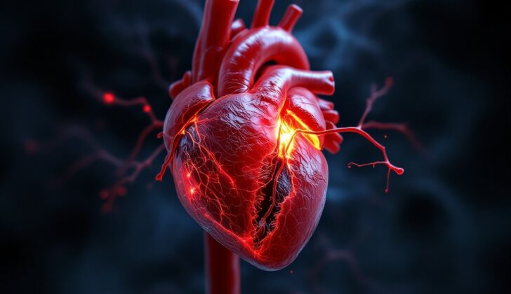
What is Left Ventricular Rupture?
Left ventricular rupture, or the tearing of the heart’s left lower chamber, is a devastating but common complication in patients suffering from a severe heart attack. Although it happens in less than 1% of cases after a severe heart attack, the chances of death are extremely high. This kind of rupture can also occur due to severe heart injury, heart infections, a tear in the body’s main artery, heart tumors, and diseases that invade the heart itself. Also, in some medical procedures involving the heart, accidental damage could lead to this issue. Additionally, it has been reported in patients with a unique heart condition known as Takotsubo cardiomyopathy.
Early diagnosis and quick surgery are essential to save the patient’s life because the chances of death are extremely high without any intervention. However, with the right care, patients can significantly improve. Latest techniques that don’t use stitches showed promising results, and they even managed to prevent the need for a procedure called a cardiopulmonary bypass, which is used to take over the function of the heart and lungs during surgery.
What Causes Left Ventricular Rupture?
Left ventricular rupture, a tear in the heart’s main pumping chamber, often happens after a heart attack. Certain factors increase the risk of experiencing this, such as not having any previous symptoms of heart disease like chest pain or heart attacks, higher levels of a heart damage indicator (CK-MB) in the blood, being female, being over 70, having a heart attack in the front of the heart that affects the full thickness of the heart muscle, and having your first heart attack. Having a certain amount of fluid build-up in the sac that surrounds the heart also increases the risk of rupture.
On the other hand, an enlarged heart, congestive heart failure (a condition where the heart doesn’t pump blood as well as it should), a history of previous heart attacks, long-term ischemic heart disease (when the heart doesn’t get enough blood and oxygen), using beta-blockers (drugs to lower blood pressure) early on after a heart attack, and swift medical intervention can protect against such rupture.
Sometimes, left ventricular rupture can be caused by an injury during certain medical procedures, such as heart catheterization, heart valve replacement via a catheter, placement of temporary or permanent wires for a pacemaker, balloon valve repair, certain heart surgeries, and removal of fluid from the sac around the heart.
Abscess (a collection of pus) in the heart muscle caused by infection can also burst and lead to rupture. It can occur in conditions like bacterial heart valve infection, tuberculosis, and echinococcal cysts (small tapeworm-caused cysts). Certain heart tumors, like lymphoma and acute myeloblastic leukemia (a type of blood cell cancer) can cause the heart muscle of any chamber to burst. One rare cause is a disease called sarcoidosis, which can form non-damaging clumps of inflammatory cells in the heart muscle walls.
Risk Factors and Frequency for Left Ventricular Rupture
Between 2 to 4% of heart attacks can lead to a dangerous situation known as myocardial rupture. This complication is more common in older people and those with high white blood cell counts. Women are also slightly more at risk than men. However, such a heart complication is seen in less than 10% of all traumatic injuries. Motor vehicle accidents are a common cause of this, and men are more prone to this type of rupture. It is most often seen in the 15 to 63 age group.
Good news is there has been a decline in both the occurrence rates and death rates related to this issue over the past few decades: From 3.3% to 2.8% to 1.7% for occurrence rates, and from 96% to 56% to 50% for death rates. This improvement is due to better healthcare and timely treatment.
- Between 2 to 4% of heart attacks can lead to a complication called myocardial rupture.
- It’s more common in older people and those with high white blood cell counts.
- Women are slightly more at risk than men.
- This heart complication occurs in less than 10% of traumatic injuries, often linked to motor vehicle accidents.
- Men are more likely to experience this rupture due to car accidents.
- People between the ages of 15 to 63 are more commonly affected.
- The occurrence and death rates have been decreasing over the past few decades.
- The decline is thanks to improvements in healthcare and early treatment.
Signs and Symptoms of Left Ventricular Rupture
The main cause of shock or a sudden decrease in blood flow following a heart attack typically within 24 hours to three weeks is a rupture in the heart muscle. This usually happens within 3 to 5 days after the heart attack, particularly after a small, first heart attack that showed no complications. Those more at risk include people with high blood pressure, older women, and patients who suffer from chest pain after a heart attack.
Left ventricle (LV) rupture, a type of heart muscle rupture, can cause sudden death. Some patients might first experience low blood pressure. In the case of a rupture induced by physical injury, it could happen soon after the injury and simultaneously affect parts of the heart like the ventricular septum, papillary muscle, pericardium, and the diaphragm.
Patients with heart muscle rupture due to injury usually experience cardiogenic shock (a condition where your heart can’t pump enough blood) or hypovolemic shock (a severe fluid or blood loss). Symptoms can include shortness of breath, chest pain, low blood pressure, cold limbs, and occasional changes in mental status. Some individuals may also experience brain or body-wide clots from pseudoaneurysms, a false aneurysm that forms as the blood collects in a pocket. This can lead to the formation of ventriculopulmonary fistulas, presenting as a cough with blood.
- Shortness of breath
- Chest pain
- Low blood pressure
- Cold limbs
- Changes in mental status
Some individuals may manifest signs of pericarditis, a swelling of the pericardium (the sac-like covering of the heart), like chest pain that worsens with deep breaths or lying down and a friction rub before the LV rupture. Tamponade, a condition where the pericardium fills with blood or fluids hindering the heart from functioning, may appear with a sudden onset of slow heartbeat, clear lung fields, and swollen neck veins. Abnormalities like the Kussmaul sign (an increase in venous pressure on inspiration), muffled heart sounds, and pulsus paradoxus (a drop in blood pressure during the inspiration phase of breathing) may be noted in a physical examination. Hypovolemic shock may manifest as low blood pressure, increased heart rate, cool extremities, and paleness. Friction rubs, a scraping or grating noise heard with a stethoscope, may be detected in pseudoaneurysms.
Testing for Left Ventricular Rupture
Doctors should be very alert if they suspect a heart muscle rupture after a heart attack. An early diagnosis and quick surgery can push survival rates up to 75%. All injuries to the heart should be considered in accidents with high-speed deceleration impact.
Using a bedside heart ultrasound (transthoracic echocardiography or TTE) is the best way to diagnose these conditions. It can show abnormal heart wall movement or heart muscle injury. If the sac around the heart is filling with fluid (tamponade), which is dangerous, it can be seen as changes in the heart chambers and changing blood flow speeds.
Sometimes doctors may also notice a muscular tear near the valves of the heart. It might look like a split in the head of the muscle or a mobile echo density that dips into the left atrium during heartbeat.
The Focused Assessment with Sonography in Trauma (FAST) ultrasound is the quickest way to diagnose heart injuries if someone has had a blunt trauma to the chest.
If the heart is forming a false aneurysm – which happens when there’s a leak in the heart muscle – it might show up as an echo-free space that enlarges with every heartbeat and communicates with the main pumping chamber of the heart (Left Ventricle or LV) through a narrow neck.
A chest X-ray might show an enlarged heart with clear lung fields in cases of LV rupture and pseudoaneurysm. If the sac around the heart tears, it might show up as blood in the chest cavity (hemothorax). If the main blood vessel that leaves the heart (the aorta) tears, it might show up as a widened space in the chest.
A CT scan or an MRI can be used when the heart ultrasound images aren’t clear enough. These imaging tests are usually reserved for stable patients.
An electrocardiogram (ECG, a heart rhythm test) might show a continued elevation of the ST segment after a heart attack, which can indicate a high possibility of heart muscle rupture. If the wall of the LV ruptures, it might cause the heart rhythm to go haywire and suddenly slow down.
In stable heart attack patients, before planning for surgery, the doctors may need to do a heart catheterization, angiography, and ventriculography. These tests can determine the degree and extent of the lack of blood supply to the heart muscle.
Treatment Options for Left Ventricular Rupture
In the event of a LV (left ventricular) rupture, which is a serious tear or break in the wall of the heart’s main pumping chamber, surgery is the primary course of treatment, while medicines can help support this process. As soon as a LV rupture is diagnosed, a heart surgeon should be consulted right away, because the patient needs to be taken to the operating room as soon as possible.
In some cases, the surgical team may need to drain accumulated blood from the pericardium, which is the sac that surrounds the heart. Some doctors may suggest the use of devices known as intra-aortic balloon pumps for the LV rupture. Pericardiocentesis, a procedure where a doctor uses a needle to remove extra fluid from the pericardial sac around the heart, is generally advised against as it could possibly make the rupture worse. The patient should be advised to rest completely and to avoid eating or drinking anything.
Before surgery, medical treatment can provide vital support, helping to stabilize a patient’s condition. Swift administration of fluids can help increase the amount of blood received by the heart thus improving the heart’s performance. Specialized medications, known as inotropes, may be needed to strengthen the heart’s contractions.
During surgery to repair the LV rupture, the damaged area of the heart is removed and sealed using synthetic or biological materials. If a false aneurysm, also called a pseudoaneurysm, has developed, it requires surgical treatment even if it doesn’t show symptoms, due its high potential for sudden rupture. If a hole (known as a ventricular septal defect or VSD) has developed in the wall separating the heart’s lower chambers because of the rupture, it is sealed with a patch. Problems with the papillary muscles, which control the valves of the heart, may need to corrected with a mitral valve replacement. While treating these heart complications, a coronary artery bypass surgery, a procedure that improves blood flow to the heart, may also be necessary.
What else can Left Ventricular Rupture be?
There are several heart conditions that can cause various symptoms. Here are a few:
- Right Ventricular Infarction: This can lead to heart failure. Symptoms include low blood pressure, swollen neck veins, and clear lung fields.
- Cardiogenic Shock: Individuals with this condition may experience low blood pressure, lack of low body fluid levels, and signs of poor blood circulation.
- Hemorrhagic Shock: This is characterized by low blood pressure, a rapid heartbeat, reduced urine production, and confusion or disorientation.
- Cardiogenic Pulmonary Edema: This condition can occur after a papillary muscle in the heart ruptures, or a hole in the heart (VSD) can be spotted in a chest X-ray. Patients with this condition may have fluid in the lungs, and it can even occur in people who have no symptoms.
- Mitral Regurgitation (MR): This can happen if a papillary muscle in the heart ruptures. Symptoms include rapid heartbeat, rapid breathing, respiratory distress, crackling sounds in the lungs due to fluid accumulation (pulmonary rales), and low blood pressure, along with signs of MR.
- Cardiac Tamponade: This can occur as a result of a ruptured left ventricle in the heart, and it’s more common with stab wounds. Symptoms include difficulty breathing, rapid heart rate, and high pressure in the neck veins (JVP).
What to expect with Left Ventricular Rupture
The outlook of a health condition is influenced by its cause, size, and how it affects blood flow. Because of this, early diagnosis and quick surgical response are crucial.
A significant number of hospital deaths following a heart attack (referred to as an ‘AMI’) are caused by a rupture in the heart muscle. However, the death rate decreases over the first two weeks after the heart attack. If a patient with a rupture due to a severe, blunt trauma (like a car accident) gets immediate medical attention, their survival rate increases notably.
Factors such as the type and size of the injury, how quickly the patient gets to the hospital, the patient’s stability in terms of blood circulation, and how soon they receive treatment all play a critical role in whether they will survive this hospital stay. As one might expect, a rupture in the left ventricle (or ‘LV’) of the heart typically has a higher mortality rate than one in the right ventricle (‘RV’).
Severe consequences of an LV rupture can include a pool of blood in the chest cavity (hemothorax), pressure on the heart due to fluid buildup (cardiac tamponade), and sudden death. Therefore, these cases need immediate medical intervention.
Possible Complications When Diagnosed with Left Ventricular Rupture
The primary complications following the rupture of the left ventricle, or LV, of the heart, are cardiac tamponade and hemothorax. Cardiac tamponade is a serious medical condition that happens when blood accumulates in the pericardial space, the area surrounding the heart. This accumulation of blood can reduce the heart’s ability to pump blood effectively, leading to possible lung congestion, shock, or even death.
Similarly, hemothorax occurs when blood collects in the pleural space, the area between the lungs and the chest wall. The usual treatment for hemothorax involves either a chest drainage tube (thoracostomy) or a small surgical procedure with a camera, known as video-assisted thoracoscopic surgery (VATS). In extreme cases, when the hemothorax isg severe or the bleeding persists, a thoracotomy, which is a larger surgical procedure, may be necessary. In some patients, the first sign of LV rupture might tragically be sudden death.
Key Risks and Treatments:
- Cardiac Tamponade – Potential congestion of the lungs, shock, or death
- Hemothorax – Treated with thoracostomy or VATS, in extreme cases thoracotomy may be required
- The possibility of sudden death due to LV rupture
Recovery from Left Ventricular Rupture
After surgery, patient care includes resting in bed, managing blood pressure and heart rate, providing inotropic support (drugs that alter the force or speed of the heart’s contractions), and ensuring the patient remains calm. Resting in bed helps prevent high blood pressure, while staying calm supports this. Managing the heart rate helps lower the heart’s need for oxygen, which helps it to recover.
Inotropic support is needed to ensure the body’s tissues receive enough blood flow. If an intra-aortic balloon pump (a device that helps your heart pump more blood) is in place, it can increase the blood flow to the heart and prevent low-cardiac output syndrome, which happens when your heart can’t pump enough blood to the rest of your body after heart surgery.
Preventing Left Ventricular Rupture
After surgery, it’s crucial for patients to understand their care instructions to aid in their recovery. This involves knowing about different aspects of the surgery aftercare process, how it’s explained to them, and how often they receive this education. The way that these instructions are individualized and delivered also has an impact, such as personalizing the content, using interactive resources, giving one-on-one education, and strengthening the benefits seen in different care settings.
Your doctor or nurse not only treats you but also teaches you some important points about your recovery. They play a crucial role in your education about your surgical aftercare. Recognizing your unique needs and characteristics can help bring better outcomes. They’ll guide you on how to care for your surgical dressing, and how to watch out for signs of infection. This knowledge will help you know when you need to call their attention. They will also provide guidelines about your diet, medications, and physical activity to ensure that you are properly participating in your own recovery.