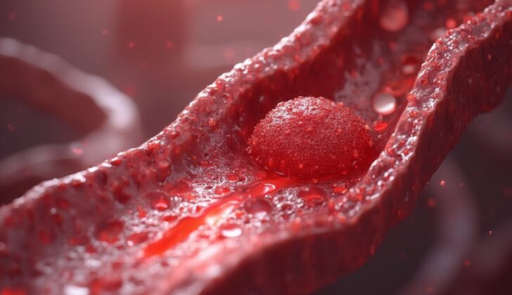
What is Mural Thrombi?
Mural thrombi are blood clots that stick to the walls of blood vessels or the inside of the heart. They are unusual in healthy or minimally clogged arteries unless there’s a greater than normal tendency for the blood to clot, or if there’s inflammation, infection, or a genetic predisposition to diseases affecting the aorta, the main blood vessel in your body. These blood clots can appear in larger vessels like the heart and aorta, and they can limit blood flow. Usually, they are found in the part of the aorta that descends from the heart, and less often, in the aortic arch—the top part of the aorta—or the part of the aorta in the abdomen. Mural thrombi can also infiltrate any part of the heart. A frequent complication after a heart attack is a blood clot forming in the left ventricle (the heart’s main pumping chamber), particularly at the tip of the heart. This clot can dislodge from the heart and move through the arteries, blocking any blood vessels.
What Causes Mural Thrombi?
The “Virchow triad” is a term used to describe the three main factors that cause a thrombus, or a blood clot, to form in our body. The first factor is an injury to the lining of our blood vessels. This could be due to an accident or injury causing damage.
Secondly, abnormal blood flow can contribute to the clot formation. This might occur if the blood is flowing in a chaotic or turbulent manner, rather than smoothly, like in arteries. Conditions that can cause this include inflammation of heart valves or an abnormal bulge in a blood vessel, known as an aneurysm.
The third factor is a high tendency for the blood to clot, a condition called hypercoagulability. This may be induced by persistent conditions like leukemia or coagulation disorders, which mess up the normal balance of clotting in our body.
These three elements work together in a sequence of reactions that eventually leads to the growth of a blood clot that could block our blood vessels and create complications.
Risk Factors and Frequency for Mural Thrombi
In the past, before we had urgent heart treatments, 25% to 40% of people who had their first major heart attack in the front and tip of their heart would develop a clot in the left chamber of their heart. However, since the development of treatments that can quickly restore blood flow, this has become less common. The biggest risk of having a clot in the left chamber of the heart is that it can break off and cause a stroke or damage a major organ.
Historically, this type of clot was most likely to cause a problem in the first two weeks after the heart attack, with the risk decreasing over the next six weeks. After this time, the clot was generally covered with a layer of cells that reduced its risk of causing harm. Nowadays, we have drugs that can dissolve clots and prevent new ones from forming. This has brought down the frequency of people developing these clots, but not dramatically so.
Signs and Symptoms of Mural Thrombi
A mural thrombus, a clot in the blood vessels, can either cause symptoms based on its location or be discovered unexpectedly during medical testing in patients without symptoms. If it moves to the brain, it can cause cerebrovascular events – conditions affecting blood supply to the brain. In the gut, it can cause mesenteric ischemia, a condition where there is inadequate blood flow to the intestines. Other potential consequences include damage to the kidneys, heart, and lungs. If it is found in the lower part of the vessel, it can cause lack of blood flow (ischemia), which can lead to limb loss.
- Can cause cerebral events if it moves to the brain
- Can lead to mesenteric ischemia (inadequate blood flow to the intestines) in the gut
- Can cause damage to kidneys, heart, and lungs
- If located in lower part of vessel, can lead to lack of blood flow (ischemia), potentially causing limb loss
Testing for Mural Thrombi
To help diagnose mural thrombi, or blood clots that form in the heart’s walls, different medical imaging tools can be used. The preferred methods are CT (computed tomography) or MRI (magnetic resonance imaging) angiography, which are types of scans that look at your blood vessels. These tests are top choices because they can help determine the exact location and extent of the blood clots. Even though these tools are a bit expensive, they are valuable because they help guide the treatment of the disease.
Another helpful test is transoesophageal echocardiography (TEE), which is relatively noninvasive and can safely offer useful images for diagnosis. TEE uses sound waves to create pictures of your heart and its blood vessels. Also, transthoracic echocardiography (TTE), which is another type of ultrasound test that takes place at your bedside, can be performed. This is a low-risk procedure and less costly than CT or MRI angiography. TEE is particularly useful in diagnosing a blood clot in the left ventricle of the heart and in the aorta, especially in its ascending part.
However, both MRI and CT scans have proven to be more sensitive than TEE in detecting blood clots anywhere in the entire thoracic aorta, which is the large blood vessel that carries blood from your heart to your body. These tests are usually easy to tolerate for patients.
Newer testing tools like intravascular ultrasound or optical coherence tomography are changing the way blood clots are identified and studied. However, they may not be the first choice for everyone.
Treatment Options for Mural Thrombi
Thrombi, which are blood clots, can increase the risk of heart attacks, strokes, and blood clots in the lungs (known as pulmonary embolisms). Because there are no standardized rules for treating thrombi, doctors often use two drugs – heparin and warfarin – to help break down existing clots and stop new ones from forming.
Heparin and warfarin work by inhibiting two enzymes that contribute to the formation of blood clots. Heparin is commonly used as the first choice to break down the clots. If this approach isn’t successful after two weeks, doctors might consider a surgical procedure.
Another possible treatment is thrombolytic therapy, which involves using drugs like streptokinase, urokinase, reteplase, and tenecteplase to help dissolve the clots. These drugs are typically given through an IV, directly into the bloodstream.
Surgery might be considered for younger patients, those who have a low risk of complications from surgery, those who haven’t responded to initial medical treatment, and those with very mobile blood clots that pose a high risk of causing other health problems.
The surgical procedures that can be used to treat thrombi include removal of the clot (thrombectomy), resection of a section of the aorta, thromboaspiration (suctioning out the clot), and endoluminal stent-grafts, which involves using a graft to reinforce the inside of the blood vessels. Each of these approaches has its pros and cons, with no one method being definitively the best choice. The least invasive of these procedures is endoluminal stent-grafting, but it does run a high risk of causing further blood clots downstream due to the manipulation of the wires and the deployment of the graft.
What else can Mural Thrombi be?
Clumps of blood cells, known as “mural thrombi,” can appear similar to a heart tumor when they form within the heart cavity. When these clumps occur in the wall of the aorta, one of the main vessels supplying blood to the body, they can give the impression of a blood-filled swelling (mural hematoma), or even a tear in the wall of the aorta (aortic dissection).
What to expect with Mural Thrombi
If we do not treat this condition, patients could potentially face a risk of ’embolization’. This refers to the blockage of an artery by a clot or foreign matter in the bloodstream, which could lead to a stroke, the most dreaded complication in this scenario. Typically, with right treatment, the outlook for people with ‘mural thrombi’ (blood clots that form on the wall of the heart) is good.
Possible Complications When Diagnosed with Mural Thrombi
Blood clots in the left part of the heart can break free and travel to the brain or other parts of the body, and this can sometimes be fatal. Clots in the wall of the main blood vessel from the heart – the aorta – also have a high likelihood of travelling elsewhere in the body, and can result in losing a limb. Other less common but dangerous complications include a decrease in blood supply to the intestines, kidney damage, loss of sight, or a heart attack.
- Blood clots in the left part of the heart
- Clots in the wall of the main blood vessel from the heart
- A decrease in blood supply to the intestines
- Kidney damage
- Loss of sight
- Heart attack
Recovery from Mural Thrombi
Taking blood-thinning medication is a proven method to stop and prevent the growth of blood clots along the inner walls of the heart or blood vessels.
Preventing Mural Thrombi
If a blood clot, known as a mural thrombus, forms in your heart, it can harmfully affect your health and well-being. It could even increase the risk of a heart attack or death. Spotting this early makes a real difference because you can start taking blood thinners right away to stop more clots. This can help avoid a lot of complications.


