What is Pericardial Effusion?
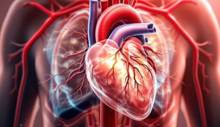
Pericardial effusion is a condition that involves the build-up of extra fluid in the tissue sac around the heart. To understand this more clearly, imagine that the heart is wrapped up in a thin sack. This sack typically contains a small amount of fluid (between 15 to 50 mL) to keep everything working properly. However, sometimes too much fluid gathers in this sack, which is what we refer to as pericardial effusion.
This sack around the heart is made up of two layers. The inner layer (which touches your heart) is a thin one made up of a single layer of cells, while the outer layer, which attaches to the lungs, diaphragm, sternum, and blood vessels, is thicker and made up of collagen and elastin. These layers together make up the pericardial sac.
What Causes Pericardial Effusion?
The root cause of pericardial effusion, or fluid build-up around the heart, can be attributable to a wide range of factors that fall into several categories:
– Infectious: Certain types of infections, from viruses, bacteria, fungi, or parasites, may cause pericardial effusion.
– Inflammatory or Rheumatologic: Certain autoimmune diseases, like systemic lupus erythematosus, rheumatoid arthritis, and Sjogren syndrome, which are conditions where the body mistakenly attacks its own tissues, can lead to this fluid buildup.
– Neoplastic: Pericardial effusion can also take place as a result of various types of cancer, including primary heart tumors or metastatic disease (cancer spread from other areas). Lung cancer is notably the most common cause of cancer-related pericardial effusion.
– Trauma: Injuries to the heart, aorta, or coronary vessels—particularly from blunt force, puncture wounds, or surgical procedures—can cause blood to collect within the sac around the heart.
– Cardiac: It can also occur following a heart attack (known as Dressler syndrome), heart surgery, or a tear in the heart wall.
Additionally, issues like aortic dissection (a severe condition in which the inner layer of the aorta tears), a number of still unexplained reasons, or other causes like radiation exposure, chronic kidney disease, heart failure, liver cirrhosis, hypothyroidism, ovarian hyperstimulation syndrome (a response to excess hormones), and certain drug reactions can lead to pericardial effusion.
Because pericardial effusion can come from so many different causes, it’s essential for doctors to conduct a thorough clinical evaluation and use appropriate diagnostic tests. Understanding these varied causes is key for providing effective treatment.
Risk Factors and Frequency for Pericardial Effusion
Pericardial effusion is a condition that can affect anyone, regardless of their age or population group. This condition is defined by fluid build-up around the heart, and its causes, or ‘etiologies’, can vary depending on factors such as age, location, and existing health issues. It’s tricky to provide exact figures on how common the condition is due to limited data. However, we do know that it’s often caused by viral pericarditis in developed countries, whereas in less developed regions, a bacteria called Mycobacterium tuberculosis is a common cause. Bacterial and parasitic causes are less common.
Certain types of cancer can also result in noninflammatory pericardial effusions. Around 12% to 23% of pericardial effusion cases involve a malignancy. Among patients with human immunodeficiency virus (HIV), pericardial effusion occurs in 5% to 43% of cases, with 13% of these reported as moderate to severe. A study involving children identified the following major causes:
- Postcardiac surgery (54%)
- Cancer (neoplasia) (13%)
- Renal conditions (13%)
- Idiopathic or viral pericarditis (5%)
- Rheumatologic conditions (5%)
These statistics show that both inflammation-causing and non-inflammation-causing conditions can lead to pericardial effusions, with the causes varying significantly among different groups of people.
Signs and Symptoms of Pericardial Effusion
Pericardial effusion, a condition where fluid accumulates around the heart, can have a range of symptoms. These can differ from person to person depending on how quickly the fluid accumulates. In some people, as little as 100 mL of rapidly accumulating fluid can cause problems with the heart’s ability to fill with blood, leading to decreased blood flow from the heart. On the other hand, slow, gradual accumulation of fluid may lead to around 1 to 2 liters of fluid around the heart, but without significant impacts on heart function.
For cases where pericardial effusion leads to inflammation of the heart lining (known as pericarditis), typical symptoms include chest pain and shortness of breath. These symptoms often get better when sitting up and worse when lying down. Additionally, individuals may show non-specific symptoms like shortness of breath, swelling, and tiredness. It’s also important for doctors to consider if there are any other underlying conditions in the patient, such as recent illnesses, cancer, tuberculosis, autoimmune disorders, chronic kidney disease, kidney failure, heart failure, an underactive thyroid, and liver disease.
Doctors also look for signs of pericardial tamponade, a dangerous condition where fluid pressure around the heart impedes its ability to pump blood. This condition should be considered if the patient shows abnormal vital signs such as low blood pressure and a fast heart rate.
In cases of a potential pericardial effusion or cardiac tamponade, the doctor will likely take a look at the patient’s health history, and do a physical examination which may include checking for a drop in systolic blood pressure during a deep breath, known as pulsus paradoxus. This might indicate that the right part of the heart is collapsing the left part, causing a decrease in blood pressure. Besides these, the doctor may also use imaging tests such as an echocardiogram for further confirmation.
With this summary, you can understand that pericardial effusion and the potential complication of cardiac tamponade can manifest through various signs and symptoms:
- Rapid fluid accumulation can cause decreased heart function even with as less as 100 mL fluid
- Slow fluid accumulation may result in up to 1-2 liters of fluid without significant symptoms
- Patients typically report chest pain and difficulty in breathing, which improve when sitting and worsen when lying flat
- Patients may also report general symptoms such as breathlessness, swelling, and fatigue
- Conditions like recent illnesses, cancer, tuberculosis, autoimmune disorders, heart and liver diseases can coexist
- Signs of cardiac tamponade include low blood pressure and high heartrate
- Physical examination and echocardiogram assist in confirming diagnosis
Testing for Pericardial Effusion
If your doctor suspects that you might have pericardial effusion, which is a buildup of fluid around your heart, they will use several tests. These tests don’t individually confirm pericardial effusion, but collectively, they help the doctor make an accurate analysis.
The first of these tests is a chest X-ray. However, this X-ray might not directly indicate a pericardial effusion. But if your heart appears boot-shaped or if it looks like a ‘water bottle,’ it could suggest a chronic pericardial effusion. Your X-ray might also show lung congestion or excess fluid in the lungs or chest, but these signs aren’t always indicative of pericardial effusion.
Another tool your doctor might use is an electrocardiogram (ECG), which measures your heart’s electrical activity. The ECG results can vary, showing normal readings or changes in the ST-segment, a part of the ECG readout, especially for small effusions. When the effusion is large or leads to ‘tamponade,’ a dangerous compression of the heart, the ECG might show ‘electrical alternans.’ This term refers to signals of different sizes, which could suggest that your heart is swinging back and forth within the fluid-filled sac enveloping your heart, known as the pericardium. If the pericardial effusion is due to inflammation of the pericardium (pericarditis), your ECG might show PR depression or diffuse ST elevation. Remember, ECG results vary, and one single reading can’t confirm pericardial effusion.
When it comes to diagnosing a pericardial effusion, echocardiography is the most preferred tool. It’s a type of ultrasound that can look at your heart in motion. The doctor may use either transthoracic echocardiography (TTE), which involves placing the ultrasound probe on your chest or transesophageal echocardiography (TEE), where the probe is inserted down your throat into your esophagus. With echocardiography, the doctor can view the fluid around your heart (pericardial effusion) and any signs that your heart is being compressed by this fluid (cardiac tamponade). The fluid appears black (anechoic) on the echocardiogram, and sometimes, if there are clots or debris in the fluid, they may appear grey (hypoechoic).
Many criteria help the doctor diagnose cardiac tamponade using echocardiography. The doctor will look for things like collapse or change in the shape of different heart chambers, increased motion of the wall dividing the heart chambers, a dilated inferior vena cava – a large vein that carries deoxygenated blood into your heart, and changes in the blood flow across different heart valves. Some of these findings are particularly indicative of cardiac tamponade, others less so.
Similarly, computerized tomography (CT) or magnetic resonance imaging (MRI) of the chest or heart can identify pericardial effusion, but they are not as commonly used as the echocardiography in evaluating for pericardial effusion.
Treatment Options for Pericardial Effusion
The treatment of pericardial effusion, which is a buildup of excess fluid around the heart, varies depending on the cause and severity. Smaller instances of this condition that don’t affect heart function might just be monitored periodically by an imaging technique called an echocardiogram, or in some cases, require no additional follow-up at all. Larger fluid buildups might be drained, either to discover their cause or to provide relief from symptoms like shortness of breath, chest discomfort, swelling in the legs, or reduced physical ability.
If the fluid buildup happens quickly, or grows to the point where it starts to impact heart function, immediate treatment is required. The approach to draining the fluid can include using a needle (called a pericardiocentesis), inserting a special drainage tube, or even performing surgery. The exact method preferred will depend on what’s causing the buildup, the patient’s condition at the time, and their expected course of recovery.
It’s important to note that patients with large pericardial effusions and underlying heart dysfunction carry a risk of developing a serious complication called pericardial decompression syndrome (PDS) after pericardiocentesis. This condition, which is rare but life-threatening, can cause instability in blood flow or fluid accumulation in the lungs after fluid is drained from around the heart. To prevent this, it’s crucial to closely watch patients, especially those who have fluid drained because of a suspected buildup due to cancer.
A good approach may be to avoid removing large amounts of fluid at once, especially in cases of large pericardial effusions. The best treatment plan often involves draining enough fluid to relieve the problem causing the fluid buildup (known as cardiac tamponade), which can be guided by monitoring vital signs or using an echocardiogram. After that, draining of more fluid can be done over a longer period until the amount of fluid being removed falls below 30-50 mL per day, which shows that the problem has been alleviated.
What else can Pericardial Effusion be?
When trying to identify the cause of fluid build-up around the heart (pericardial effusion), a doctor will need to rule out multiple potential conditions. These include:
- Acute pericarditis (an inflammation of the pericardium)
- Cardiac tamponade (pressure on the heart caused by fluid)
- Cardiogenic pulmonary edema (fluid leakage into the lungs)
- Constrictive pericarditis (hardening/thickening of pericardium)
- Dilated cardiomyopathy (weak and enlarged heart chambers)
- Effusive-constrictive pericarditis (combined inflammation and hardening of pericardium)
- Myocardial infarction (heart attack)
- Pulmonary embolism (blood clot in the lungs)
To make the correct diagnosis and treatment plan, the doctor will need to use careful examination and specific diagnostic tests.
What to expect with Pericardial Effusion
Pericardial effusion, or the buildup of excess fluid around the heart, can happen on its own or be tied to certain underlying health issues like autoimmune or cancerous diseases. An ultrasound of the heart, or echocardiography, is essential in diagnosing pericardial effusion, measuring the amount of fluid, and regularly checking how this fluid affects the heart when it is relaxing and filling with blood.
If the ultrasound doesn’t provide clear results, more advanced forms of imaging such as computed tomography (a type of X-ray that gives a 3D image) or cardiac magnetic resonance imaging (an imaging method that uses powerful magnets and radio waves) can be used.
When it comes to treating patients, doctors follow the 2015 European Society of Cardiology’s guidelines for diseases of the pericardium (the outer covering of the heart). The first step involves examining if the effusion is affecting the heart’s ability to pump blood or if it is because of cancerous or infected fluid. The next step involves checking the levels of C-reactive protein in the blood, a substance that increases when there’s inflammation in the body. Doctors also test for conditions known to cause pericardial effusion.
Lastly, doctors consider the size and how long the effusion has been present. The treatment plan is personalized based on these things. The outcome of long-term pericardial effusions largely depends on what is causing them.
Recent studies suggest that people with unexplained, long-term (over 3 months), large (more than 2 cm), and symptomless pericardial effusions usually have a good outlook. Careful monitoring is often more sensible and cost-effective than routinely draining the fluid, which was the prior standard treatment.
Possible Complications When Diagnosed with Pericardial Effusion
A recent research study compared two techniques for managing excess fluid around the heart, known as pericardial effusion: pericardiocentesis and the pericardial window procedure. Both procedures had high success rates, with pericardiocentesis achieving 95% success and the pericardial window procedure achieving a full 100%. However, this difference in success rates was not statistically significant.
Patients who had the pericardial window procedure tended to stay in the hospital more than 7 days, compared to those who had pericardiocentesis. Specifically, 47% of the pericardial window group had this extended hospital stay, versus 17% of the pericardiocentesis group.
But in terms of fluid reaccumulation within 30 days after treatment, the pericardiocentesis group had a significantly higher rate (34%) compared to the pericardial window group (0%). Lastly, the pericardial window procedure had a higher rate of serious complications such as major bleeding and infection. The mortality rate between the two procedures was not significantly different.
Procedures Comparison:
- The success rate is almost the same for both procedures, with a slightly higher success for the pericardial window
- Patients undergoing a pericardial window procedure tend to stay longer in the hospital
- Fluid reaccumulation is significantly higher in patients undergoing pericardiocentesis
- Major complications, like heavy bleeding and infection, were higher in the pericardial window procedure
- There is no significant difference in the death rate between the two procedures
Preventing Pericardial Effusion
Doctors need to always be alert for the chance of a severe build-up of fluid around the heart – known as a large pericardial effusion – that’s on the verge of causing life-threatening pressure on the heart, known as tamponade. This can happen in patients even if they’re not showing any symptoms, or if their initial health concerns seemed unrelated. Patients who have this major fluid build-up around their heart can rapidly worsen, no matter when they first started showing symptoms. So, it’s especially crucial for doctors to watch closely for any major fluid changes in patients.


