What is Pulsus Bisferiens?
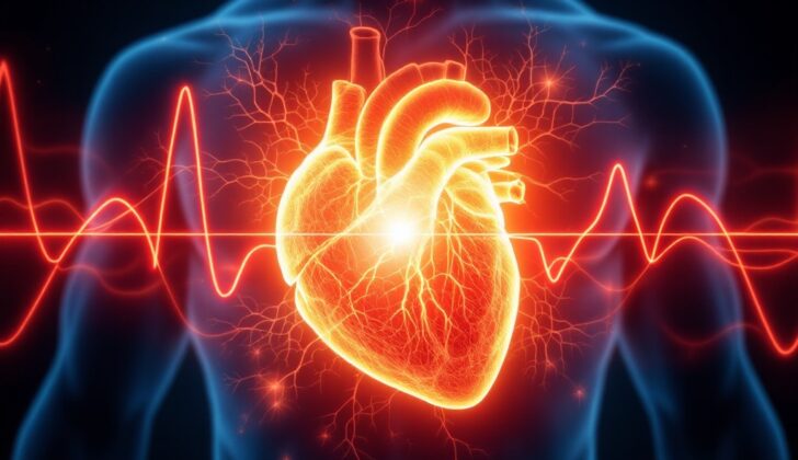
A pulse is like a rhythmic wave that’s created when the main pumping chambers of your heart, called ventricles, contract. Sometimes, you may feel a double pulse during each heartbeat. This kind of pulse is known as pulsus bisferiens, Latin for “strike twice”. It’s also referred to as a biphasic wave, meaning that it has two phases. This phenomenon was first identified in 1952.
Pulsus bisferiens is usually connected with severe aortic disease, a condition that affects the main artery in our body, the aorta. It is associated with aortic regurgitation, where the aortic valve doesn’t close tightly and allows some blood to leak back into the heart, and hypertrophic obstructive cardiomyopathy (HOCM), a disease where the heart muscle becomes abnormally thick. This double pulse can often be misinterpreted by doctors as a condition called a dicrotic pulse. However, what sets pulsus bisferiens apart is you’ll notice two peaks in a heartbeat instead of one. On the other hand, a dicrotic pulse has one peak in the heartbeat and another peak in the pause between heartbeats. A dicrotic pulse usually occurs when the heart isn’t pumping enough blood, during sepsis, an infection that can trigger inflammation throughout the body, or cardiac tamponade which is fluid buildup around the heart.
To define it, pulsus bisferiens is a single heartbeat with two peaks that are separated by an observable dip. The first peak occurs due to the rapid ejection of blood from the left ventricle of the heart, while the second peak corresponds to the bounce-back wave created by the main arteries.
Throughout history, there have been many discoveries regarding the pulse. Some notable examples include:
* Galen, in 1932, was the first to observe a double-pulse.
* In 1872, Mahomed was the first to label the terms “percussion wave” and “tidal wave.”
* Broadbent, in 1900, noted pulsus bisferiens in relation to aortic valve disease.
* Bramwell, in 1937, proposed the theory of the origin of the abnormal pulse.
* Fleming, in 1957, explained the mechanism of pulsus bisferiens.
What Causes Pulsus Bisferiens?
“Pulsus bisferiens” is a condition where you feel a double pulse when you take a person’s heartbeat. Here are the most usual reasons for it:
– Combination of diseases that affect the aortic valve (the valve that controls the flow of blood out of the heart), including:
– Infective endocarditis – an infection of the inner lining of the heart
– Rheumatic heart disease – damage to the heart caused by rheumatic fever
– Marfan syndrome – a genetic disorder affecting the connective tissue
– Bicuspid aortic valve – a heart defect where the aortic valve only has two leaflets instead of three.
– Hypertrophic cardiomyopathy with obstruction (HOCM): a disease that makes the heart muscle abnormally thick, making it harder for the heart to pump blood.
And here are some less typical reasons:
– Severe aortic regurgitation: When the aortic valve doesn’t close tightly, causing blood to leak into the heart.
– Large PDA (patent ductus arteriosus) or arteriovenous fistula: these are conditions where there is an abnormal connection between arteries and veins.
– Significant mitral valve prolapse: A condition where the door-like flaps of the heart’s mitral valve do not close correctly.
– Sometimes in normal people where there is a high blood flow round the body.
Risk Factors and Frequency for Pulsus Bisferiens
Pulsus bisferiens is often related to severe aortic regurgitation and hypertrophic cardiomyopathy. Here is a simplified explanation:
- Severe aortic regurgitation usually happens in people between their fourth and sixth decade and becomes more common with age, especially in older adults.
- It’s more common in men (13%) than in women (8.5%).
- In the United States, about 4.9% of the population have aortic regurgitation and 0.5% have severe aortic regurgitation according to the Framingham study.
- Hypertrophic obstructive cardiomyopathy, or HOCM, is the most common heart disease that can be inherited. It is found in the US in about 1 out of every 500 people (0.2%) and in Germany in about 1 out of every 1,372 people (0.07%).
- HOCM affects males more than females.
Rheumatic heart disease and bacterial endocarditis are more common in developing countries like Asia and Africa. Aortic diseases are seen at a rate of 10 to 374 cases per 100,000 people, particularly in the Pacific and Australia.
Signs and Symptoms of Pulsus Bisferiens
Age can provide valuable information when diagnosing certain illnesses. For children, if a double-pulsed heartbeat can be felt, this could indicate a condition known as Patent Ductus Arteriosus (PDA) with a left-to-right blood flow. On the other hand, younger patients might hint towards conditions like obstructive hypertrophic cardiomyopathy or a two-part heart valve disease. For the older population, mixed conditions involving the aortic valve are more common. A pulse that seems more pronounced when performing a specific breathing exercise (Valsalva maneuver) is a key sign.
Common symptoms of these conditions could include fainting, difficulty breathing, chest pain, and a racing heart. In some cases, people could have these conditions for a long time without showing any symptoms before suddenly experiencing symptoms of lung fluid overload and heart failure. It’s noteworthy to mention that obstructive hypertrophic cardiomyopathy can lead to unexpected cardiac arrest and life-threatening irregular heart rhythms, notably in younger athletes.
During a physical examination, doctors check for a double-pulsed heartbeat by feeling the patient’s arm pulse (radial artery). Other signs that might be checked for include:
- Bigger pulse pressure, which could indicate aortic regurgitations
- Distinctive pressure observed in neck veins, typically seen in obstructive hypertrophic cardiomyopathy
- The heart’s apex seems to be out of place in severe aortic regurgitations. In obstructive hypertrophic cardiomyopathy, doctors can feel a double heartbeat at the heart’s apex.
During a detailed heart checkup, doctors might hear various abnormal heart sounds, which could help diagnose these conditions. Some of these sounds could indicate aortic stiffening, an early diastolic murmur, or a specific type of heart murmur known as the Austin Flint murmur, or the presence of Obstructive Hypertrophic Cardiomyopathy.
Aortic regurgitation is also associated with a variety of other clinical signs. These could include repeated color changes in the face and forehead, head nodding in tune with the heartbeat, fluctuations in the nail bed’s blood vessel color, alternating pupil sizes, visible artery beats in the retina, uvula pulsations, pulsations in the liver or spleen, certain audible sounds during heartbeats, and specific types of murmurs heard over the femoral arteries.
Testing for Pulsus Bisferiens
When it comes to diagnosing bicuspid aortic valve disease, doctors often use a variety of tests. The first one is a blood test to check the levels of two specific substances: high-sensitivity troponin T (Tn-T) and N-terminal pro-B-type natriuretic peptide (NT-proBNP). If the levels of these substances are high, it points towards the disease.
The next step could be a chest X-ray. This imaging test can show if there’s enlargement of parts of the heart (left atrial enlargement and left ventricular hypertrophy or LVH) or fluid accumulation in the lungs – a condition known as heart failure or pulmonary edema.
An Electrocardiogram (ECG) is another common test that can highlight abnormalities in the heart’s electrical activity, such as irregular heart rhythms (atrial fibrillation), or changes in the heart’s size that suggest disease.
Echocardiography, also known as a 2D-Echo, is a key tool in diagnosing this disease. It uses sound waves to provide a detailed picture of the heart’s structure and function. It can observe the shape of the aortic valve, measure how much blood is flowing backwards (a condition called aortic regurgitation), and even help assess whether heart surgery might be needed. It also provides valuable information on the size and function of the heart’s chambers.
For a more detailed view, doctors may also use Cardiac magnetic resonance imaging (MRI). An MRI can give a clear picture of the heart’s structure and how well it’s pumping blood. It’s becoming an essential tool to assess the severity of valve disease and to measure changes in the heart’s size (a process known as LV remodeling). It’s also the best test to diagnose hypertrophic cardiomyopathy (HOCM), another heart condition.
A Cardiac Computed Tomography (CT) scan can provide accurate measurements of the heart and blood vessels, which is crucial for planning certain treatments such as transcatheter interventions – a minimally invasive procedure used to repair the valve.
Lastly, Nuclear Imaging and Fluoroscopy can be used to measure the severity of infection, assess the metabolic activity of the heart, and detect the presence of calcium deposits which can signal heart disease.
Treatment Options for Pulsus Bisferiens
Managing a condition known as pulsus bisferiens really depends on what is causing it. Therefore, early diagnosis and a quick referral to a heart doctor are key to successful treatment and to lowering the risk of serious complications or death. A mixed aortic valve disease and a condition known as hypertrophic cardiomyopathy with obstruction are the most common causes of pulsus bisferiens.
Medication Management
In cases of mixed aortic valve disease, vasodilators may help patients by reducing the amount of blood the heart has to pump out (preload) and increasing the heart’s overall performance (cardiac output). Medicines like ACE inhibitors and angiotensin receptor blockers, which help control heart rate and blood pressure, might also be helpful. Beta-blockers may be less beneficial for treating patients with a specific form of this disease known as aortic regurgitation.
For hypertrophic obstructive cardiomyopathy, beta-blockers are generally the first treatment method. Calcium channel blockers like verapamil or diltiazem can also be advantageous in treatment as a second resort. ACE inhibitors and ARBs, which generally act to relax and widen the blood vessels, are best avoided. Similarly, medicines that promote the widening of blood vessels (vasodilators) should also be avoided.
For a condition called patent ductus arteriosus, early use of medications like indomethacin, ibuprofen, or acetaminophen to close a birth-caused passageway in the heart could prevent further complications. Diuretics can be used to treat acute fluid build-up in the lungs.
Surgery Management
In mixed aortic valve disease situations, recent advancements in surgical techniques, especially those related to valve repair; have improved the results and patient outcomes. Valve-sparing surgical repair is becoming a popular alternative to typical surgeries. A technique known as transcatheter aortic valve replacement has revolutionized the treatment of patients with a severe aortic stenosis, providing minimally invasive treatment options for many patients.
In hypertrophic obstructive cardiomyopathy situations, in certain severe cases, a dual chamber pacemaker and potentially an implantable cardioverter-defibrillator to regulate abnormal heart rhythms could be beneficial. The surgical removal of a portion of thickened heart muscle (myomectomy) improves patient outcomes. Another procedure called septal alcohol ablation may also be effective in 9 out of 10 severe cases.
In patent ductus arteriosus situations, surgical binding is an option for newborns with a significant PDA, although its role in treatment can be controversial.
What else can Pulsus Bisferiens be?
When feeling a pulse, doctors sometimes become unsure whether it’s a “bisferiens pulse” or a “dicrotic pulse”. These two types of pulses can be confusing to tell apart. In a dicrotic pulse, you can feel two beats – one as the heart contracts (systole) and the other as it relaxes (diastole). But for the bisferiens pulse, both beats happen during the heart’s contraction or systole. Dicrotic pulse is something you often see in people who are in heart failure or who are very sick with another severe condition like sepsis. Sometimes, healthy people can also have a dicrotic pulse after exercising.
What to expect with Pulsus Bisferiens
Mixed Aortic Valve Disease generally has a good prognosis for patients with severe aortic regurgitation who show no symptoms and have a preserved LV function, which refers to the functioning of the left side of the heart. On the other hand, patients with moderate to severe aortic regurgitation might not have the same positive outcomes. In these cases, surgical intervention or aortic valve replacement can improve their chances of survival.
Hypertrophic Obstructive Cardiomyopathy (HOCM), a condition where the heart muscle becomes abnormally thick, has a very low mortality rate for patients, meaning the chances of death are low. Generally, patients with HOCM have a good long-term outlook. However, a small fraction of people with this condition are at risk of sudden cardiac death.
With Patent Ductus Arteriosus (PDA), a condition where a blood vessel called the ductus arteriosus fails to close after birth, severity scores have been developed to predict serious outcomes. Naturally, in most healthy newborns, the ductus closes on its own. Only 3% of infants weighing more than 1,000 grams may need to undertake a PDA intervention, which means a medical procedure to close the open blood vessel.
Possible Complications When Diagnosed with Pulsus Bisferiens
People with a condition known as pulsus bisferiens can face several complications such as:
- Heart failure, where the heart can’t pump blood effectively
- Stroke, a sudden disruption of blood supply to the brain
- Arrhythmias, abnormalities in the heart’s rhythm
- Infective endocarditis, an infection of the heart’s inner lining
- Sudden cardiac death, an unexpected loss of heart function
Preventing Pulsus Bisferiens
As people get older, there is an increasing number who are diagnosed with severe mixed aortic disease. This can be a serious condition, but the appropriate treatments can really help. Aortic disease is a condition that affects the aorta, the main artery in your body. It can cause the walls of the aorta to bulge and weaken.
Optimal treatment methods for patients with severe aortic disease, hypertrophic obstructive cardiomyopathy (HOCM), or patent ductus arteriosus (PDA) can greatly improve their overall health. HOCM is a condition where the heart muscle becomes thick and can block blood flow, while PDA is a condition in which the ductus arteriosus, an artery in the heart, doesn’t close properly after birth.
An echocardiogram, which is a type of ultrasound test that uses sound waves to capture images of the heart, can be used as a screening tool. This can help doctors diagnose these diseases early and determine how severe they are. Screenshots are especially beneficial because they can detect any complications or damage caused by the disease.
If someone in your family is diagnosed with HOCM, it’s recommended that all first-degree relatives also get screened. This is because HOCM is an inherited disease, meaning it can be passed down from parent to child.


