What is Sinus of Valsalva Aneurysm?
A Sinus of Valsalva aneurysm or SOVA, is a rare heart condition where the root of the aorta (the main blood vessel supplying oxygenated blood to our body) unusually expands. This happens because of a weakness at the spot where the aorta and the heart’s fibrous ring connect. Under normal conditions, these ‘sinuses’ in the aorta help prevent blockages in the artery supplying the heart when our heart beats and the aortic valve opens. On average, in men, these sinuses are less than 4 cm and in women, less than 3.6 cm in diameter.
These aneurysms usually occur in isolation, but rare cases involve growths in two or three sinuses. The most common location for a SOVA is the right coronary sinus, and it often has no symptoms until it bursts. In such a case, the grown sinus can burst into a nearby heart chamber based on its position. The right, left, and non-coronary sinuses are close to different parts of the heart, such as the wall separating the two lower chambers of the heart, the wall of the left lower chamber, and the wall separating the two upper chambers of the heart, respectively. Should a rupture occur, it creates a passage between the aorta and the heart chamber, causing the heart to fail progressively. Without adequate treatment, this can lead to grim health outcomes and high death rates, which is why it’s crucial to intervene as soon as possible. Anyone diagnosed with SOVA should consult a heart surgery specialist immediately.
About 0.2% to 0.9% of all heart surgery patients may have SOVA. The condition can either be congenital – present at birth, or acquired – developed later in life. Congenital SOVA often happens in men from Asian descent. It can occur due to abnormal development of certain heart areas, and often, coexist with other heart conditions such as holes in the wall separating the two lower chambers of the heart, leaky aortic valve, or a two-leaflet aortic valve instead of the usual three. Acquired SOVA can occur due to earlier surgeries, narrowed arteries due to fat and cholesterol deposits, inflammation of the heart’s inner lining, syphilis, and other types of trauma.
What Causes Sinus of Valsalva Aneurysm?
As mentioned before, Sinus of Valsalva Aneurysms (SOVAs) can either be present from birth (congenital), or develop later in life (acquired). Congenital SOVAs are due to abnormal development of a part of the heart during birth, leading to weak spots in the aortic wall. This kind of SOVA is often associated with different heart problems, with ventricular septal defect (VSD) – a hole in the wall separating the heart’s main pumping chambers – being present in 30% to 60% of cases. Other commonly linked conditions include aortic valve regurgitation (backflow of blood due to a leaky heart valve), bicuspid aortic valve (having two instead of three parts in the aorta), pulmonic stenosis (narrowing of the valve from the heart to the lungs), coarctation of the aorta (narrowing of the large blood vessel), atrial septal defect (hole in the wall between the upper chambers of the heart), and sometimes, coronary artery anomalies. Also, aneurysms that affect the parts connecting the aortic and mitral valves of the heart are known to be associated.
Acquired SOVAs, in contrast, are caused by factors that weaken the aortic wall over time, including problems in the connective tissues and infectious diseases. The Sinus of Valsalva forms as a sort of pocket due to pressure on the root of the aorta. This pocket can stretch and become an SOVA because of weaknesses in the connective tissues from disorders like Marfan syndrome and Ehlers-Danlos syndrome. This is also commonly associated with having a bicuspid aortic valve. Other connective tissue issues include chronic changes due to atherosclerosis (hardening of the arteries), chest injury, and accidental damage during heart valve replacement surgery. Additionally, vasculitides (inflammation of the blood vessels) affecting the beginning portion of the aorta like Takayasu arteritis (inflammation and damage to the large blood vessels of the heart) and inflammatory aortitis (inflammation of the aorta tissue) can lead to the formation of SOVA. Infections that weaken the tissue and lead to acquired SOVA include syphilis, tuberculosis, and infective endocarditis (infection of the inner lining of the heart).
Risk Factors and Frequency for Sinus of Valsalva Aneurysm
Sinus of Valsalva aneurysms (SOVAs) are quite rare, and only affect a very small portion of people who undergo heart surgeries. These aneurysms are even less common in the wider population, and often don’t get detected until a serious issue like a rupture happens. SOVAs account for a minor proportion of all the birth defects that involve the heart. People of Asian descent and males are at a higher risk of developing these types of aneurysms, suggesting that certain genetic factors might be involved. Because they are often present at birth, they are more commonly found in younger patients. However, older adults can also acquire SOVAs due to other risks such as heart disease from hardened arteries and past infections. It’s estimated that a majority of these aneurysms occur in the right section of the upper part of the heart known as the right coronary sinus. Unfortunately, these persons often don’t show any symptoms until an aneurysm ruptures, which can be very dangerous if not treated quickly.
- Sinus of Valsalva aneurysms (SOVAs) are extremely rare, affecting only 0.2% to 0.9% of individuals undergoing heart surgery.
- The occurrence of SOVAs is even less in the general population.
- Many cases go unnoticed until a rupture occurs.
- SOVAs constitute up to 3.5% of all heart defects present at birth.
- Men, especially those from Asian populations, are more likely to have these aneurysms, hinting at possible genetic factors.
- Young people tend to have congenital SOVAs more frequently due to their presence at birth.
- Older adults can also acquire SOVAs from risk factors like hardening of the arteries and previous infections.
- About 70% of SOVAs are found in the right coronary sinus, an upper part of the heart.
- Many SOVAs don’t show symptoms until a rupture happens, which can cause severe problems if not addressed immediately.
Signs and Symptoms of Sinus of Valsalva Aneurysm
A Sinus of Valsalva aneurysm, or SOVA, is a condition that may not always show symptoms. Sometimes, it’s discovered by accident when imaging studies are done for other health reasons. However, if someone with an intact SOVA does have symptoms, they might experience a range of symptoms such as chest pain, difficulty breathing, and irregular heartbeats.
- Chest pain: This can be confusing as it may be mistaken for angina or other heart-related conditions.
- Difficulty breathing: This can happen due to the aneurysm causing pressure on heart structures, and in severe cases, this could lead to heart failure.
- Irregular heartbeats: The aneurysm might affect the electric system of the heart causing these.
If the SOVA bursts open (ruptures), the symptoms become more severe and sudden:
- Severe chest pain: This is often described as a tearing or ripping sensation.
- Sudden difficulty in breathing: This could be due to a quick onset of heart failure.
- Fainting: Sudden loss of consciousness resulting from major instability in blood flow.
People might also report having had earlier heart surgery, trauma, or health conditions that could make them more prone to getting an acquired SOVA, such conditions could be an inflammation of the heart’s inner lining(endocarditis) or the sexually transmitted infection syphilis.
The physical exam findings in people with SOVA could range from not noticeable in cases without symptoms, to very obvious in cases where it has ruptured. These findings include:
- Heart murmurs: This is abnormal heart sounds that occur if the SOVA has ruptured into a heart chamber, causing unusual blood flow patterns. They could also be indicative of an aortic valve affected by the aneurysm leading to aortic regurgitation.
- Signs of heart failure: Things like a rise in jugular venous pressure, swelling in the peripherals of the body, and crackling sounds in the lungs that could point to fluid buildup.
- Pulse pressure: The difference between the systolic and diastolic blood pressure might be seen to increase, particularly if there’s significant aortic regurgitation.
In cases where the SOVA has ruptured, more findings could be observed:
- Low blood pressure (hypotension): This could happen due to acute loss of blood and disruption in blood circulation (hemodynamic collapse).
- Rapid heart rate (tachycardia): This is the heart’s response to low blood pressure.
- Bluish skin or mucous membrane discoloration (cyanosis): This results from inadequate oxygen supply.
Testing for Sinus of Valsalva Aneurysm
When it comes to diagnosing and treating a Sinus of Valsalva Aneurysm (SOVA), a type of heart condition, doctors use a mixture of physical assessments, imaging studies, and diagnostic tests. These help confirm the presence of the aneurysm, understand its size and location, and plan the best course of action for treatment.
There are several types of imaging tests doctors might use:
Echocardiography
– Transthoracic echocardiography: This technique allows your doctor to see the root of the aorta (major blood vessel), identify the aneurysm, check its size, and spot any related issues like aortic regurgitation (leakage of the aorta valve) or VSD (hole in the wall between the heart’s two lower chambers).
– Transesophageal echocardiography: This technique gives more detailed images, so it’s really good at showing the aneurysm’s size and how it affects structures around it. Color Doppler, a specific method, can even show a continuous blood flow if SOVA has ruptured.
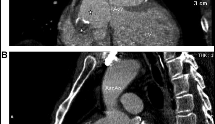
Computed tomography angiography
This test offers high-resolution images of the aortic root and sinuses of Valsalva (pouches between the aorta and heart). It is the best test for measuring the aneurysm’s size and shape and is especially valuable when preparing for surgery.
Cine cardiac magnetic resonance imaging
This is considered the top-notch method for diagnosing SOVA, but it isn’t required if other imaging tests have provided enough information. It offers excellent contrast in soft tissues and detailed anatomical information, making it useful for patients who can’t have computed tomography due to allergies or kidney issues.
Cardiac Catheterization
– Angiography: An invasive procedure that gives detailed images of the coronary arteries and the aortic root. This method is particularly useful for measuring the aneurysm’s impact on blood flow if surgery is planned. Patients with a moderate or high risk for coronary artery disease generally have this procedure to evaluate the need for bypass grafting during cardiac surgery.
Lab tests are an essential part of the process too:
Blood tests
– Complete blood count: This test looks for signs of infection or anemia, which could make treating SOVA more complicated.
– Inflammatory markers: If markers like C-reactive protein or erythrocyte sedimentation rate are high, it might indicate ongoing inflammation or infection.
– Syphilis serology: Because syphilis has historically been associated with aortic aneurysms, testing for syphilis may be necessary.
Additional testing might include an electrocardiogram, which looks for any electrical abnormalities or evidence of insufficient blood supply to the heart that might result from the aneurysm.
Treatment Options for Sinus of Valsalva Aneurysm
When it comes to managing SOVA (Sinus of Valsalva Aneurysm), a type of bulge in your aorta, the treatments offered are usually to help keep the patient stable until a more permanent solution can be provided. This can be achieved through surgery or a procedure called transcatheter intervention. For cases where the aneurysm hasn’t ruptured, monitoring its size and progress through regular scans such as an echocardiography, becomes part of the treatment. Managing blood pressure is also crucial, with medications like beta-blockers, angiotensin-converting enzyme inhibitors and calcium channel blockers used to lower stress on the aorta. Patients are also advised to avoid heavy lifting and strenuous physical activity which could increase the pressure inside the chest and potentially cause the aneurysm to burst.
In the unfortunate event that the SOVA ruptures, immediate stabilization is a must, involving careful management of fluid levels, blood pressure control, and dealing with symptoms of heart failure or shock until surgery becomes possible.
Definitive treatment for SOVA often involves surgery, especially for large, symptomatic, or rapidly expanding aneurysms, and all those that have ruptured. The principal aim is to prevent a rupture and restore the normal function of the aorta and heart. The decision to opt for surgery would largely depend on the severity of symptoms and the speed at which the aneurysm is expanding. Specific size guidelines have been outlined in the 2010 American Guidelines for Thoracic Aortic Disease and a growth rate exceeding 0.5 cm per year is considered significant. This surgery typically involves employing a method called cardiopulmonary bypass and then closing the SOVA.
The surgical procedure provides direct access to the heart and the chosen method depends on several factors including the size, location of the aneurysm and whether the aortic valve is involved. It could either involve replacing the affected part of the aorta with a synthetic graft, which might also include preserving the valve or replacing it, or a less invasive technique called “patch repair” might be carried out for smaller aneurysms. Urgent surgical intervention becomes a necessity in case of a ruptured SOVA, due to the high risk of death. This would typically involve similar techniques but under more urgent circumstances. Follow-up care in an intensive care unit postoperatively is necessary to monitor and manage potential complications such as bleeding, infection or heart failure.
An alternative, minimally invasive procedure to treat ruptured and unruptured aneurysms is called transcatheter closure of SOVA. This technique may be particularly beneficial for those considered at high risk for open-heart surgery due to age, comorbidities, or other factors. The process involves obtaining vascular access, usually through the femoral artery or vein, and under guidance through imaging techniques, a closure device is navigated to the site of the aneurysm, which seals it and eliminates the abnormal communication between the aorta and the heart chambers. Transcatheter closure typically results in a faster recovery and a shorter hospital stay compared to open-heart surgery. Regular follow-up scans are required to ensure the closure remains intact and helps detect any complications related to the device early.
While this technique is becoming more popular for SOVA management, it’s important to note that more studies are required to define the best practice approach in managing these rare conditions.
What else can Sinus of Valsalva Aneurysm be?
When trying to diagnose SOVA, or Sudden Onset Ventricular Arrhythmia, doctors might consider these conditions because they share similar symptoms:
- Aortic stenosis
- Atrial septal defect
- Atrioventricular block
- Dilated cardiomyopathy
- Hypertrophic cardiomyopathy
- Isolated coronary artery anomalies
- VSD surgery
What to expect with Sinus of Valsalva Aneurysm
The outlook for patients with Sinus of Valsalva Aneurysm (SOVA) — a rare abnormal dilation or bulging in the heart— is generally good, especially when surgery is performed on unruptured aneurysms. A recent study showed that most surgeries on unruptured SOVAs went well, with a success rate of 96%. However, in 4% of the cases, patients didn’t survive — one passed away during surgery and another within 48 hours after the surgery due to multiple organ failure. The time patients spent in the hospital after surgery varied between 4 and 21 days, depending on their recovery speed.
Follow-up information for 31 patients, gathered over a period of 5 days to 9 years, revealed that most patients were symptom-free and had fully healed. Their aortic root (the beginning of the aorta, the main artery in the body) returned to normal, and the heart valve worked perfectly well in 94% of the cases. However, a small number of patients experienced complications. One patient needed additional intervention due to a detected leak, and though the aneurysm did not completely heal after surgical repair, it had almost fully healed within 2 months after the intervention. Another patient had a recurrence of the aneurysm 9 years after the initial surgery and had to undergo additional surgery. But all in all, patients with SOVA, especially those who undergo surgery for unruptured aneurysms, generally have a positive long-term outlook.
The prognosis is, however, less promising for patients with ruptured SOVA if they don’t receive immediate surgical intervention. The bursting of the aneurysm can lead to the sudden onset of severe symptoms such as severe chest pain, difficulty breathing, and instability of blood pressure and/or pulse rate. This rupture can also result in significant blood loss, swift development of heart failure, and the potential for multiple organs to fail due to inadequate blood flow. Without immediate surgical repair, the survival rate is low. Early medical attention is essential, as delays can lead to death. Surgical intervention doesn’t guarantee survival due to the complicated nature of the condition, and the patient’s overall health and other existing health conditions can significantly impact the results.
Surgical repair of a ruptured SOVA can help stabilize the patient’s condition and improve long-term outcomes, but the time immediately after the surgery is crucial. The success of the surgery depends on several factors, such as the size and location of the rupture and the state of the patient’s blood pressure and pulse rate at the time of presentation. Other existing heart conditions can also play a role. After surgery, patients usually need close monitoring in an intensive care setting to manage potential complications such as bleeding, infection, and any remaining or recurring aneurysms. Regular check-ups are crucial to monitor for recurrence, assess heart function, and ensure the surgical repair remains intact. Despite the risks, successful surgical intervention can significantly improve the prognosis for patients with ruptured SOVA and enables many of them to achieve long-term, stable health.
Possible Complications When Diagnosed with Sinus of Valsalva Aneurysm
The complications of SOVA (Sinus of Valsalva Aneurysm) can be severe and even fatal, particularly if the aneurysm bursts. Here are the main complications:
- Rupture: This is the most critical and life-threatening complication. A rupture or break can cause abnormal communication between the aorta (the main blood vessel) and adjacent heart chambers or the sack around the heart, leading to a sudden drop in blood pressure. This can manifest as sudden intense chest pain, shortness of breath, low blood pressure, and possibly a life-threatening situation known as cardiac tamponade if blood accumulates in the sac around the heart. If not rapidly treated with surgery, rupture can be fatal.
- Heart failure: Chronic or acute overload of blood due to aortic regurgitation or rupture can cause heart failure. You might see symptoms like shortness of breath, tiredness, swelling, and a reduced ability to exercise.
- Aortic insufficiency: The aneurysm can distort the aortic valve, causing it to be insufficient. This may cause symptoms like breathlessness, fatigue, and heart flutter due to blood flowing back from the aorta into the heart’s left ventricle.
- Endocarditis: The abnormal structure of the aneurysm can become a hotspot for bacterial infection, leading to an infection of the heart’s inner lining, known as endocarditis. Symptoms for this include fever, chills, night sweats, and signs of widespread embolization or blockages in the blood-stream.
- Arrhythmias: The aneurysm can disrupt the heart’s normal electrical pathways. Irregular heartbeats can cause symptoms like heart flutter, dizziness, fainting, and potentially sudden heart failure (if a severe arrhythmia occurs).
- Thromboembolism: Blood stagnation in the aneurysm can cause clots. These clots can travel to different parts of the body, causing strokes or other loss-of-blood-flow events.
- Pressure on nearby structures: A large aneurysm can apply pressure on adjacent heart parts or heart arteries. This can present as chest pain due to heart artery compression or shortness of breath and difficulty swallowing due to pressure on other parts.
- Recurrence: Even after surgical repair, there is a risk of the aneurysm coming back, requiring ongoing check-ups. Regular imaging tests should be done to catch and manage any recurrence early.
Preventing Sinus of Valsalva Aneurysm
To manage and prevent complications from SOVA, a type of heart aneurysm, it’s important for patients to be fully informed and constantly monitored. This includes regular heart checks or echocardiograms, as well as check-ups with a heart specialist, to keep an eye on the size and progression of the aneurysm.
Another crucial aspect is managing risk factors such as high blood pressure and avoiding infections. This could be done with preventive measures like taking antibiotics when needed. It’s also advisable to avoid hard activities such as contact sports and heavy lifting, as these could increase chest pressure and risk breaking the aneurysm.
If a SOVA is hereditary, genetic counseling is recommended, especially for those with family history of related conditions like disorders affecting the connective tissue or heart aneurysms.
Patients should completely understand what SOVA is, what causes it, its symptoms, and the potential complications that might ensue. Timely detection and treatment are crucial, so patients should be taught how to spot any symptoms that might indicate the progression or rupture of the aneurysm. It’s also key for patients to regularly take their prescribed medicine, adopt a heart-friendly diet, quit smoking, and manage their stress levels. Formulating an emergency plan in case of acute symptoms is also vital.
If patients undergo surgery to repair their SOVA, clear instructions on how to care for themselves afterwards, what to look out for in case of complications, and regular follow-up visits are essential. Doctors may also recommend limiting certain activities during recovery and slowly reintroduce them as the healing progress allows.
Supporting patients through patient groups and involving family members in learning sessions can help create a nurturing home environment. This can assist patients in sticking to their treatment plans and leads to better management of SOVA. By taking these measures, patients can avoid complications, improve their quality of life, and help prevent their condition from worsening.


