What is Venous Leg Ulcer?
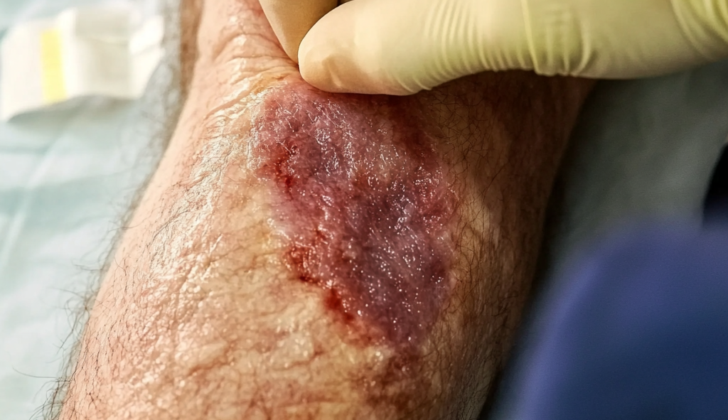
When veins in the legs don’t function as they should over a long period, they can lead to venous leg ulcers (VLUs). VLUs are open sores or wounds on your leg that are difficult to heal. They can be an indication of chronic venous insufficiency (CVI) or high blood pressure in the veins. Normally, the calf muscles and tiny valves inside the veins make sure blood keeps flowing towards the heart, stopping it from flowing backwards. However, when there’s a backflow of blood, a blockage, or both, it results in chronic high blood pressure in the veins. This chronic high blood pressure can subsequently cause skin and blood vessel complications, eventually leading to a venous leg ulcer (see the image titled ‘Venous Leg Ulcer’).
How serious are VLUs? Not only are they a significant drain on global healthcare budgets, but they also have a large human cost. In the United States, the annual costs of treating patients with venous leg ulcers amount to around $18,986 for Medicare patients and around $13,653 for those with commercial insurance. Together, this adds up to a staggering $14.9 billion annually for U.S healthcare payers, a sharp increase from the previous estimate of $1 billion. As the population ages and cases of obesity and sedentariness rise around the world, we can anticipate an increase in the incidents of CVI and VLUs. This growing issue underscores the urgent need for a coordinated and interdisciplinary response from all those concerned with the problem.
What Causes Venous Leg Ulcer?
Chronic venous insufficiency (CVI), a condition where blood doesn’t flow properly through your veins, is often connected to the formation of Venous Leg Ulcers (VLUs), which are open sores that can appear on your leg. However, these ulcers only occur in about 5.1% of CVI cases. This condition might develop due to improper blood flow, a blockage, or both, causing issues with your body’s circulatory system.
The problem begins when pressure builds in your veins, leading to protein leakage and the forming a sort of ‘fibrin cuff’. These chains of proteins can block oxygen and other important growth factors and trigger an inflammatory response within your body.
At the cellular level, a lot of processes contribute to changes in your veins and the formation of varicose veins. Some factors perpetuate the situation by maintaining an inflammatory environment, driving further issues like inefficient blood flow and blood clot formation, leading to further complications. All these inflammatory processes can prevent your body’s natural healing process, leading to the formation of ulcers if a wound appears.
Many of the known risk factors for VLUs are things that people can’t change, such as family medical history, age, sex, history of blood clots or lung clots, multiple pregnancies, certain skin conditions, and muscle and joint diseases. There are also controllable risk factors like obesity and a sedentary lifestyle.
Genetics might also play a role, with certain traits being passed down in families in a way that might make a person more likely to develop these conditions. However, we don’t yet know the exact genes that might be responsible.
Interestingly, there are differences in gene expression between VLUs that heal and those that don’t. Non-healing VLUs show increased activity of certain genes involved in inflammation control, cell growth, and other important processes, while genes responsible for skin repair and collagen formation are less active.
In one study, scientists found that treating VLUs with a special bioengineered skin graft seemed to shift the gene expression profile from a chronically inflamed, non-healing state to one that promotes healing. This shows how chronic inflammation can hinder wound healing.
Risk Factors and Frequency for Venous Leg Ulcer
Chronic wounds on the lower part of the body, known as VLUs, are quite common. It’s estimated that between 1 to 3% of older people in the United States and Europe have these wounds. In a study that included Asia, Eastern Europe, Latin America, and Western Europe, it was found that 2.21% of almost 100,000 patients with CVI, a condition that causes these wounds, had either a healing or an active VLU. The rates varied between the regions, ranging from 1.27% to 3.97%.
It appears that these types of wounds are more common in women than in men, but the specific numbers are hard to determine—the results often vary depending on the study group and location of the study. On average, it takes about 5 years from the time a person is diagnosed with CVI for a wound to occur. One report showed a 4.49% risk of getting a first wound within 3 years for people with CVI, which increased slightly to 4.93% at the 5-year mark.

considerable slough and scarce granulation tissue. The borders are irregular but
well-defined. The surrounding skin shows hyperpigmentation, and there is notable
leg edema.
Signs and Symptoms of Venous Leg Ulcer
It’s important to identify risk factors if someone has a non-healing wound on their lower extremity. This can help differentiate a venous leg ulcer (VLU) from other potential causes. People having chronic venous insufficiency (CVI) or VLUs may often experience itching, sometimes accompanied by a rash, aching pain around the ankles, swelling in the feet in the evening, and cramps during the night.
When examining for early signs of CVI, the first symptoms typically detected include small blood vessels visible just beneath the skin (telangiectases) and veins appearing blue or green just beneath the surface of the skin (reticular veins). More advanced indicators of CVI can include varicose veins, orange-brown skin color changes, chronic swelling in the legs, skin irritations related to poor blood flow (stasis dermatitis), white scars surrounded by capillaries (atrophie blanche), and a type of skin hardening called lipodermatosclerosis.
VLUs, in general, occur towards the lower part of the legs, most frequently over the inner side. They often look like shallow, irregular, well-defined ulcers with yellowish stringy material on the base.
The clinical, etiological, anatomical, and pathophysiological (CEAP) classification records these signs for treatment and research purposes. There are also other metrics like the venous clinical severity score and the Widmer classification, but these are not used as often.
Testing for Venous Leg Ulcer
When checking out ulcers on your skin, doctors will look at the ulcer’s size, how deep it goes, its edges, the base of the wound, any signs of infection, and if there are any changes in the skin around the wound.
Your doctor will also check the pulses in your feet and use a test known as the ankle-brachial pressure index (ABPI) to see if enough blood is getting to your feet. Blood flow is important because about 20% of people with these ulcers also have a problem with their arteries. The ABPI is calculated by dividing your ankle blood pressure by your arm blood pressure while you’re laying down. If your ABPI is between 1.00 to 1.3, that’s normal. Any lower, and it could be a sign of disease, which could affect how your doctor decides to treat you. ABPI values above 1.3 might show up in people with hardened arteries, a condition known as vascular calcification, which would require them to see a specialist.
Based on ABPI, the doctor will decide whether or not to apply compression to the wound to treat it. Here is how the scoring works:
- ABPI between 1 – 1.3: Normal blood flow, compression can be applied.
- ABPI between 0.8 – 1.0: Mild problem, compression should be used cautiously.
- ABPI between 0.8 – 0.6: Serious problem with arteries, modified compression should be used cautiously and you will need to see a specialist.
- ABPI less than 0.5: Extremely serious problem, do not compress and you urgently need to see a specialist.
An ultrasound that visualizes blood flow, known as a color-flow duplex ultrasound, is another not-too-expensive diagnostic test that doesn’t hurt. It’s great for getting a look at the veins near the surface of the skin. This test can help find clots and identify malfunctioning valves, which are identified if blood flows backward for more than 0.5 seconds.
CT scans and MRIs are preferred for looking at vessels that are deeper in the body, as these might be difficult or impossible to check with ultrasound. Other tests such as phlebography, plethysmography, and phlebodynamometry are less commonly used because they are not as accurate and come with more risks.
Treatment Options for Venous Leg Ulcer
Venous leg ulcers (VLUs), or wounds on the leg that are difficult to heal, are typically treated with two strategies: compression therapy and direct wound care.
Compression therapy aims to improve blood flow in the legs, which can help reduce swelling and aid wound healing. This is usually the most effective and practical treatment for VLUs, and it involves using special types of socks or bandages to help apply pressure on the legs. These can be elastic or non-elastic and may consist of single or multiple layers. Elastic materials have been shown to effectively apply pressure during both rest and physical activity. Multi-component, stretchy systems have superior outcomes in treating venous ulcers compared to single-layered bandages.
In addition to compression, there are some additional therapies that can be useful such as manual lymphatic drainage (a type of massage) and extracorporeal shockwave therapy (using sound waves to stimulate healing), although these have less scientific evidence supporting their use.
Certain medicines, such as pentoxifylline and micronized purified flavonoids, can also assist in healing when added to a treatment plan involving compression therapy. They have shown positive results in several scientific reviews, improving healing rates. Other systemic treatments, such as aspirin, zinc, and some other drugs, don’t currently have enough evidence to be generally recommended for treating VLUs.
Direct care of the ulcer involves cleaning, debriding (removing dead or damaged tissue), controlling infection, and applying dressing and topical agents. Cleaning should be done with a substance that won’t harm healthy tissue. Debriding is highly recommended and can be done surgically or with special dressings or agents that encourage the body to naturally remove the dead tissue, although these methods may take more time.
While leg ulcers are often found to have lots of bacteria, a severe infection is less common. In cases where infection is suspected, systemic antibiotics should be considered, guided by local tissue culture (a sample taken of the wound to identify pathogens).
Dressings can help maintain moisture, provide protection and support healing. Various materials can be used depending on the state of the wound, whether it is dry, moist, or infected. The specific type of material used will also depend on its purpose, such as acting as an antiseptic (e.g., iodine), antimicrobial (silver sulfadiazine), or debriding agent (e.g., collagenase, hydrogels).
For ulcers that aren’t improving after 4-6 weeks of standard treatment, additional therapies involving skin grafting may help. This involves applying a laboratory-grown, two-layered skin patch over the wound, which may be more effective than conventional dressings, especially when used alongside compression therapy.
In more severe instances, procedures to eliminate the faulty veins causing the problem might be necessary. These can include various methods like injecting a solution to close off the veins, surgical intervention, radiofrequency (using heat to close off veins), and endovenous procedures. These more invasive strategies might be considered when other treatments have been insufficient. In one large study, it was found that patients who underwent early vein treatment healed faster and spent more time ulcer-free.
What else can Venous Leg Ulcer be?
The ulcers found on the legs and feet can be caused by different conditions, which can generally be identified by their specific characteristics and locations:
- Arterial ulcers are usually deep, dry sores that show up on the toes, top of the foot, or front of the leg. They happen when tissues don’t get enough blood and oxygen (ischemia). You might see abnormal pulse rates in the lower leg and foot, the ankle-brachial pressure index (ABPI), and feel that your leg is colder than usual.
- Neuropathic or diabetic ulcers often result from nerve damage (peripheral neuropathy) and poor circulation. They appear as deep sores surrounded by hard skin, usually on the bony parts of the bottom of the foot.
- Pressure ulcers, also known as bedsores, usually show up on body parts like the lower back (sacrum), tailbone (coccyx), heels and hips, where they’ve been under pressure for a long time or rubbed a lot.
If an ulcer has a lot of new tissue growth, raised edges, and doesn’t get better with normal treatment, it might not be related to blood vessel problems. It could be various types of skin cancer, or a condition called pyoderma gangrenosum. A tissue sample (biopsy) would be needed in these cases. Lower leg ulcers can also be due to conditions like calciphylaxis, vasculitis, sickle-cell disease and other infectious and connective-tissue diseases.
What to expect with Venous Leg Ulcer
According to medical studies, the size and age of the wound are fundamental factors determining its healing possibilities. For venous leg ulcers (VLUs), if the ulcer is less than a year old and smaller than 10 square cm when first seen, there’s a 29% chance it might not heal by the end of six months. This chance rises to 78% if the ulcer is more than a year old and larger than 10 square cm. Other factors that may decrease wound healing include advanced age, non-white race, high body weight, leg muscle weakness, poor blood return from veins, blood clots, deep vein involvement, and no strong compression treatment.
A study involving multiple medical facilities discovered that VLUs which increased at least 3% in size during the first month of treatment had a 68% chance of not healing by the end of six months. Similarly, a study group indicated that if the ulcer’s size didn’t reduce by at least 30% during the first month of treatment, it had a low chance of healing by the third month. However, predicting healing based on size or percent reduction is a subject of ongoing debate and review in medical circles.
It’s important to note that healed ulcers can occur again, but the chances considerably decrease with sustained compression therapy (a treatment that helps boost blood flow in the veins) and vascular surgery (reworking the blood vessels to improve circulation).
Possible Complications When Diagnosed with Venous Leg Ulcer
The common complications that come with any long-term or chronic wound, such as Venous Leg Ulcers (VLUs), generally include infection and pain. Managing these complications well is important to enhance the wound healing process and ensure patients stick to their treatment plan. It is however less common, but possible, for skin cancer to develop in wounds that don’t show signs of improvement over an extended period.
Common Complications of Chronic Wounds (like VLUs):
- Infection
- Pain
- Skin cancer (less common, in long-lasting wounds)
Preventing Venous Leg Ulcer
People suffering from venous leg ulcers (VLUs), which are long-lasting sores on the legs, may find their quality of life affected, particularly due to disability. Additionally, these ulcers can sometimes produce a lot of fluid (referred to as ‘heavy exudates’) which can cause a bad smell, leading to feeling isolated socially and emotional distress that can greatly impact their life quality.
It’s important for these individuals to strictly follow the treatment plan, as this is key to improving their condition. Patients should understand that VLUs are long-lasting, and require ongoing evaluation and care, even after the wound has healed.
Making certain changes to diet and lifestyle may help reduce the chance of the ulcers coming back, but there isn’t enough evidence to confirm this. Nonetheless, these steps can promote a healthier life overall, which may indirectly aid their condition.