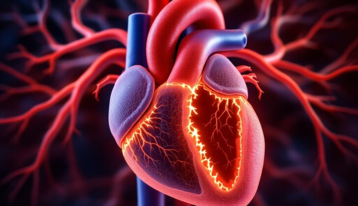
What is Ventricular Septal Rupture?
In 1847, a doctor named Latham first introduced the diagnosis of a condition called ventricular septal rupture (VSR). The ventricular septum is a part inside the heart that splits the lower heart chamber into two parts, known as the right and left ventricles.
The ventricular septum has two parts: one muscular and one membranous. The muscular part is the larger of the two, is located below the membranous part, and is thick. It comes from a structure called the bulboventricular flange. The membranous part is the smaller and thinner of the two, located above the muscular part, and it comes from a group of cells known as the neural crest cells.
A ventricular septal rupture is a rare but extremely serious complication of a heart attack, also known as an acute myocardial infarction. The condition has now become less common due to quick and effective treatment strategies for heart attacks. Yet, if it happens, it can be deadly. A VSR could occur in any part of the ventricular septum. The size of the rupture influences the patient’s outcome, with smaller ruptures and stable bodily functions generally having better outcomes.
In most cases, VSR tends to happen within a week following a heart attack. Often, there is a rapid worsening in the heart’s function, potentially leading to a life-threatening condition known as cardiogenic shock, which happens when the heart can’t pump enough blood to meet the body’s needs. VSR is a medical emergency that requires immediate surgery in patients showing symptoms. The operation involves the closing of the VSR and ‘re-plumbing’ part of the heart through a process known as coronary artery bypass grafting.
The surgery is generally done through the area of the heart attack and utilizing synthetic material to close the septum and the ventricular wall to avoid additional strain on the heart. However, over time, with the advancement of surgical techniques and better drugs and mechanical support, the outcomes of VSR treatment have improved significantly.
What Causes Ventricular Septal Rupture?
The ventricular septum is a wall in the heart that separates the lower chambers, or ventricles. When a hole, or rupture, happens in this wall, it’s often due to a severe heart attack known as a full-thickness myocardial infarction. This heart attack affects the entire wall of the heart and can happen in one of a few key arteries:
1. The left anterior descending coronary artery, which provides blood to a large part of the front of the septum that separates the chambers of the heart. If a heart attack happens here, it could lead to the rupture happening near the top (apex) of the heart.
2. The dominant right coronary artery. This artery gives blood to the lowest part of the septum. A heart attack here can lead to the rupture happening near the base of the heart.
3. The dominant left circumflex artery. This artery provides blood to the back of the heart, and a heart attack here might also lead to a rupture.
Less severe heart attacks, such as non-ST elevation myocardial infarction or unstable angina, which only affect part of the heart wall, can also increase the chance of a rupture in the ventricular septum.
Risk Factors and Frequency for Ventricular Septal Rupture
Ventricular septal rupture is a heart-related condition whose incident rates have significantly decreased from around 2% to 0.31% thanks to advanced therapies and quicker interventions with medical procedures. The Global Registry for Acute Coronary Events study found out that ventricular septal rupture incidents are lower (0.7%) in patients who underwent a specified heart procedure than in those receiving thrombolytic therapy (1.1%).
- The rate of this condition also depends on the type of heart attack, with the rate being higher in patients suffering from ST-segment elevation myocardial infarctions (0.9%).
- Meanwhile, the rate is 0.17% in non-ST-segment elevation heart attack patients and 0.25% in cases of unstable angina.
Notably, the occurrence of ventricular septal rupture is not influenced by the location of the heart attack — whether at the front (anterior infarct) or at the bottom side (inferolateral infarct) of the heart.
Signs and Symptoms of Ventricular Septal Rupture
One of the complications that can occur a few days after a heart attack is called flash pulmonary edema. In severe cases, this can lead to a state of medical emergency known as cardiogenic shock. A key sign of this condition is a loud heart murmur that doctors can hear through a stethoscope. This heart murmur is a common feature in most serious heart problems, and it can be heard all over the chest. In some people, the doctor may be able to feel a vibration, known as a thrill. Other signs might include an unusually loud second heartbeat, blood flowing back into the heart through the tricuspid valve, or a third heart sound. Almost all patients report recurring chest discomfort. Sometimes, the onset of the heart murmur can lead to a sudden change in the body’s normal functions.
There are certain risk factors that can increase a person’s chances of experiencing a ventricular septal rupture after a heart attack. These include being a woman, being older in age, having your first heart attack, severe heart attack with ST-segment elevation, a high GRACE risk score (which predicts the probability of death in patients with heart attack), and chronic kidney disease. If a patient has a heart attack and subsequently presents with low blood pressure or signs of unstable bodily functions, it should be considered that they might have a ventricular septal rupture, which is a medical emergency.
Testing for Ventricular Septal Rupture
Chest x-rays can show if your heart’s left side is enlarged or if fluid has built up in your lungs. Another test used to detect a condition called ventricular septal rupture, where a hole forms in your heart’s wall, is two-dimensional echocardiography with Doppler. This test shows how blood moves across the part of your heart called the ventricular septum.
Echocardiograms can also show if the right side of the heart is enlarged and if there’s high blood pressure in the lungs because more blood is flowing to the right side of the heart. Color Doppler echocardiography, another version of this test, can help doctors evaluate the size of a rupture in your heart.
When it’s tough to get a clear picture of the heart muscle from a regular echocardiogram, we might use a transoesophageal echocardiogram. We generally use this test for patients who have difficulty in imaging due to large body size or those on a mechanical ventilator.
An electrocardiogram (ECG), which measures the electrical activity of your heart, is necessary to rule out a reoccurrence of a heart attack. It could also reveal elevated ST segments, a sign that a ventricular aneurysm might be present. Ventricular aneurysm is a condition where a weak spot in your heart muscle balloons out and can lead to complications. In some patients, around 30%, a heart block might be detected through this test.
We only perform cardiac catheterization, an invasive test to examine how the heart is working, on patients who are stable, as it requires a careful assessment. This test can help distinguish between ventricular septal rupture and mitral regurgitation, a condition where leakage occurs in one of the heart valves. In the case of a ventricular septal rupture, the catheterization test will show a significant increase in oxygen levels from the right atrium to the pulmonary artery.
Treatment Options for Ventricular Septal Rupture
Ventricular septal rupture, a serious heart condition, typically demands a team of specialists, including a heart surgeon and heart disease specialist, for effective treatment. The primary way to treat this condition is through surgical repairs. However, before the surgery, the doctor needs to ensure adequate blood flow to the area affected by a heart attack, especially if the right part of the heart is involved.
In order to make the patient stable before surgery, doctors commonly use medications that help dilate the blood vessels. Such medicines not only improve blood flow to the heart muscle but also help manage some symptoms of ventricular septal rupture. Certain patients with low cardiac output may require medicines to boost the pumping action of their heart. However, some of these medications might increase the pressure against which the heart needs to pump blood, worsening some symptoms of the condition. An intra-aortic balloon pump, a device placed into the large blood vessel called the aorta, may be used temporarily in such patients. This device decreases the pressure on the heart and facilitates the blood flow in the heart vessels. Once the patient’s condition becomes stable, the doctor will move forward with surgery. It’s important to note that surgery may be postponed when the patient:
* Shows no signs of cardiogenic shock (a deadly condition when the heart can’t pump enough blood)
* Has good blood circulation and heart output
* Displays minimal or no signs of heart failure
* Needs minimal use of medications that constrict blood vessels
* Shows no signs of fluid accumulation
* Has normal kidney function
Regarding surgical closure, there are two main techniques – the Daggett procedure and the David procedure. The Daggett procedure is done by suturing a patch over the defect in both heart chambers, while the David procedure uses stitches to secure a patch only in the left heart chamber. Repairing a posterior ventricular septal rupture (on the back part of the ventricular septum) can be more challenging than an anterior rupture (at the front), due to the nearby papillary muscles. Surgery becomes even trickier if the rupture develops within 24 hours of a heart attack because differentiating healthy tissue from the damaged one is hard, coupled with the weak heart muscle that may not hold onto sutures.
In addition to these techniques, researchers have made advancements in percutaneous closure, a minimally invasive procedure to close the rupture. This might still be under constant improvement, but it has seen some success in patients, leading to its increased utilization. While using this technique, doctors may temporarily plug the hole using a balloon catheter in some cases. However, it’s critical to understand this is a temporary fix. Post-surgery, doctors typically recommend anticoagulant drugs that prevent blood clot formation due to the large quantity of prosthetic material used to patch up the ventricular septal defect.
Despite undergoing treatment, the ventricular septal defect may persist in approximately 10%-25% of cases. Depending on the size of the defect and the severity of the condition, doctors may monitor the patient or suggest returning for another surgery.
Currently, these surgical interventions carry significant risk. If the surgery is performed within 24 hours of a heart attack, the risk of death is over sixty percent. Otherwise, the risk of death without any treatment is somewhere between 40% and 80%. While late surgical intervention can be more successful, it might not always be possible especially for those suffering severe symptoms. For those needing surgery within one week of developing the condition, the mortality rate is 54.1%, but if surgery is performed after one week, the mortality drops to 18.4%.
For ventricular septal defects located at the front and measuring less than 1.5 cm in diameter, percutaneous techniques could offer a biological repair. Still in development, this method carries promise for the future treatment of ventricular septal rupture.
What else can Ventricular Septal Rupture be?
Here are some heart conditions that might have similar symptoms:
- Acute mitral regurgitation due to papillary muscle rupture
- Free wall rupture
- Tricuspid regurgitation
- Congenital ventricular septal defect
- Atrial septal defect
- Acute flash pulmonary edema
What to expect with Ventricular Septal Rupture
Post-heart attack ventricular septal rupture (VSR) is a life-threatening condition that often results in high death rates. This condition involves the septum, the wall separating the chambers of the heart, getting damaged or ruptured. Early repairs might be risky as the weakened heart tissues might not hold the surgical stitches properly. If the repair is delayed, it allows for scar tissue to form, which makes stitching easier.
Death rates are also high in individuals suffering from cardiogenic shock, a condition where the heart suddenly can’t pump enough blood to meet the body’s needs. Patients with a rupture in the front part of the heart, known as an anterior VSR, have slightly lower death rates compared to those who have a rupture at the back side, known as a posterior VSR.
Other factors that contribute to a poor prognosis are old age, failure of multiple organs, and advanced classification according to the New York Heart Association (NYHA), which determines the stage of heart disease. The following circumstances could also predict an increased death rate within 30 days:
- Shock during surgery
- Kidney failure
- Need for emergency intervention
- Severe coronary disease, specifically disease in the right coronary artery and the circumflex artery
- Lengthy surgery
- Long durations of cardiopulmonary bypass time, which is the time during which the heart-lung machine takes over the functions of the heart and lungs during surgery
- Incomplete revascularization, which means not all blocked arteries were successfully opened
However, if the rupture size is small and the patient is stable during surgical repair, the outlook can be positive.
Possible Complications When Diagnosed with Ventricular Septal Rupture
Possible complications of heart disease include:
- Cardiogenic shock: a serious condition where the heart can’t pump enough blood to meet the body’s needs
- Ventricular aneurysm: a bulge in the wall of the heart’s main pumping chamber
- Thrombus formation: the formation of a blood clot inside a blood vessel
- Ventricular arrhythmias: irregular heart rhythms originating in the lower chambers of the heart
- Free wall rupture: a tear in the heart wall
- Death
Recovery from Ventricular Septal Rupture
The patient will need to follow a long-term program to help improve their heart health, also known as cardiovascular rehabilitation. It is recommended that they join a guided exercise program to aid in their recovery.
Preventing Ventricular Septal Rupture
Important advances in treatment methods, such as the use of medication to dissolve blood clots (thrombolytic therapy) and other improved ways to restore normal blood flow to the heart (reperfusion therapies) have greatly reduced the incidence of a serious complication known as ventricular septal rupture (VSR) after a heart attack. VSR is the sudden rupture or splitting of the wall separating the two lower chambers of the heart. But these advances have also resulted in more complex and severe cases.
Only patients with extremely weakened heart muscle and complicated heart artery disease progress to this severe complication. As this condition often results in death, it’s crucial to identify patients unable to protect against VSR early on and to aggressively treat heart failure before it develops into a more severe condition called cardiogenic shock – a potentially fatal condition where the heart can’t pump enough blood to meet the body’s needs.
The only available preventive strategy seems to be the prompt treatment of heart attacks to limit the damage to the heart muscle. If VSR does occur, rapid and aggressive treatment of heart failure to prevent cardiogenic shock can help avoid bad outcomes. A carefully considered treatment plan for VSR can also improve survival rates.


