What is Bacterial Endophthalmitis?
Endophthalmitis is a serious eye infection that can lead to quick and permanent vision loss just hours or days after symptoms start to show. This condition involves an infection of the eye’s inner fluids (vitreous and/or aqueous humor), usually caused by bacteria or fungi. However, eye infections caused by viruses or parasites are classified under uveitis, not endophthalmitis.
The origins of endophthalmitis can be either exogenous, coming from the outside, or endogenous, coming from inside the body. The more common type is exogenous endophthalmitis, where germs are introduced to the eye from the eye’s surface or another external source. Depending on specific risk factors, exogenous endophthalmitis is labeled into different subtypes such as post-cataract surgery, post-traumatic, and bleb-related endophthalmitis. These subtypes have their own unique symptoms, bacteria types, and predictions for vision recovery, which necessitates accurate categorization for the best treatment and management.
On the other hand, endogenous endophthalmitis comes from germs spreading through the bloodstream during episodes of bacterial or fungal infection in the blood.
Changes over time can influence the prevalence of endophthalmitis types. For example, since the U.S. Food and Drug Administration’s approval of intravitreal anti-vascular endothelial growth factor medications in 2004, there has been increased use of these and other eye injections. As a result, some centers have seen cases of endophthalmitis after injections outnumber cases after surgery.
It is critical to diagnose and treat bacterial endophthalmitis quickly with the right antibiotics, as patients can recover completely and maintain their vision. Because time is of essence in treating bacterial endophthalmitis, medical professionals need to be able to recognize this eye disorder quickly and get patients on the path to recovery as soon as possible.
What Causes Bacterial Endophthalmitis?
Normally, the healthy eye’s internal fluids, called vitreous and aqueous humor, are free from bacteria. However, bacteria can still find their way into the eye from outside sources or from the body itself. More common is the case where bacteria enters the eye from the outside during incidents like eye injury, surgery, or injections in the eyeball. Although not very common, bacteria can also travel in the bloodstream from infections elsewhere in the body, or due to the use of intravenous drugs.
Up to 80% of all infections in the eye, known as endophthalmitis, can be traced back to eye surgeries, especially cataract surgery. Here, most infections occur due to Staphylococcus bacteria. Injections into the eyeball and eye trauma fall next in line as causes of endophthalmitis. Other bacteria and even fungi also can cause infections in cases like these.
An infection related to an eye bubble, keratitis where the cornea gets disrupted, and blood-borne infections are other possible causes of endophthalmitis. In these cases, different bacteria and fungi can be the culprits. However, these causes are less frequent.
When considering postoperative endophthalmitis, most infections are caused by coagulase-negative staphylococci. Other bacteria are also responsible. Eye injuries can lead to an increased chance of endophthalmitis. Nowadays, we see a shift in the bacteria involved – it’s becoming more common to see other kinds of staphylococcal bacteria and Bacillus cereus bacteria in these cases.
Endophthalmitis can also occur when infections spread to the eyes from elsewhere in the body. Though not as common as after surgeries or injuries, such a spread presents a chance of infection in both eyes. These infections might be due to fungi or bacteria. Particularly, fungal and gram-negative bacterial infections are of concern due to their association with poor outcomes and difficulties in treatment.
Risk Factors and Frequency for Bacterial Endophthalmitis
Endophthalmitis, an eye condition, is most commonly caused by cataract surgery and intravitreal injections, although it is quite a rare condition thanks to antibiotics. Out of those getting cataract surgery, around 0.1% experience endophthalmitis. Also, eye injuries that penetrate the eye can result in endophthalmitis in 1% to 18% of cases. This condition is even rarer in those using IV drugs, people with diabetes, those with weakened immune systems, those with cancer, or those hospitalized for a long period, with a 0.04% to 0.4% chance.
In the last 20 years, postoperative endophthalmitis has become more common. In the 1990s, only about 0.1% or 1 in 1000 people developed this condition after cataract surgery. But by the early 2000s, about 0.2% or 1 in 500 people developed it after the same procedure. Out of all eye surgeries, cataract surgery is the most likely to cause postoperative endophthalmitis. Globally, postoperative and post-traumatic endophthalmitis are most common, with 40% to 80% of endophthalmitis related to surgery, and 2% to 15% related to trauma.
In some parts of the world like Egypt, India, and China, endophthalmitis resulting from eye trauma is even more common, making up 40% to 60% of all cases. Cataract surgery is the biggest single cause of bacterial endophthalmitis.
There are various risk factors tied to endophthalmitis before, during, and after cataract surgery:
- Preoperative Risk Factors
- Blepharitis or abnormalities in the lid
- Use of 2% xylocaine gel before using povidone-iodine
- Diabetes
- Old age
- Weakened immune system
- Intraoperative Risk Factors
- Poor sterile technique
- Tear in the back of the lens capsule
- Loss of vitreous from the eye and wound leaking
- Use of certain drugs and dyes during surgery
- Medication contamination
- Postoperative Risk Factors
- Leaking surgical wound
- Type of intraocular lens that’s not made of silicon
Endophthalmitis that originates within the body is often associated with infections in the liver, lungs, heart, brain, and urinary tract. Those with diabetes, with compromised immune systems, or those who have been recently hospitalized or had surgery are at increased risk.
Signs and Symptoms of Bacterial Endophthalmitis
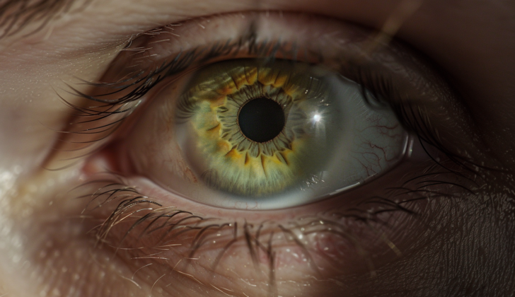
Bacterial endophthalmitis is an eye condition that requires immediate diagnosis and treatment to avoid lifetime blindness. The most usual symptom is a decrease in vision. Other symptoms may include eye pain, discharge, or redness. However, it’s important to note that these symptoms may not be present in every case of bacterial endophthalmitis. It’s also beneficial to understand when these symptoms began, so doctors can determine whether it’s a bacterial or a fungal infection.
Generally, bacterial endophthalmitis shows symptoms promptly, unlike fungal endophthalmitis that gets worse over days or weeks. If you suspect bacterial endophthalmitis, the doctor may ask about any recent eye surgery or injury, as these can potentially lead to this condition. Symptoms such as fever, chills, recent infections, recent surgery, hospitalization for sepsis, antibiotic use, and IV drug use should also be shared as they can indicate endophthalmitis.
- Decreased vision
- Eye pain
- Eye discharge
- Red-eye
- Ocular surgery or injury
- Fever
- Recent infections
- Recent surgery
- Hospitalization for sepsis
- Recent antibiotic use
- IV drug use
Testing for Bacterial Endophthalmitis
Exogenous endophthalmitis, which is an infection inside the eye, manifests in many ways. This usually affects one eye and symptoms fluctuate from no symptoms at all to evident signs such as eye redness, sensitivity to light, clouding of the cornea, inflammation inside the eye, floating particles in vision, or reduced vision. During an eye exam, eye doctors may observe a buildup of white blood cells in the front part of the eye, which is common in most bacterial cases and in up to 80% post-cataract surgery cases. To diagnose bacterial from fungal endophthalmitis, doctors look for patterns of inflammation inside the eye, where bacterial infections often cause widespread inflammation, while fungal ones result in cluster-like inflammations. An ultrasound can also be helpful to spot inflammation or infiltrate. A surgical procedure to remove some fluid from the eye often provides a better chance for diagnosis than a needle biopsy.
Endogenous endophthalmitis, on the other hand, primarily affects one eye, although it can sometimes affect both. Diagnosis of this condition is quite challenging as it often requires multiple clinic visits and produces many false negatives. Symptoms might vary from none to indications such as a buildup of white blood cells in the front part of the eye, inflammation inside the eye, redness, clouding of the cornea, inflammation of the iris, inflamed cells in the anterior chamber, or diminished vision. Drastic changes, like white infiltrations stemming from the choroid and bulging into the jelly-like vitreous of the eye, serve as key diagnostic indications for endogenous endophthalmitis.
A study conducted in 2015 categorized the likelihood of having endogenous endophthalmitis based on certain symptoms. The prognosis and responsiveness to treatment for both types of endophthalmitis can greatly vary and is affected by numerous factors. These include the severity of the causing microorganism, how it entered the eye, when treatment started, patient’s age, how susceptible the pathogen is to treatment, and the pre-existing condition of the eye. Generally, delay in the treatment can lead to poor vision, prompting speedy and broad-spectrum treatment. Typical treatment includes a combination of antibiotics, usually delivered directly to the vitreous part of the eye to achieve fast-acting, high-dosage exposure, which is crucial for rapid elimination of the pathogen and reduction of damage and inflammation, especially for severe cases.
Treatment Options for Bacterial Endophthalmitis
Bacterial endophthalmitis, an infection in the eye, requires an immediate treatment plan that typically involves the use of antibiotics, hospital care and possibly a surgical procedure known as pars plana vitrectomy. After medical professionals have collected the necessary culture samples to help identify the infection’s source, patients are typically put on IV, topical, and possibly intravitreal antibiotics. Additionally, patients may be given a topical medication like atropine to help relax the eye muscles and another called prednisolone acetate to help decrease inflammation.
Intravitreal antibiotics, which are injected directly into the eye, offer a high concentration directly at the site of infection. These can be effective if they are appropriately matched against the type of infection present. Several commonly used ones include ceftazidime, vancomycin, and amikacin.
The decision to use a specific antibiotic is typically based on what is known about the antibiotic’s effectiveness against common eye infection organisms. For instance, an antibiotic called vancomycin is often chosen due to its wide-ranging ability to fight against gram-positive bacteria, while another called ceftazidime can tackle a broad spectrum of gram-negative bacteria. However, some bacteria may become resistant to these antibiotics, which could reduce their efficacy.
Fluoroquinolones are another class of antibiotics that show promise in treating endophthalmitis due to their broad-spectrum activity against various eye pathogens. However, antibiotic resistance is a growing concern with this class too. Despite this, they may still be beneficial in certain situations due to their ability to cross the eye’s defensive barriers without needing an intravitreal injection.
On top of antibiotics, it might be beneficial to use anti-inflammatory drugs such as corticosteroids to control inflammation, a natural reaction to infection that, if excessive, can lead to further damage. However, the use of intravitreal corticosteroids, like dexamethasone, is controversial, as studies show conflicting results regarding their effectiveness in managing endophthalmitis.
If the infection becomes severe, a surgical procedure called pars plana vitrectomy may be necessary to remove infected and inflamed material from inside the eye. This can help hasten visual recovery, and newer, minimally invasive techniques have been developed to lower the risks associated with this procedure.
Despite these treatment options, outcome can unfortunately still be poor in some cases of endophthalmitis, with some patients requiring the removal of the eye. Further research is needed to develop better treatment strategies and improve patient outcomes.
What else can Bacterial Endophthalmitis be?
Bacterial endophthalmitis is an urgent eye condition where inflammation and infection occur within the eye. It’s very important to distinguish it from other illnesses that look similar, so the right treatment can be chosen.
Some conditions that can seem like bacterial endophthalmitis include:
- Sterile endophthalmitis: This looks like infectious endophthalmitis but happens due to a non-infectious reaction to treatments inside the eye or surgery. There are no organisms growing in this condition.
- Fungal endophthalmitis: This condition has similar symptoms but progresses more slowly. It’s more prevalent in patients with weaker immune systems, extended use of antibiotics, and those who use intravenous drugs.
- Viral retinitis: This is caused by specific viruses, resulting in retinal damage and bleeding. It may not be as painful as bacterial endophthalmitis.
- Non-infectious uveitis: This is an inflammatory condition without an infection, usually linked to autoimmune diseases.
- Acute retinal necrosis: This syndrome causes damage to the retina, typically due to a virus, and usually leads to retinal detachment.
- Toxic anterior segment syndrome (TASS): It’s caused by a non-infectious substance entering the anterior segment during cataract surgery.
In contrast, some examples of conditions that can also look like bacterial endophthalmitis but occur due to different causes include:
- Intraocular foreign bodies: Sometimes, after an eye injury, a foreign object can be inside the eye, which can seem like endophthalmitis.
- Ocular ischemic syndrome: This presents with similar symptoms but is caused by problems with blood flow, not by an infection.
- Neoplastic conditions: Certain tumors like retinoblastoma, uveal melanoma, or intraocular lymphoma can present symptoms similar to endophthalmitis.
To diagnose bacterial endophthalmitis accurately, it’s essential to get a detailed medical history, carry out a comprehensive eye examination, and use certain laboratory tests including cultures and PCR testing. This careful diagnosis helps pinpoint the best treatment for the patient, whether it be antibiotics, antivirals, or treatments to suppress the immune system, depending on what’s truly causing a patient’s symptoms.
What to expect with Bacterial Endophthalmitis
Research shows that endophthalmitis caused by the bacterium S pneumoniae often results in poor vision. In a study on this type of infection, it was found that about 11.11% of cases ended up needing an evisceration, a surgical removal of the eye. Similarly, endophthalmitis caused by Pseudomonas bacteria is associated with a high rate of eye removal, with one study suggesting up to 64% of instances lead to this outcome.
A number of factors are strongly linked with the need for eye removal, including: endophthalmitis that originates from within the body (endogenous endophthalmitis), an ulcer on the cornea, old age, initially poor vision, being female, and late treatment onset. However, people who developed endophthalmitis after an eye injury or surgery were less likely to require eye removal.
The prognosis, or future outcome, of bacterial endophthalmitis can vary depending on several important factors. The quicker the infection is diagnosed and treated, the better the result – waiting too long can cause rapid, irreversible damage to the eye and loss of vision. Some factors that influence outcomes include:
- Pathogen virulence: Highly aggressive bacteria (like Bacillus species) often lead to worse outcomes due to rapid progression and severe damage. On the other hand, less aggressive bacteria, like coagulase-negative staphylococci, could lead to better vision if treated quickly and effectively.
- Initiation of therapy: Starting treatment as soon as possible, especially with the right antibiotics, can lead to better outcomes for your eyesight.
- Surgical intervention: The necessity and timing of a vitrectomy (a surgical procedure involving the removal of the jelly-like substance inside your eye), can affect prognosis.
- Host factors: The patient’s overall health status, especially for those with compromised immune systems or diabetes, can affect prognosis. These individuals might experience more severe infections and complications, leading to worse outcomes.
- Complications: The development of complications, such as retinal detachment or ongoing eye inflammation, can result in poorer vision.
- Visual acuity at presentation: People with better initial vision typically have a better prognosis.
- Rehabilitation potential: Access to and the effectiveness of rehabilitation services including low vision aids, can help maximize vision outcomes and improve post-infection life quality.
Despite advancement in diagnosis and treatment, bacterial endophthalmitis can still potentially lead to serious outcomes. A team approach to patient care, involving eye doctors, infectious disease specialists, and rehabilitation services can help optimize patient outcomes. Ongoing research into new treatment strategies and diagnostic tools is also crucial to improve future outcomes for this serious eye infection.
Possible Complications When Diagnosed with Bacterial Endophthalmitis
The challenges of managing complications may necessitate additional procedures, including surgery. Spotting the issues early and promptly treating them is essential in reducing potential risks. Here are some complications that can arise after ocular surgery:
- Permanent visual impairment: This is the most critical complication, it ranges from partial vision loss to total blindness.
- Retinal detachment: Inflammation and pressure on the retina can cause it to detach, possibly needing surgical repair.
- Vitreous hemorrhage: Bleeding into the area at the back of the eye can blur vision and complicate managing an infection.
- Corneal edema and opacification: Inflammation may result in damage that might become permanent and need corneal transplantation.
- Cystoid macular edema (CME): Accummulation of fluid in the macula can distort the central vision.
- Glaucoma: The pressure inside the eye may rise suddenly due to inflammatory debris or harm to the mesh-like structure in the eye.
- Phthisis bulbi: A shrunken, non-functional eye resulting from severe injury or illness.
- Choroidal effusion: Accumulation of fluid in the choroid can cause retinal detachment and decreased vision.
- Endophthalmitis-related uveitis: Inflammation in the uveal tract can cause persistent discomfort and light sensitivity.
- Ocular hypotony: Reduced eye pressure can occur due to dysfunction or detachment of the ciliary body.
- Epiretinal membrane formation: Scar tissue on the retina can cause visual distortion.
- Optic atrophy: Serious and ongoing inflammation leads to irreversible vision loss due to optic nerve damage.
- Proliferative vitreoretinopathy (PVR): Formation of scar tissue could lead to retinal detachment and pose treatment challenges.
- Subretinal abscess formation: If the infection goes beyond the retina into the retinal tissue, it can lead to this.
- Sympathetic ophthalmia: A rare but severe condition affecting the non-infected eye triggered by the immune system.
- Panophthalmitis: An extreme form of endophthalmitis that affects all eye layers and structures, leading to total loss of vision and a painfully non-functional eye. Eye removal might become necessary.
- Corneal ulcer: Infection progression may involve the cornea, leading to ulceration. These ulcers can advance quickly, resulting in perforation and further infection spread and might need corneal grafting if the structural integrity is compromised.
- Orbital cellulitis: The infection can spread beyond the eye into the orbital tissues; it is a severe condition that can cause pain and swelling and lead to abscess formation, vision loss, and life-threatening complications if the infection spreads to the central nervous system.
Recovery from Bacterial Endophthalmitis
After eye surgery, it’s vital to constantly check for positive signs of recovery or any signs that the condition might be worsening. This includes looking out for symptoms such as eye pain, redness, discharge, or changes in vision. Antibiotics delivered directly into the eye may be required, depending on the type and severity of the infection.
Anti-inflammatory drugs, like corticosteroids, may also be needed to help reduce inflammation within the eye. These drugs can be applied topically, around the eye, or directly injected into the eye. It’s also important to regularly monitor the eye pressure as it can both increase and decrease after surgery, requiring appropriate treatment.
The guidelines from the endophthalmitis vitrectomy study can help doctors make decisions about further surgeries like vitrectomy – a specific type of eye surgery.
Post-surgery, patients may require visual aids or services to assist in restoring their vision. It would be beneficial for these patients to be evaluated and guided by a low-vision specialist. Furthermore, it’s crucial to educate patients about the symptoms of complications that need immediate medical intervention. Endophthalmitis can be a traumatic experience, so counselling might be helpful to deal with potential vision loss and the associated impact on quality of life.
In severe cases, where significant vision loss has occurred, physical training on orientation and mobility may be needed to help patients stay safe and independent. Regular eye check-ups are imperative in the long term, to watch out for late-onset complications like cataract formation, detachment of the retina, or glaucoma.
The aim of meticulous post-surgery care and rehabilitation in bacterial endophthalmitis is to maximize the patient’s vision and improve their quality of life as much as possible.
Preventing Bacterial Endophthalmitis
Using a certain medication (cefuroxime) inside the eye during cataract surgery can help lower the chances of getting an infection called endophthalmitis after the operation. Other factors that could increase the risk of this infection include specific types of corneal incisions and use of silicone lenses. One study, involving over 45,000 cataract surgeries, showed that if patients were given antibiotics and anti-infection medication before surgery, the chances of getting endophthalmitis after the operation dropped by 72%. This method was confirmed effective via another study involving over 6,000 cataract surgeries.
It’s hard to study how to prevent endophthalmitis after cataract surgery because it doesn’t occur often. However, proper wound care, combined with antibiotics and a type of antiseptic medicine (povidone-iodine), can be effective in lowering the risk of this serious but rare infection.
Preventing and managing endophthalmitis requires patient education and taking preventative steps. Here are a few key points:
- Understanding endophthalmitis: It’s important for patients to understand how serious this infection is, what causes it, its symptoms, and why it’s crucial to detect and treat it early.
- Preventive measures: Patients should be reminded to protect their eyes properly to avoid trauma, a major risk factor for this infection. They need to understand how careful cleaning before and after eye surgery and proper contact lens hygiene can prevent infections.
- Early symptom recognition: Educating patients to recognize early signs of infection, such as redness, pain, vision changes, or discharge, could save their eyesight. They should seek immediate help if they notice symptoms.
After surgery care: It’s crucial that patients understand and follow after-surgery care instructions, including using antibiotic eye drops. For patients with chronic eye diseases or who use specific types of eye drops (corticosteroids), they should be aware of their higher risk and the need for regular eye doctor visits. Similarly, advising patients about the possibility of getting endophthalmitis from other body infections and the importance of managing health conditions like diabetes is essential. Regular follow-up visits to keep an eye on recovery after eye surgery or when symptoms appear are important too.
Patient empowerment: Promoting a proactive approach to eye health, like seeking immediate help for eye injuries or suspected infections, is important.
All these patient education efforts aim to lower the chances of endophthalmitis and improve outcomes for those who get it. All healthcare providers involved in a patient’s care must reinforce these educational messages.


