What is Echinococcus Granulosus (Tapeworm Infection)?
Echinococcosis is a disease caused by a parasite named Echinococcus that can infect both animals and humans all over the world. The World Health Organization (WHO) estimates that the global cost of handling this disease surpasses three billion US dollars each year.
There are many types of Echinococcosis, but here are the four that affect humans the most:
1. Echinococcus granulosus: This parasite causes a condition known as cystic echinococcosis or hydatidosis, where fluid-filled sacs (or cysts) develop in the body.
2. Echinococcus multilocularis: This results in alveolar echinococcosis, a more serious disease that affects many parts of the body.
3. Echinococcus vogeli and Echinococcus oligarthrus: These both lead to a disease called polycystic echinococcosis, where multiple cysts form in the body.
This summary will primarily focus on the infections caused by Echinococcus granulosus.
What Causes Echinococcus Granulosus (Tapeworm Infection)?
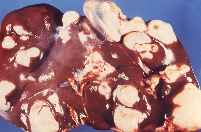
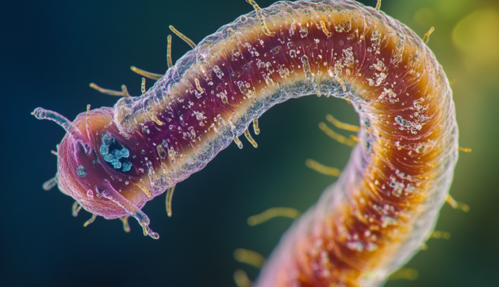
Hydatidosis is a disease caused by the larvae of a type of tapeworm called Echinococcus granulosus. This illness is known for the growth of unique cysts called hydatid cysts in the internal organs of infected individuals, including humans.
The tapeworm typically resides in carnivores like dogs. Humans and other hosts become infected when they consume the eggs or sections of mature tapeworms that are found in the feces of these animals.
Risk Factors and Frequency for Echinococcus Granulosus (Tapeworm Infection)
Hydatidosis, a disease caused by certain pathogens, tends to be more widespread in communities that raise dogs for the purpose of protecting and herding livestock. Although this disease can be found all over the world, it is most common in certain regions including the Mediterranean, Russia, China, North and East Africa, Australia, and South America.
Signs and Symptoms of Echinococcus Granulosus (Tapeworm Infection)
People who are infected with a type of parasite that forms cysts might not show any signs or symptoms for months or even years. Some might have these cysts for a long time without even noticing. In some cases, the cysts might burst and disappear completely on their own or due to an injury.
If a cyst keeps growing, however, it can start to cause symptoms by putting pressure on the surrounding tissues. Showing sudden signs and symptoms typically indicates a burst cyst rather than the growth of the cyst.
If a cyst ruptures, it might cause a severe allergic reaction, with symptoms such as hives, swelling inside the mouth and throat, and skin redness. This reaction can be dangerous and potentially life-threatening.
A leaking or ruptured cyst can also release small parasitic larvae into the body, causing a condition called secondary hydatidosis.
The type of signs and symptoms a patient experiences largely depends on where the cyst is located. It’s most common for cysts to form in the liver (in about 65% of cases) or lungs (about 25% of the time), but they can also form in other areas like bones, the spleen, brain, and heart. Most people have just one cyst, but around 20% to 40% of patients end up with cysts in multiple organs.
Even though people can get infected during childhood, it typically takes a long time for the slow-growing cysts to cause symptoms. For this reason, most cases only become apparent later in life. However, cysts in the brain or eyes can cause symptoms early on and thus often present in childhood.
Common signs of a liver cyst include:
- Abdominal pain
- Loss of appetite
- Enlarged liver
- Noticeable mass in the abdomen
- Bloating
Common signs of a lung cyst include:
- Chronic cough
- Chest pain
- Shortness of breath
In assessing someone for these types of cysts, it’s important to check for potential risk factors for echinococcosis, especially exposure to dogs and cattle.
Testing for Echinococcus Granulosus (Tapeworm Infection)
Diagnosis of echinococcosis, or infection with tapeworm larvae, often begins with discussing your detailed medical history, particularly if you’ve recently travelled to or lived in areas where the disease is commonly found. Following this, a combination of blood tests and imaging studies is used to confirm the diagnosis.
Blood tests, including liver function tests, aren’t always reliable in determining how bad the disease is. These tests are only abnormal in about 40% of patients. In these cases, levels of an enzyme known as alkaline phosphatase are often elevated, whereas other measures such as AST, ALT, and bilirubin – elements found in the liver – usually stay within a normal range. You may also have a blood count test done, which could show an increased number of a type of white blood cell known as eosinophils.
Another common method for diagnosis is testing for Echinococcus antibodies, a sign your body is fighting the infection, in the blood. This often begins with a screening test known as ELISA. If you test positive, a more specific test known as an immunoblot assay is done to confirm it. However, it’s important to know that not all patients with echinococcosis produce antibodies, and the strength of your immune response can depend on various factors such as the health and integrity of the cysts caused by the infection.
Imaging tests play a significant role in identifying and monitoring the disease, especially in cases where blood tests don’t show antibodies. Ultrasound is the preferred method for looking at infections in the liver and the abdominal cavity, but the accuracy of these tests can vary based on who conducts them. Nevertheless, ultrasound has many advantages, such as being widely available, portable, and useful in tracking the disease after treatment.
For liver infections specifically, the World Health Organization developed an ultrasound classification system to identify the stage of the disease:
- CE 1: a single fluid-filled pocket with a “double line” sign.
- CE 2: a so-called “honeycomb” or “rosette-like” pattern of many smaller pockets.
- CE 3A: a fluid collection with a detached membrane, known as the “water lily” sign.
- CE 3B: multiple smaller “daughter” cysts within a solid mass.
- CE 4: hypoechoic/hyperechoic matrix – basically a complex mixture of solid and liquid elements without smaller cysts within it.
- CE 5: a fully solid wall around the cyst.
Stages CE1 and CE2 indicate an active disease; stage CE3 signifies a transitional stage where the cyst is damaged, and CE4 & CE5 indicate an inactive disease.
Radiographs or X-rays can be used to see if there are areas of hard tissue, known as calcifications, which are present in up to 30% of cases. The calcifications are often ring-like and can progress throughout all stages of the disease. CT scans, which provide more detailed images than X-rays, are excellent for detecting complications like cyst rupture, underlying infection, and involvement of the bile ducts or blood vessels. They’re particularly helpful when ultrasound is difficult, such as in people who are obese.
Other diagnostic tools include using a thin needle to collect fluid from the cyst under ultrasound guidance (in cases where blood tests don’t show antibodies and imaging is inconclusive), and an endoscopic retrograde cholangiopancreatography. This last test is primarily used to diagnose and treat echinococcosis that affects the bile ducts – the tubes that carry bile from the liver to the gallbladder and small intestine.
Treatment Options for Echinococcus Granulosus (Tapeworm Infection)
There are many ways to manage liver cysts, from simply monitoring them to performing surgery. The best method depends on the patient’s overall health and the size of the cyst, which is typically determined via ultrasound.
In some situations, a “watch and wait” approach might be best, especially for uncomplicated cysts. This involves regular monitoring using ultrasound imaging to keep an eye on the cyst’s development or changes.
Medication treatment with a class of drugs called benzimidazoles (BMZ) can be used for smaller liver cysts or cysts found in multiple organs, or if surgery isn’t an option. These medicines can also be used after surgery or minimally invasive procedures to prevent the cyst from returning. However, these medications should not be used if the cyst is at risk of rupturing or during early pregnancy.
Surgery is often the preferred treatment for more complicated cysts. This includes large cysts, cysts that could burst from trauma, infected cysts, cysts causing pressure on other organs, and cysts that are communicating with the biliary tree, a part of the body’s system for digesting food. A minimally invasive, ultrasound-guided procedure called PAIR (Puncture, Aspiration, Injection, Re-aspiration) is also an option. This procedure is best for certain types of cysts and is performed in combination with medication treatment. However, certain types of cysts aren’t suited for this procedure, for example, those that might burst.
No matter what treatment is chosen, it’s important to monitor the patient regularly after treatment. This can be done by performing blood tests and conducting ultrasound scans. Initially, these check-ups might occur every three to six months, then yearly once the patient’s condition is stable.

granulosus, Pathology, Parasitology
What else can Echinococcus Granulosus (Tapeworm Infection) be?
Hydatidosis is a medical condition that can look like a lot of different illnesses, depending on where the cysts are located in the body.
In particular, when it concerns the liver, there are several other conditions it could be mistaken for:
- A liver abscess
- Liver cysts
- Budd-Chiari syndrome (a condition affecting the veins in the liver)
- Biliary colic (pain caused by gallstones)
- Biliary cirrhosis (scarring of the liver due to bile buildup)
- Tuberculosis
- Primary hepatic carcinoma (a type of liver cancer)
In order to detect hydatidosis and rule out these other possibilities, doctors need to perform a careful review of the patient’s medical history and a thorough physical exam. This typically includes specific blood tests and imaging studies.
What to expect with Echinococcus Granulosus (Tapeworm Infection)
The outlook is usually positive with the right treatment. However, if cysts develop in areas which are challenging for surgery like the heart and spine, the prognosis might be less favorable. In some instances, there can be a recurrence or return of the cyst in the same location or other areas of the body.
Possible Complications When Diagnosed with Echinococcus Granulosus (Tapeworm Infection)
When liver or lung cysts burst, whether on their own or due to an injury, it can lead to particular complications. For instance, if a liver cyst bursts into the bile duct, this can cause a blocked bile duct, an infected cyst, and inflammation of the membrane that lines the abdominal cavity. On the other hand, if a lung cyst ruptures into the intake pathway of the lungs, it can cause inflammation of the lung tissue, build-up of air or gas within the space between the lung and chest wall, fluid in lung spaces, and inflammation of the protective layers of the lung.
Potential Complications :
- Blocked bile duct (from liver cyst rupture)
- Infected cyst (from liver cyst rupture)
- Inflammation of the membrane lining the abdomen (from liver cyst rupture)
- Inflammation of lung tissue (from lung cyst rupture)
- Build-up of air or gas in chest wall (from lung cyst rupture)
- Fluid in lung spaces (from lung cyst rupture)
- Inflammation of protective layers of lungs (from lung cyst rupture)
Preventing Echinococcus Granulosus (Tapeworm Infection)
In areas where a certain disease is common, it’s particularly important for people to understand the disease and how it spreads. This is especially crucial for those who work with cattle and dogs. These workers should be taught about how the disease can be transmitted and the correct methods for handling sheep organs.
Doing large-scale health check-ups with ultrasound can help keep track of how common the disease is in these areas. This information can lead to public health conversations on how to control the disease and keep it from spreading.


