What is Eumycetoma?
Mycetoma is a slow, chronic infection that affects the skin and tissue directly beneath it. There are two types of mycetoma: “eumycetoma”, caused by fungi, and “actinomycetoma”, caused by certain bacteria. Thus, eumycetoma is a deep skin and subskin infection that happens due to string-like fungi. This deep infection provokes inflammation in the skin layers and results in the formation of small hard particles called grains, which can damage under-the-skin tissues, muscles, bones, joints, and tendons.
The World Health Organization (WHO) acknowledges mycetoma as a serious but neglected tropical disease, which primarily strikes individuals in hot and humid climates who touch soil frequently. Feet are particularly susceptible to this fungal infection because they often come into contact with fungi present in the soil, especially when there are skin injuries. Much less commonly, other body parts such as legs or hands may also get affected, as these fungi usually get inside the body through skin injuries. In extremely rare occasions, these infections can spread through the bloodstream to other parts of the body.
The most common fungus that causes eumycetoma is named Madurella mycetomatis. They inhabit soil and can embed into the skin after minor injuries. Following this, a slow skin swelling begins and, over time, multiple lumps form that turn into pus-filled lesions with numerous draining passages. Through these passages, clusters of the offending organism can emerge. Treating eumycetoma is a significant challenge and involves a lengthy treatment course combining antifungal medications which usually exceeds 6-month duration, along with surgery.
If left untreated, these infections can lead to serious damage to tissues, highlighting the importance and the complexities associated with treating eumycetoma. In places where eumycetoma is common, it can lead to significant social and economic consequences for the patients and their families.
What Causes Eumycetoma?
Eumycetoma, a type of fungal infection, is mostly caused by a pathogen called M mycetomatis. The pathogens responsible for eumycetoma can be sorted according to the type of grain they form. These grains can be black, white or pale unstained, or yellow-to-yellow-brown.
Let’s see what sort of pathogens can cause each:
* Black grains: The pathogens responsible for creating black grains include M mycetomatis, Trematosphaeria grisea (used to be known as M grisea), Exophiala jeanselmei, Medicopsis romeroi (formerly known as Pyrenochaeta romeroi), Falciformispora senegalensis (used to be known as Leptosphaeria senegalensis), F thompkinsii, and Curvularia lunata.
White or pale unstained grains: Pathogens such as Acremonium spp, Fusarium spp, Neotestudina rosatii, Aspergillus nidulans, and A flavus, along with Microsporum ferrugineum, M audouinii, and M langeronii are usually linked to the creation of the white or pale unstained grains. Moreover, Scedosporium apiospermum and S boydii (previously termed Pseudallescheria boydii) are also associated with these types of grains.
* Yellow-brown grains: Yellow-brown grains are usually caused by pathogens such as Nocardia brasiliensis, N otitidiscaviarum (formerly known as N caviae), Actinomadura madurae, and Streptomyces somaliensis.
Yellow grains: Yellow grains can be linked to a pathogen known as Pleurostomophora ochrac.
While these organisms are mostly found in tropical regions, cases have been reported in the United States and other countries too.
Risk Factors and Frequency for Eumycetoma
Mycetoma, a tropical disease, affects many people, especially in tropical and subtropical countries such as India, Senegal, and Sierra Leone. It was added to the World Health Organization’s list of neglected tropical diseases in 2016. This illness impacts those who often walk barefoot and is most common in areas called the “Trans-African Belt” or “mycetoma belt”, spreading from Sudan to Senegal. While it is rare in developed countries, it can be seen in people who have migrated from affected areas.
Eumycetomas, a type of mycetoma, are mainly seen in adult men. People who have more exposure to soil or a history of injuries to their feet or other areas exposed to soil, such as farmers, homemakers, and animal breeders have a higher risk of developing this disease. However, only around 20% of people recall having a traumatic event before the condition began, such as injuries by splinters or thorns or physical exertion.

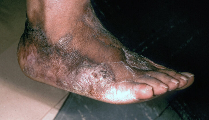
patient with progressing eumycetoma, showcasing deep-tissue penetration in the
right foot.
Signs and Symptoms of Eumycetoma
Eumycetoma is a disease primarily affecting the feet, but also the hands and legs. It causes painless plaques, tough swelling, and the discharge of small granules from under the skin. Other symptoms include scarring and hyperpigmentation of the surrounding skin. This is especially common amongst people who walk barefoot in Africa and Southeast Asia.
- Painless plaques
- Hard swelling
- Discharging sinuses
- Granules vary in color from white to yellow to black
- Scarring and darker skin color
The classic signs of eumycetoma are tumor growth, sinus tracts, and discharge with grains. Once the causative agent enters the body, it gradually leads to a subcutaneous fungal infection over several months to years, indicating a chronic condition.
Testing for Eumycetoma
In simpler terms, doctors often diagnose the disease by observing three main signs: a lump, hollow passage ways in the body, and discharge containing grains. This is especially true in low-resource areas where advanced tests may not be available. However, sometimes additional tests like culture tests, ultrasounds, or fine needle aspiration might be needed. A culture test is a method used to grow and identify organisms (like bacteria and fungi) that may cause disease, but it can take up to two months for final results. Fine needle aspiration is a simple procedure in which a thin, hollow needle is inserted into the mass to extract cells or fluid for examination.
In some situations, a punch biopsy may be conducted. This is a procedure where a doctor uses a special circular blade to remove a small section of skin tissue. The tissue is then examined under a microscope to look for signs of disease. This test is typically used for diagnosing eumycetoma, a type of fungal infection. This test might not be necessary in all cases but can be helpful if no drainage material can be analyzed.
Determining the exact cause and extent of the infection can help in tailoring the most effective treatment. Sometimes, doctors might use imaging tests like x-rays, ultrasound, CT scans, or MRI to see the extent of the disease in your body. CT scans can show the eumycetoma infection in more detail than traditional x-rays. In the situations where your doctor thinks that your bone or soft tissues could be involved, they might use an MRI. MRI is a type of imaging that uses a magnet and radio waves to create detailed images of the inside of your body.
Additional tests include surgical biopsy, histopathological examination, and fungal tissue cultures, which all help in identifying the specific causative organism. Molecular techniques such as species-specific polymerase chain reaction (PCR), enzyme-linked immunosorbent assay (ELISA), and counter immunoelectrophoresis could also be used for further characterization. These are special laboratory techniques used to detect specific types of organisms or proteins found in the samples.
Treatment Options for Eumycetoma
Treating a condition called eumycetoma isn’t straightforward because there aren’t any strict guidelines to follow. The current treatment plans are based on a mix of previously published individual case reports and group studies. Usually, the treatment involves combining antifungal medications and surgeries.
Most often, patients are first given antifungal medication for around six months. Then, they undergo surgeries that aim to remove as much of the disease as possible. This is followed by another six months on antifungal treatment. In a few cases, some patients have responded well to just the antifungal medication. In areas where resources are limited, medical therapy may be the only available option because surgical procedures might not be possible.
Generally, there are several different types of antifungal medications that can be used to treat eumycetoma.
The first type is known as azoles, which are considered the best treatment option. The most commonly used one is called itraconazole. Note that long-term use of this medication can lead to liver toxicity. Because of this, patients often need treatment that spans from six months to three years, as longer treatment has been found to have better outcomes.
There are newer azole antifungals, including voriconazole and posaconazole, that have also been used and have shown good results.
Another type is amphotericin B. However, it is not commonly used because its toxicity levels require hospitalization, and due to its harmful effect on the kidneys. When it’s been administered directly into lesions, it has worked well for those with a single lesion but is not effective for patients with multiple lesions.
The final type is terbinafine. After having twice daily doses of it for 24 to 48 weeks, 80% of patients saw a significant improvement.
When it comes to surgey, the current proposal is to start with preoperative treatment of eumycetoma using itraconazole. This is because antifungal medication causes the eumycetoma to encapsulate, making it easier to localize and surgically remove. This approach lessens the need for broader, more complicated, and potentially disfiguring surgeries.
What else can Eumycetoma be?
When looking for a possible diagnosis related to unusual skin conditions, doctors may consider the following possibilities:
- Lobomycosis: A long-term skin fungus infection found mainly in Central and South America. It first presents as small lumps and then develops into bigger, smoother lumps generally found on the hands, ears, and ankles. Unlike mycetoma, lobomycosis does not create sinus or fistula.
- Mycobacterium marinum: Sometimes called “fish tank granuloma”, this is a less common bacterial infection picked up from contaminated water like swimming pools or lakes. Symptoms include painful red lumps on the fingers or hands.
- Chromoblastomycosis: A skin fungus infection found in hot, humid climates. This is often seen in middle-aged men and features crusty lesions on the lower part of the body.
- Histoplasmosis: A fungal infection which forms dimpled lumps, hard lumps or open sores. Swollen lymph nodes are common, and risk is higher in people with a weakened immune system.
- Cutaneous tuberculosis: This varies in the way it appears clinically and shows as non-healing lumps, sores, plaques, or draining lymph nodes. It is more common in patients from areas where tuberculosis is common.
Doctors may also consider other conditions like deep fungal infections, unusual bacterial infections, podoconiosis, leishmaniasis, skin infections around hair follicles (folliculitis), cysts, foreign body reactions, and botryomycosis.
What to expect with Eumycetoma
If eumycetoma, a type of fungal infection, is left untreated, it can slowly but surely get worse. In certain situations where it’s not immediately identified or treatment isn’t accessible or affordable, eumycetoma can evolve from being painless to causing serious damage to a limb and might even lead to the need for amputation.
Several factors have been identified in past studies that may increase the chances of a person’s recovery from this condition. These include ongoing treatment over a long period and no previous instances of the condition showing up again. Interestingly, when the infection is between 5 and 10 cm, or even greater than 10 cm, and managed with a combination of medication and surgery, this can also lead to a better outcome.
However, it’s quite common for eumycetoma to show up again, particularly in people who’ve had the condition for more than ten years or in parts of the body other than the feet. Those who’ve had surgery in the past to remove the infection, have a family history of the condition, or work in occupations other than farming, are found to be at a greater risk of the condition reappearing.
Possible Complications When Diagnosed with Eumycetoma
If not treated on time, the infection can travel along the layers of connective tissue and attack the muscle and bone. This can lead to advanced complications such as bone infections (osteomyelitis) or infecting deeper parts of the body, making it tougher to get rid of the infection. In some cases, this disease can lead to serious visible changes to the body’s appearance which can greatly affect a person’s emotional and social life.
- Infection spreading along the connective tissue layers
- Infection affecting muscles and bones
- Development of bone infections, known as osteomyelitis
- Infection extending into deeper body parts
- Resistance to treatment
- Significant visible changes to the body
- Negative impact on emotional and social life
Preventing Eumycetoma
Since the cause of this disease can be linked to injuries, wearing shoes is an effective method of prevention, particularly against eumycetoma. This is why it is suggested to avoid walking barefoot, especially in areas where this disease frequently occurs. Shoes are a great way to protect your feet while walking or working in places where you might come into contact with water and soil that contain fungal elements.
Furthermore, identifying and addressing the disease at an early stage, before eumycetoma spreads deeper under the skin, can help lessen the detrimental effects associated with the condition and improve the final health outcomes. So it’s always best to seek medical help immediately if you suspect any symptoms related to this disease.


