What is Fascioliasis (Liver Fluke Infection)?
Fascioliasis is a somewhat rare infection caused by two types of flatworms, Fasciola hepatica, or Fasciola gigantica, also widely known as “liver flukes.” This infection mostly affects the liver and bile duct system. Fasciola gigantica primarily thrives in tropical climates, while Fasciola hepatica is commonly found in areas with a temperate climate. These flatworms are parasites that usually lodge in animals who eat plants, and their young ones, known as larvae, are found on water-based plants.
What Causes Fascioliasis (Liver Fluke Infection)?
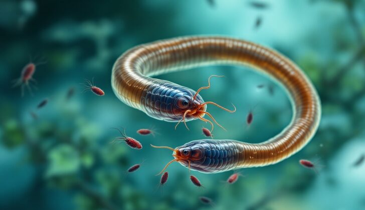
Fasciola hepatica, or liver flukes, are flatworms that can grow up to the size of 30 mm by 15 mm. They are brown, spiny, leaf-shaped creatures easily visible to the human eye. On one end, they have two suckers. The larger one, found on the bottom, allows the fluke to stick to the wall of the bile duct. This keeps it in place while the smaller sucker feeds on bile. Another fluke, Fasciola gigantica, behaves similarly but can grow up to 75 mm in length.
No matter if the host is a human or a cow, the life cycle of these flukes is the same. The adult liver flukes release their eggs into the host’s bile ducts. These eggs then get passed out of the body through the host’s feces. When the feces reach a water source, the eggs hatch into miracidia, which are free-swimming. These miracidia then find a host snail and crawl into its tissues. Inside the snail, the miracidia transform into various life stages until they become cercariae. These cercariae then exit the snail and become free-swimming again. They attach themselves to aquatic plants, where they are then eaten by their final host.
People can get infected by these flatworms by eating plants that these flatworms have attached themselves onto. When eaten, the flatworms excyst, or come out of their protective covering, in the small intestine. They then burrow into the peritoneum, which is the lining of the abdominal cavity. From there, they crawl head-first into the liver through its tissue and settle in the bile ducts. The flatworms develop into adults in 3 to 4 months, laying more than 20,000 eggs per day which are excreted through the host’s feces. If left untreated, they can live in the human body for up to 13 years.
The flatworms can cause damage to the liver tissue while migrating and settling in. This triggers inflammation and immune responses, which can result in a wide range of symptoms. Adult flukes may block the bile ducts, causing fibrosis or scarring, and leading to the enlargement and dilation of the proximal or nearest part of the biliary tree. Usually, the more the parasites in the body, the more damage it can cause to the liver.
Risk Factors and Frequency for Fascioliasis (Liver Fluke Infection)
Fasciola infection, often underestimated and under-detected, affects between 2.4 and 17 million people worldwide. In the United States, it is mostly seen in travelers and immigrants, although it is more common in cattle and other livestock, varying by location. The two types of fasciola, Fasciola hepatica and Fasciola gigantica, are found in different parts of the world. Fasciola hepatica is common in Europe and Asia, occasionally seen in Northern Africa, Central and South America, and the Middle East, with sporadic cases in the United States and the Caribbean. On the other hand, Fasciola gigantica usually infects livestock in Asia, the Pacific Islands, and some parts of Northern Africa.
- The two species are sometimes found in the same areas in Africa and Asia, making their symptoms hard to distinguish.
- The most affected hosts are cattle and sheep, but various other grazing animals can also get infected in the wild.
- People living in areas with prominent cattle and sheep industries who consume raw water plants, especially watercress, are at the highest risk of catching the parasite.
Signs and Symptoms of Fascioliasis (Liver Fluke Infection)
Fascioliasis is an infection that usually occurs in two stages, separated by different symptoms and impacts on the body.
The first phase, also known as the acute or hepatic phase, typically begins between 6 to 12 weeks following ingestion of metacercariae from contaminated water. Patients might initially experience a high fever, pain in the upper right portion of their abdomen, an enlarged liver, and occasionally a yellowing of the skin or eyes (known as jaundice). Additional symptoms may include muscle pain, skin rash, nausea, loss of appetite, and diarrhea. These symptoms result from the flatworms moving through the liver, triggering inflammation and immune responses. Lab tests may reveal anomalies such as anemia, elevated proteins in the blood, and unusual liver enzymes. Serious complications can develop in cases with a high number of parasites, such as accumulation of fluid in the abdominal cavity, internal bleeding in the bile ducts, and even severe liver necrosis – but these situations are rare. Most people’s acute symptoms typically resolve within 6 weeks, after which the infection moves into the chronic phase.
Some early non-liver symptoms are linked to how the body’s immune system reacts to the infection. A particular type of lung inflammation is a common reaction to helminth infections, including fascioliasis. An intense immune response can lead to inflammation of the blood vessels. This can cause issues like myocarditis (inflammation in the heart leading to abnormal heart rhythms) or cerebral vasculitis (which could lead to neurological issues like seizures). However, these are quite rare. Some patients may also present with generalized swollen lymph nodes.
The chronic or biliary phase usually begins about 6 months after the acute phase. This occurs when the flukes have settled in the bile ducts and can last up to a decade or more. Often, this phase is without symptoms, but sometimes, patients might experience chronic pain in the upper middle or right abdomen, nausea, vomiting, diarrhea, an enlarged liver, jaundice, and even malnutrition. Liver infection can sometimes lead to chronic conditions like blockages in the bile duct, recurring jaundice, pancreatitis, and even serious conditions like ascending cholangitis. Over longer periods, the infection can cause serious damage to the liver and bile ducts – leading to conditions like chronic cirrhosis, sclerosing cholangitis, and even cholangiocarcinoma (bile duct cancer).
Sometimes, the parasites may get lost and end up outside the digestive and liver systems. In this case, the infection occurring is known as ectopic fascioliasis. This can lead to inflammation and secondary tissue damage from the immune response. They can sometimes be found under the skin, in the lungs, heart, brain, or even the eyes, causing pain, swelling, and possibly infection. Another rare form of this condition is Halzoun syndrome or pharyngeal fascioliasis, typically seen in areas where people eat raw liver. In this case, the flukes attach themselves to the upper respiratory or digestive tract, causing inflammation. Severe swelling in these areas can sometimes lead to breathing difficulties.
Testing for Fascioliasis (Liver Fluke Infection)
Fascioliasis, which causes belly pain and changes in liver enzymes, is very rare in the United States but it might be suspected in people who have travelled to parts of Europe, Asia, or the Pacific. It’s even more likely if they’ve eaten watercress or raw vegetables that were washed in dirty water. Since the disease is so rare and its early symptoms are quite vague, it can often take some time to confirm a diagnosis, even in areas where it’s more common.
A blood test called serology has become the quickest and most effective way to test for fascioliasis. This test measures the body’s response (levels of specific proteins called ‘antibodies’) to the parasite’s presence. The specific proteins that this test looks for appear around two to four weeks after exposure to the parasite. The ‘ELISA’ technique is now often used instead of microscope examination of stool samples because it’s faster and more reliable.
Blood tests also show how severe the infection is, as high levels of these specific proteins suggests a larger number of parasites. Five to seven weeks after infection, the parasites reach maturity and start to produce eggs. Up until this point, stool samples will not show any trace of the infection because no eggs have been laid. The level of these proteins in the blood drops off once the parasites have been successfully treated, but some of these proteins may still be at a detectable level for years afterwards, even without any evidence of active infection.
Microscope examination of stool samples or fluids taken from the ducts in the gut can still be useful for diagnosing chronic infections in areas where test resources may be limited. The parasite lays thousands of yellow-brown, egg-shaped eggs per day, although not consistently, so a single negative sample doesn’t necessarily rule out a diagnosis.
Occasionally, adult parasites might be found during certain procedures to check out the ducts in the abdomen (‘biliary tree’) of people with blockages. Sometimes irregular lumps can be spotted on the liver during keyhole surgery, but knowledge of the patient’s medical history is typically needed to make a diagnosis. Generally, liver biopsy is not recommended because blood tests are much more reliable and cost-effective.
Scans such as CT (Computerized tomography) may show small, branch-like lesions in the liver, which are the tracks left by the migrating parasites. Areas of dead tissue might also be visible on scans if dye has been injected into the blood vessels (‘IV contrast’). Other potential findings include collections of blood under the liver capsule, thickening of the capsule, or hardening of the liver tissue.
Ultrasound, cholangiogram, and ERCP scans can be useful for visualizing the mobile, leaf-shaped parasites in the ducts of the gallbladder. They are often associated with the presence of gallstones. Other indicators of the disease, particularly in its early phase, include irregular thickening of the common bile duct wall, enlarged liver or spleen, or swollen lymph nodes around the liver.
Treatment Options for Fascioliasis (Liver Fluke Infection)
The best treatment for fascioliasis, a disease caused by parasites, is a medication called triclabendazole, which is taken by mouth. This medication is taken after a meal and should be used for two days. Triclabendazole works by preventing the formation of cellular structures in the parasite, thereby taking away their ability to survive and reproduce. This medicine is effective against all stages of the parasite’s development and has a success rate of over 90%. Other medications like ivermectin, praziquantel, artesunate, mebendazole, or albendazole are not effective against this parasite.
When a patient is diagnosed with fascioliasis, it is advised that their family or other house members are also tested. Fascioliasis can be detected by testing for specific antigens, which are substances in the blood that the body’s immune system responds to. If a person tests positive, they should take the treatment, even if they do not have any symptoms. This is important as the disease can lead to serious complications in the future.
What else can Fascioliasis (Liver Fluke Infection) be?
Conditions like acute hepatitis, autoimmune hepatitis, and shock liver can cause a significant increase in transaminases, typically rising to levels in the thousands. This is much higher than what we would usually see with fascioliasis, where levels are usually in the hundreds.
There are some conditions, including toxocariasis, acute schistosomiasis, ascariasis, and strongyloidiasis, which can be tough to distinguish from fascioliasis during the acute phase. This becomes particularly tricky when signs such as high eosinophils level in the blood (peripheral eosinophilia) and lung symptoms are present.
To definitely determine the presence of these diseases, a type of blood test called serologic testing is needed.
What to expect with Fascioliasis (Liver Fluke Infection)
If you’re diagnosed with fascioliasis, which is an infection usually caused by a worm-like parasite, the odds of a good outcome are highly favorable. The medicine Triclabendazole is known to be very effective against all stages of this infection, achieving a cure rate of over 90%. So, with the correct diagnosis and treatment, you have a very high chance of recovering successfully from fascioliasis.
Possible Complications When Diagnosed with Fascioliasis (Liver Fluke Infection)
Long-lasting infections can become more severe due to a condition called ascending cholangitis, which typically needs antibiotics delivered through an IV (directly into the veins) and often surgery. If this condition happens because of parasites causing a blockage, it becomes an emergency and a procedure called endoscopic retrograde cholangiopancreatography (ERCP) is needed. This procedure is used to directly remove the worms from the common bile duct if it’s possible. If these infections persist for a long time, it can lead to chronic inflammation and damage in the bile ducts. This can progress to conditions like sclerosing cholangitis, biliary cirrhosis, and cholangiocarcinoma.
Possible Complications of Chronic Infections:
- Ascending cholangitis
- Biliary obstruction by parasites
- Emergencies requiring endoscopic retrograde cholangiopancreatography (ERCP)
- Chronic biliary inflammation
- Sclerosing cholangitis
- Biliary cirrhosis
- Cholangiocarcinoma
Preventing Fascioliasis (Liver Fluke Infection)
After treatment, the medical team will assess if there’s been any improvement in the patient’s condition. This is achieved by checking for a reduction in serologic titers, which is a measure of antibodies in the blood, an indication of the infection level. The medical team will also look out for a decrease in higher-than-normal levels of a type of white blood cell called eosinophils (referred to “resolution of eosinophilia”). Another way to measure improvement is to check for the absence of parasite eggs in the patient’s stool.
Typically, if any tests were positive before treatment, they should be repeated three months after treatment to make sure the infection has been truly eliminated. If the patient initially had obstructions in their bile duct due to the infection, follow-up ultrasound scans can help check if things have gotten better, despite possible lingering issues like scar tissue and thickened ducts, which might continue to cause discomfort.
If someone is found to have fascioliasis, a parasitic infection typically obtained from eating contaminated plants or water, it’s important to test everyone living in the same household. Anyone who tests positive ought to be treated, even if they’re not showing any symptoms. This measure is taken to prevent the possibility of future health complications.
One of the best ways to prevent this infection is by advising patients to avoid eating raw freshwater plants, especially in places where the parasite is commonly found. While previously attempts have been made to control the snail population, which serves as an intermediary host for the parasite, this has proven impractical. Currently, no vaccines are available, and given the rarity of this disease among humans, developing one doesn’t seem economically feasible.


