What is Herpes Zoster Ophthalmicus (Shingles Involving the Eye)?
Varicella-zoster virus (VZV), more commonly known as chickenpox, often affects children and can spread through the air or physical contact. Herpes zoster, also known as shingles, happens when the VZV virus, which has been dormant in nerve cells, becomes active again. This usually occurs many years after the original chickenpox infection. The disease often shows up as a one-sided rash with bumps or blisters in a pattern that follows nerve paths on the skin.
When the virus affects the first division (V1) of the trigeminal cranial nerve, it’s defined as Herpes zoster ophthalmicus (HZO). It’s important to note that having HZO doesn’t always mean that the eyes are affected, but studies show about 50% of HZO cases result in eye problems. These can range from simple inflammation of the eye’s surface or inner layers to more serious conditions damaging the cornea or retina. This disease is seen as a serious emergency in eye care as any delay in identifying and treating it could lead to loss of vision.
What Causes Herpes Zoster Ophthalmicus (Shingles Involving the Eye)?
The Varicella-zoster virus (VZV), which causes chickenpox, usually stays in our body without cause for alarm after the first infection, hiding out in specific nerve cells. This is possible because our immune system, made stronger by the initial infection, keeps the virus in check. But sometimes, when our immune systems are weakened for various reasons, the virus can wake up and cause shingles, also known as herpes zoster.
Herpes Zoster Ophthalmicus (HZO) is when the virus comes back to life in the V1 division of the trigeminal nerve, a nerve that had been its dormant home. The V1 nerve has three sub-parts: the frontal nerve branch, the nasociliary nerve branch, and the lacrimal nerve branch. The virus can affect any or all of these, resulting in eye and facial skin sores. Unfortunately, these can also lead to more severe complications.
Risks for this condition increase in people over the age of 50 and those with compromised immune systems. This includes individuals with HIV, autoimmune diseases taking steroids or other immunosuppressant drugs, those who’ve had organ or bone marrow transplants, or individuals undergoing chemotherapy. Other long-term illnesses, acute illnesses, and stress, both physically and emotionally, can also trigger HZO.
Risk Factors and Frequency for Herpes Zoster Ophthalmicus (Shingles Involving the Eye)
In the United States, herpes zoster, also known as shingles, affects about 1 in 1000 people every year. However, for people over 60 years old, the chances of getting it are higher, about 1 in 100. It’s interesting to note that taking certain vaccinations significantly lowers the risk for older adults. Unfortunately, about 8 to 20% of people with shingles will have complications like Herpes Zoster Ophthalmicus (HZO), which can affect the eyes.
Signs and Symptoms of Herpes Zoster Ophthalmicus (Shingles Involving the Eye)
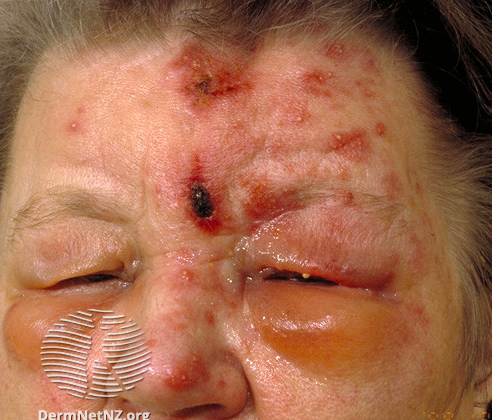
Herpes zoster ophthalmicus (HZO) is a condition that often starts with a unilateral intense pain along the forehead, followed by a reddish, blister-like rash in the same area. The pain can be described as “burning” or “shooting” with occasional numbness or tingling. Often, there might be symptoms such as fever, fatigue, malaise, or headaches even before the rash appears. Herpes sores around the tip of the nose, known as the Hutchinson sign, indicate a higher risk for eye involvement.
During a medical examination, the doctor would examine the spread of the lesions, including the eyelids and scalp. Initially, the rash might appear as small, raised red spots before turning into blisters and eruptions that eventually crust over. While the rash usually occurs in one localized area on one side, patients with weakened immune systems may have a more widespread occurrence, possibly affecting both sides. The doctor would pay special attention to the eyelids, as their involvement often signifies disorders like blepharitis, conjunctivitis, or episcleritis. Drooping of the eyelid (ptosis) is unusual but can occur when there is severe inflammation. Furthermore, a detailed eye exam is necessary to check visual acuity, ocular tonometry, and a dilated fundus examination.
Corneal involvement in HZO includes both epithelial keratitis and stromal keratitis. Epithelial keratitis can show up as dotted keratitis, where specific areas of the cornea’s surface stain or pseudodendritic keratitis, non-erosive mucous patches that mildly stain without any terminal bulbs. Stromal keratitis, which may develop after epithelial keratitis, can either be anterior or deep. Anterior stromal keratitis is an immune response to the virus and shows up as circular granules in the superficial stroma. Deep stromal keratitis, often happening later in the disease’s course, leads to severe corneal swelling and might be coupled with anterior uveitis.
Uveitis, inflammation of the eye’s middle layer, may be anterior, posterior, or panuveitis. Anterior uveitis is seen as floating white blood cells and proteins, often referred to as “cell and flare”, in the eye’s anterior chamber, shown in a slit-lamp examination. Posterior uveitis involves white blood cells in the vitreous cavity or white areas on the retina signifying inflammation and tissue death in severe cases. In some cases, it’s associated with optic neuritis, inflammation of the optic nerve.
All people diagnosed with HZO should get ocular tonometry as a part of their examination, to measure the pressure inside the eyes. This intraocular pressure might increase due to HZO, often secondary to the inflammation of the trabecular meshwork (trabeculitis) caused by the herpes zoster virus. The pressure may also increase due to the obstruction of the trabecular meshwork from the resulting inflammatory cells and proteins or the formation of adhesions as a consequence of extensive intraocular inflammation. If not addressed promptly, a persistently raised intraocular pressure could potentially lead to secondary glaucoma, a condition damaging the optic nerve.
Testing for Herpes Zoster Ophthalmicus (Shingles Involving the Eye)
Diagnosing herpes zoster ophthalmicus (HZO) is usually based on personal health history and recognizable signs found during a physical and slit-lamp eye examination. There are other procedures, like ocular tonometry (eye pressure test) and corneal esthesiometry (sensitivity test of the eye’s front surface), that may be done to check for any complications.
Other tests like viral cultures, polymerase chain reaction (PCR) tests, and antibody tests are usually not needed to confirm a HZO diagnosis. If a patient has widespread herpes zoster (affects multiple areas of the skin), severe illness, or significant risk factors, an HIV test may worth considering. Typically, additional lab tests and imaging are rarely needed.
Treatment Options for Herpes Zoster Ophthalmicus (Shingles Involving the Eye)
The treatment for Herpes Zoster Ophthalmicus (HZO) involves starting antiviral medications for all affected patients as quickly as possible. Antiviral medicines are the main treatment, while other treatments such as pain management, antibiotics, steroid treatments, or a procedure to remove damaged surface tissue from the cornea (epithelial debridement), may be considered based on individual patient circumstances.
Breifly, here are the key elements of treatment:
– Symptom support: This can involve using artificial tears, cold compresses, and pain relievers.
– Antiviral medicines: The best results are seen when these treatments start within 72 hours of the disease beginning. This shouldn’t be delayed, even if you’re waiting for a definitive diagnosis. Antiviral eye drops can be considered, but their effectiveness for HZO isn’t well-established.
– Antibiotics: Doctors often prescribe antibiotic eye drops or ointments, like erythromycin, to prevent secondary bacterial infections.
– Steroid treatments: Steroids, both applied directly to the eye or given systemically (like a pill), can be beneficial for managing HZO. However, they’ve had mixed results in clinical trials, and the possible risk of side effects should be carefully weighed against benefits.
– Removal of affected corneal surface tissue or Debridement: This can be considered in situations where the cornea, the clear front layer of the eye, is affected.
Keep in mind, the dosage and specific medication used will depend on whether a patient is immunocompromised or not and may need adjustments based on factors like kidney function. As with all medical treatment, an ophthalmologist should oversee the specifics of your care.
What else can Herpes Zoster Ophthalmicus (Shingles Involving the Eye) be?
When a doctor starts suspecting eye-related complications due to HSV, or Herpes Simplex Virus, they’ve to make sure it’s not one of the other potential conditions that can show similar symptoms. These are:
- HSV or Herpes Simplex Virus related skin inflammation
- Other types of pink eye caused by different viruses or bacteria
- Eye allergies leading to pink eye
- Pink eye or cornea inflammation due to exposure to harmful elements
- Acute angle-closure glaucoma, a serious eye disease
- A corneal ulcer, which is an open sore on the cornea
- Corneal abrasion, or scratches on the cornea
- Impetigo, a contagious skin infection
- Cellulitis, an infection that can cause severe pain and swelling
- Bites from insects
- Contact dermatitis, a type of skin inflammation caused by direct contact with an allergen or irritant
Accurate diagnosis is key to determining the right treatment plan. Accordingly, the physician will perform thorough examinations and possibly some tests to exclude these potential diseases.
What to expect with Herpes Zoster Ophthalmicus (Shingles Involving the Eye)
The outcome of Herpes Zoster Ophthalmicus (HZO) can greatly differ from person to person. It depends on several factors such as the individual’s health risks, when treatment starts, and how severe the disease is. Not much is known about how often it causes severe disease or death.
However, for most patients with healthy immune systems, early treatment of herpes zoster can clear up the skin lesions within four weeks. These patients can typically be treated as outpatients, which means they don’t need to stay in the hospital.
Possible Complications When Diagnosed with Herpes Zoster Ophthalmicus (Shingles Involving the Eye)
As mentioned, a person with herpes zoster ophthalmicus (HZO) can experience various complications. These can range from inflammation of various parts of the eye including the eyelid (blepharitis), the eye’s outer layer (conjunctivitis), the cornea (keratitis), the middle layer of the eye (uveitis), and secondary high eye pressure (glaucoma). If the inflammation is prolonged, it can result in corneal scarring, which can lead to permanent vision loss.
Another possible complication is neurotrophic keratopathy, a condition where the nerves in the cornea don’t function properly. This makes the person more vulnerable to corneal abrasions, corneal ulcers, and persistent defects in the corneal epithelial cells, even without the presence of active viruses or inflammation.
Although it’s rare, a complication known as necrotizing retinitis can occur. This can lead to tears in the retina, the losening and eventual separation of the retina from its supportive layers (retinal detachment), and permanent changes in vision.
Another common development after HZO is postherpetic neuralgia (PHN), a chronic pain condition. In PHN, the pain caused by the herpes zoster virus continues more than 90 days after the initial rash outbreak. Older age and severe disease increase the risk for developing PHN, while having received a prior zoster vaccination can lower the risk.
Furthermore, patients with HZO are also prone to secondary bacterial infections.
Main complications of Herpes zoster ophthalmicus include:
- Blepharitis
- Conjunctivitis
- Keratitis
- Uveitis
- Secondary glaucoma
- Permanent vision loss due to prolonged inflammation and corneal scarring
- Neurotrophic keratopathy, which can lead to corneal abrasions, ulcers, and persistent epithelial defects
- Necrotizing retinitis, potentially resulting in retinal tears, detachment and permanent vision changes
- Postherpetic neuralgia, especially in older patients and those with severe disease
- Secondary bacterial infections
Preventing Herpes Zoster Ophthalmicus (Shingles Involving the Eye)
The U.S. Centers for Disease Control and Prevention (CDC) advises that adults over 50 should get a vaccine for herpes zoster as a preventative measure. This can reduce the chance of the virus flaring up again. There’s currently one vaccine available – a deactivated version of the virus created through genetic engineering. This vaccine was introduced in 2018 and is given to the patient in two doses.
The vaccine has shown to be 97% effective in individuals aged 50 to 70, and 90% effective in those older than 70. Even if someone had the previous live vaccine, they can still get the deactivated one. However, the safety of this vaccine in people with compromised immune systems is still being studied.
While the vaccine doesn’t fully stop herpes zoster, it usually makes the duration and severity of the disease less for vaccinated individuals. To help prevent the spread of the disease, people with an active infection should keep their rash covered and wash their hands frequently.
Extra precautions are necessary for those with a widespread infection, including keeping the virus from getting airborne. People with herpes zoster should stay away from pregnant women, babies and young children who haven’t been exposed to the virus before, and people with weakened immune systems.


