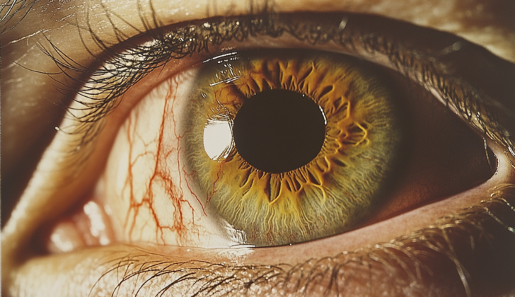
What is Ocular Tuberculosis?
Ocular tuberculosis is an eye disease caused by tuberculosis bacteria (TB). It can be transmitted in different ways and can infect virtually any part of the eye. Similar to how syphilis can look like various skin diseases, TB can also take on the appearance of other eye diseases, earning it the nickname “the great imitator” of eye conditions.
Choroidal tubercles, a particular TB eye condition, were first explained in 1855 and recognized with an eye examination tool in 1867. Shortly after the TB bacteria was discovered, in 1883, it was identified in the eye. Interestingly, in a 1950 study, an eye examination was found to be more useful in diagnosing TB than a chest x-ray. However, since then TB has become less common in Western countries and with advancements in testing labs, eye examinations for this condition have become less popular.
There are over 1.7 billion people estimated to be infected with TB worldwide. It’s the leading cause of death from a single infectious disease and tops the list of causes of death among people living with HIV. It’s crucial to recognize ocular TB as it’s a sign of extrapulmonary TB – a form of TB that affects organs and tissues outside of the lungs. An early diagnosis can lead to immediate treatment to prevent TB and ensure better outcomes for patients.
What Causes Ocular Tuberculosis?
Ocular tuberculosis, a disease affecting the eye, can develop in three ways:
1. The eye can get infected directly from an external source, such as through contact with the eyelids or eye surface (known as primary ocular TB).
2. The bacteria causing tuberculosis, M. tuberculosis, can spread through the bloodstream from a site of infection in the lungs or elsewhere in the body (secondary ocular TB).
3. Parts of the eye can have an allergic reaction after getting exposed to tuberculosis antigens, substances that trigger an immune response.
The most common way the eye gets affected is through M. tuberculosis spreading through the bloodstream from an infection in the lungs. The bacteria can spread either from a new infection or by waking up a dormant, or sleeping, infection.
M. tuberculosis spreads through tiny droplets in the air. Once inhaled, the bacteria reach small air sacs in the lungs called the alveoli, where they meet alveolar macrophages, a type of immune cell. Most people who inhale the bacteria (about 90%) never develop symptoms and have what’s termed latent TB. Of the remaining 10%, half will develop tuberculosis in the first few years after exposure, and the other half might develop symptoms later as their immune system weakens.
The alveolar macrophages try to fight off the bacteria by swallowing them up, a process called phagocytosis, and calling for backup from circulating immune cells called monocytes. However, M. tuberculosis has a trick to avoid being destroyed – it prevents the macrophage from fully activating its destructive abilities. This allows the bacteria to multiply inside macrophages that are not fully activated. These bacteria-carrying macrophages can then spread via the lymphatic system and bloodstream, reaching various parts of the body rich in oxygen, such as the peak of the lungs, various organs, and the eye.
Risk Factors and Frequency for Ocular Tuberculosis
As per the World Health Organization (WHO), tuberculosis, often known as TB, affects approximately 10.4 million people and causes about 1.8 million deaths each year. This illness is considerably more common in poorer countries, where its rate is eight to twelve times higher than in wealthy, industrialized countries. The largest number of TB cases were found in South-East Asia (44%) in 2018, specifically in countries like India (27%), China (9%), Indonesia (8%), the Philippines (6%), and Pakistan (6%).
TB can also affect parts of the body other than the lungs, such as the eyes (ocular TB). This type of TB is rare, representing less than 1% of cases in the United States, 4% in China, 6% in Italy, and 16% in Saudi Arabia.
Historically, the statistics related to ocular TB have varied widely due to the lack of specific methods for diagnosis. For instance, a 1967 study reported that out of 10,524 patients at a TB clinic, only 1.4% had ocular TB. In contrast, a study in Spain from 1997 found that 18% of 100 TB patients had ocular TB. Other studies in North India and Japan have reported that TB was the cause of inflammation inside the eyes (uveitis) in 9.86% and 7.9% of patients, respectively.

reaching the macula in a patient with TB choroiditis.
Signs and Symptoms of Ocular Tuberculosis
Those most likely to develop ocular tuberculosis are people with weakened immune systems. Most notably, this includes individuals undergoing immunosuppressive therapy, people who have both AIDS and Tuberculosis, and people with certain other health conditions such as alcoholism, chronic liver disease, diabetes, and cancer. Healthcare workers, the homeless, prisoners, and immigrants from countries where TB is common are also at heightened risk.
When trying to identify ocular TB, doctors need to ask patients about their medical history and especially their HIV status. Other information that can be useful includes symptoms of lung TB (like chronic cough, chest pain, loss of weight and appetite, fever and night sweats), previous positive test results for TB, travel to countries where TB is common, and contact with active TB patients.
- Chronic lung symptoms such as cough and chest pain
- History of travel to countries where TB is common
- Prior contact with TB patients
- Previous positive TB test results
Ocular TB can cause various eye problems and is not always easy to identify. Symptoms patients might report include inflammation in one or both eyes, headaches, seeing “flashes” or “floaters”, or redness of the eye. Some people might even report diminished visual clarity or sensitivity to light due to small growths in the eye. Notably, having no apparent visual symptoms does not necessarily mean no ocular TB, as some small growths in the outer part of the eye might cause no noticeable symptoms.
Doctors trying to diagnose ocular TB will look out for different signs. These vary depending on which part of the eye is affected and include:
- Outside the eye: patients might have eye swelling, headaches, nosebleeds, blurred vision, or a variety of other symptoms.
- Eyelids: there might be reddish-brown nodules, eyelid abscesses, chronic eyelid inflammation, or unusual styes.
- Lacrimal Gland: there might be inflammation indistinguishable from other bacterial infections.
- Conjunctiva (eye surface): chronic disease that eventually leads to scarring and presents with eye redness, discomfort, and abnormal discharge, often with swollen regional lymph nodes.
- Cornea: presenting as inflammatory nodules at the limbus or interstitial keratitis. Both conditions can cause discomfort and visual symptoms.
- Sclera (outer white of the eye): hard to diagnose outside the context of active systemic TB. Scleritis is typically chronic, not responsive to anti-inflammatory treatment, can cause tissue death, and usually presents anteriorly (towards the front); posterior sclera involvement is rare.
- Inside the eye: depending on the part of the eye affected, ocular TB can present as inflammation, nodules, intraocular abscesses, or retinitis. It might cause significant visual symptoms or none at all, and look similar to various other ocular conditions.
Ultimately, ocular TB requires expert diagnosis and treatment due to its possible serious complications and varying presentation.
Testing for Ocular Tuberculosis
Ocular tuberculosis, a form of eye infection, can be hard to diagnose due to its various symptoms. The process usually begins with thorough patient history, a physical examination, and an exam where a doctor looks at the back of your eye (fundoscopic exam). Some specific signs were identified with high specificity but low sensitivity, meaning they are strong indicators of ocular tuberculosis if present, but their absence doesn’t necessarily rule it out.
Some of the suspicious signs include broad-based posterior synechiae, a condition where the iris sticks to the lens of the eye, retinal vasculitis with or without choroiditis, inflammation of the blood vessels and layers of the eye. Other conditions like multifocal serpiginous choroiditis, occlusive retinal periphlebitis, and granulomatous uveitis, which refers to swelling and irritation in the eye, are also potential indicators of ocular tuberculosis.
There are several tests used to investigate ocular tuberculosis. One is the Tuberculin skin test (TST) or the interferon-gamma release assay (IGRA), which are used to assess the body’s immune response to tuberculosis. However, these are not always accurate; for example, TST can give a false positive if you’ve had a BCG vaccination or have other mycobacteria infections.
In contrast, IGRA tests such as T-SPOT and QuantiFERON-TB Gold tend to be more specific, but false positive can still occur — particularly in areas where the disease is not common. Additionally, chest x-rays or a high-resolution computed tomography (HRCT), a detailed type of CT scan of the chest, can show whether there are any signs of tuberculosis in the lungs.
Doctors sometimes attempt to take a biopsy, a sample of the tissue for testing, but this can be difficult and risky for patients. Instead, doctors may collect fluid samples from the front (anterior chamber) or back (vitreous humor) of the eye for testing, using a method called polymerase chain reaction (PCR) that helps detect the presence of the bacteria that causes tuberculosis. PCR has become popular due to its accuracy and quick turnaround time compared to traditional culture tests.
In some cases, The World Health Organization (WHO) recommends the use of Xpert MTB/RIF assay, a kind of automated real-time PCR. However, it is currently used with samples of sputum (a kind of mucus), and has not been approved by the U.S. Food and Drug Administration for use with ocular fluids.
Diagnosing ocular tuberculosis definitively is only possible when the tuberculosis bacteria or its DNA is detected in ocular fluids. However, in many cases, a definitive diagnosis is not achievable. If patients have eye symptoms consistent with ocular tuberculosis, a confirmed exposure to tuberculosis or evidence of a tuberculosis lesion on a chest x-ray or CT scan, a “presumed ocular tuberculosis” diagnosis is often made, and treatment is recommended.
Treatment usually begins with a trial of antituberculous therapy (ATT), with the use of medications like isoniazid, rifampicin, ethambutol, and pyrazinamide. Doctors evaluate the patient’s response after 4 to 6 weeks, and if they respond positively to this therapy, it is considered evidence of ocular tuberculosis, and the treatment continues.
Treatment Options for Ocular Tuberculosis
Treating eye tuberculosis usually follows the same approach as lung tuberculosis. The typical treatment involves a set of four different drugs taken in two stages: rifampicin, isoniazid, pyrazinamide, and ethambutol taken daily for the first two months, and then rifampicin and isoniazid taken for the subsequent four months. If the patient doesn’t show improvement after three to four weeks, the tuberculosis might be resistant to multiple drugs, and the doctor will need to manage the treatment in consultation with a specialist in infectious diseases.
Steroids, another class of medications, are also used during treatment. They can help to control the inflammation typically caused by this form of tuberculosis and can prevent an overly strong immune response to the tuberculosis organisms. These steroids, however, must be used carefully and in combination with the other tuberculosis drugs. Using steroids alone could potentially risk reactivating dormant tuberculosis or prolong its active growth in the eye. Another consideration is that rifampicin, one of the tuberculosis drugs, can decrease the effectiveness of steroids. Therefore, the doctor might need to adjust the patient’s steroid dosage if they are also taking rifampicin.
Interestingly, sometimes a patient’s condition might appear to worsen initially after starting the tuberculosis treatment. This is thought to be similar to the so-called Jarisch-Herxheimer reaction, which is an intense immune response that occurs when the treatment significantly improves exposure of the immune system to the tuberculosis organism and leads to the release of substances that promote inflammation. Co-treatment with steroids can help to manage this reaction.
In addition to these drugs, a particular type of eye inflammation caused by tuberculosis, known as Eales disease, can also be treated with laser treatment. This treatment is designed to reduce the drive for new blood vessels to form in the retina due to lack of oxygen, a process known as retinal neovascularization.
What else can Ocular Tuberculosis be?
If you have tubercular uveitis – an eye infection caused by tuberculosis – your doctor would look at several other conditions that can show up in similar ways. These include:
- Sarcoidosis (an immune system disorder)
- Syphilis (a sexually transmitted infection)
- Toxoplasmosis (parasite infection)
- Toxocariasis (parasitic worm infection)
- Fungal infections
Tubercular uveitis in kids might appear similar to a condition called juvenile idiopathic uveitis. For patients mainly suffering from retinal vasculitis – inflammation of the blood vessels in the eye – another disease to think about is Eales disease. This disease consists of three overlapping stages: inflammation of the retinal blood vessels, blockage of these blood vessels, and new blood vessels growing in the retina. The exact cause of Eales disease is not known, but it’s speculated to be a delayed eye response to M. tuberculosis.
Sometimes, the symptoms of tuberculous serpiginous-like choroiditis (an eye disease that affects the layer of blood vessels in the eye, the choroid) may resemble two other diseases. These are acute multifocal posterior placoid pigment epitheliopathy (AMPPPE) – a rare eye disease that mainly affects the macula (a part of the retina at the back of the eye) – and classic serpiginous choroiditis. AMPPPE often displays multiple flat, grey-white lesions in the retinal pigmented epithelium (the pigmented layer of cells supporting the retina). While AMPPPE and serpiginous-like choroiditis might look similar in early stages, the lesions in serpiginous-like choroiditis usually grow progressively and merge into a continuous layer of inflammation with an active leading edge.
What to expect with Ocular Tuberculosis
Starting treatment for ocular tuberculosis (a type of tuberculosis that affects the eyes) right away can lead to positive results. Most of the time, the damage caused by the disease completely goes away and leaves only a small amount of residual damage. Depending on how severe the disease is, some lesions (areas of damaged tissue) may turn into small scars on the choroid and retina, which are parts of the eye.
However, in people who have AIDS, these lesions have sometimes been observed to get worse, even with treatment for tuberculosis.
Possible Complications When Diagnosed with Ocular Tuberculosis
You should know that certain medications used to treat tuberculosis, known as ATT medications, can have side effects that impact your eyes. One such medication, Ethambutol, can cause a variety of eye problems depending on the dosage.
These problems can include:
- Optic neuritis, which is inflammation that damages the optic nerve
- Photophobia, which is a sensitivity to light
- Paresis of the extraocular muscles, which can interfere with eye movement
- Red-green dyschromatopsia, which affects color perception
- Central scotomas, which are blind spots in your central vision
- Disc edema, which is a swelling of the optic nerve at the back of the eye
If you’re taking more than 15mg/kg/day of the medication, it’s recommended that you have your eyes checked by an ophthalmic doctor every four weeks. If you experience any of these side effects, they will usually go away between three to twelve months after stopping the medication.
Preventing Ocular Tuberculosis
If you have been diagnosed with eye tuberculosis, it’s important to understand that the treatment typically lasts for six months. It’s crucial to complete the full course of treatment because not doing so can lead to serious health problems. A special treatment strategy, known as Directly Observed Treatment (DOT), is suggested by the World Health Organization. This method involves having a healthcare provider or family member watch while you take your medication. This is done to ensure you stick to the treatment plan and improve recovery.


