What is Paracoccidioidomycosis?
Paracoccidioidomycosis (PCM) is an infection that spreads throughout the body and is caused by a particular type of fungus. This fungus is most commonly found in the Americas, especially in Brazil, Venezuela, and Colombia. PCM is the most common type of fungal infection in this region.
The disease was first identified by a scientist named Adolfo Lutz in Brazil in 1908. Another scientist, Splendore, then further studied this disease and the fungus that causes it, describing its physical appearance and four patient cases. In 1930, the organism was officially named Paracoccidiodes brasiliensis by Floriano Paulo de Almeida.
This disease develops due to a specific type of fungus that can change its form with heat, belonging to the Paracoccidioides group. Other species within this group can also cause PCM, including P americana, P restrepiensis, P venezuelensis, and P lutzii. Of these, Paracoccidioides brasiliensis and Paracoccidioides lutzii are considered the main culprits, as they are identified most frequently in cases of infection.
What Causes Paracoccidioidomycosis?
Paracoccidioides, a type of fungus, belongs to the group known as Ascomycota, within the order Onygenales. This type of fungus is found in different geographic subtypes: PS1 is most commonly found in South America, PS2 is found in Brazil and Venezuela, PS3 in Colombia, and PS4 is only found in Venezuela.
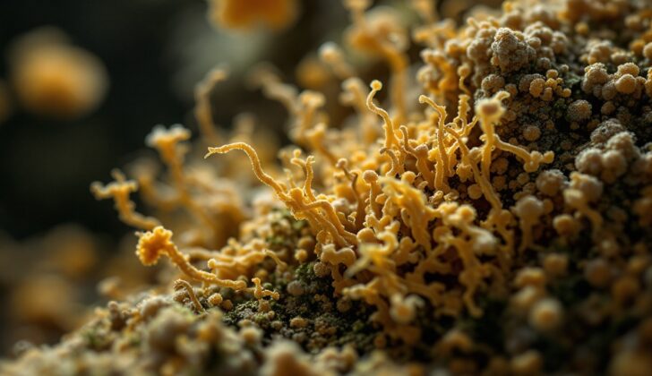
According to the book “Diagnosis and Treatment of Human Mycoses,” Paracoccidioides goes through two forms of growth. When it’s cooler (22° to 24° C), it grows as long, segmented filaments called mycelium, occasionally producing reproductive cells called chlamydospores and conidia. When it’s warmer (36° to 37° C), it grows as yeast which can be recognized by its “pilot’s wheel” appearance – like yeast cells sprouting multiple buds. Importantly, the main species of Paracoccidioides do not go through a sexual stage of reproduction.
Paracoccidioides typically grows in soil found in wet regions with regular to heavy rainfall, mild temperatures, and in areas with forests and rivers. Interestingly, Paracoccidioides infections are often reported in regions where coffee and tobacco are grown.
Usually, humans and a type of mammal known as the 9-banded armadillo are the hosts where this fungus grows. It’s important to note, however, that there hasn’t been any reported case of the fungus spreading directly from one person to another.
Risk Factors and Frequency for Paracoccidioidomycosis
The fungus, Paracoccidioides, is mostly found in certain areas of Central and South America, with around 80% of cases reported in Brazil. It has been found as far north as Mexico and as far south as Argentina, but hasn’t been noticed in Chile, Surinam, Guyana, Nicaragua, Belize, and most Caribbean islands. In regions where it’s commonly found, up to 75% of adults may test positive for it. Close to 10 million people are infected by this fungus in Latin America, but only 1 to 2% actually develop any symptoms of the disease.
- Incidence rates are between 1 to 4 cases per 100,000 people each year in most areas.
- In areas with a high amount of the fungus, like parts of the Amazon and Rondônia state in Brazil, there are 9 to 40 cases per 100,000 people each year.
- Less than 5% of patients with this disease are expected to die from it.
The disease tends to affect adult males (75% to 95% of cases), even though females are exposed to the fungus just as much. Hormones (estrogens) in the female body can apparently prevent the fungus from developing into yeast, which is what causes the disease. But if women do develop symptoms, the disease can often be more severe and spread more widely.
This disease is especially connected with people who live in rural areas and work with crops, such as coffee and tobacco, due to their increased exposure to this fungus. People who smoke or consume more than 50g of alcohol daily are 14 times more likely to develop the disease. It’s rarely linked to HIV and AIDS, but it can show up alongside tuberculosis in about 15 to 20% of patients. There have been a few reports of it appearing along with cancer and solid organ transplants.
Signs and Symptoms of Paracoccidioidomycosis
Most people who contract Paracoccydiomycosis, about 95%, do not display any symptoms and are found to have a quiet infection in the lungs, detectable only through specific medical tests. For those who do show symptoms, there are two main types of the disease: juvenile form and chronic adult form.
The juvenile (or acute) form appears in roughly 10% of cases. It’s more likely to affect young people under 30 and shows up about 45 days after exposure. Symptoms can include a fever, weight loss, tiredness, skin sores, swollen glands, sores opening onto the skin, large liver and spleen, and malfunctioning bone marrow. In this form, mouth and respiratory symptoms are rare. On a physical exam, the person might have enlarged lymph nodes, pus-filled cavities or open sores draining pus. This form of the disease tends to get worse over time.
The chronic (or adult) form is seen in 80 to 90% of cases and is more common in adult men. Symptoms relate mainly to lung infection and may include fever, tiredness, cough, and shortness of breath. Some patients may show lung damage such as scarring, air-filled spaces, and lung hypertension. This type of the disease is a reactivation that can happen months to years after the initial infection. Lung involvement is the most common symptom. Big lymph nodes are not usual, except in children. In over half of the cases, the infection spreads to the bloodstream with mucus membrane involvement.
It may include sores in the windpipe and throat resulting in altered voice, difficulty swallowing, harsh breathing sound, and sores around the mouth. Gum involvement is frequent and may cause tooth loss. Characteristic oral erosions may be associated with nose and throat ulcers.
Enlarged neck lymph nodes, as well as swelling in the armpit and groin regions, may be present. If the infection involves the lymph nodes inside the stomach, patients may experience widespread abdominal pain and yellowish skin due to bile duct compression. Partial blockage of the intestine can also be witnessed. Malabsorption syndrome, due to hardening of the lymph nodes in the stomach region, can occur.
About 25% of cases have skin sores. Skin symptoms could be ulcers, crusted sores, bumps, thickened areas of skin, and wart-like growths. Usually, these are caused by the spread of the disease from the lungs to the bloodstream. In rare cases, these lesions can occur due to direct inoculation.
Testing for Paracoccidioidomycosis
To diagnose the presence of the Paracoccidioides species of fungus, a combination of methods are used. These can include examining tissue under a microscope, culturing specimen samples, taking biopsies, examining the structure of taken tissues, and carrying out specific blood tests.
Microscopic examination, for instance, is commonly carried out using a liquid substance known as potassium hydroxide (KOH). In about 90% of cases, a positive response is yielded in either skin samples or sputum samples – a mixture of saliva and mucus coughed up from the lungs. This method allows scientists to spot yeast cells with distinctive characteristics – such as variable size, thick walls and unusual bud-like protrusions.
Culturing, or growing, the Paracoccidioides fungi for identification is another method, although rarely used as it tends to be a slow process taking 20 to 30 days, and is hence not ideal for timely diagnosis.
Using special stains such as Methenamine silver stain or periodic acid Schiff stain to view tissue samples collected, is another diagnostic method. Under the microscope, unique features reveal the existence of the fungi. For example, you might see varied cells surrounding yeast cells, abscesses in skin and mucous cells, or widespread tissue reactions in certain forms of the disease.
Blood tests are also significant for both diagnosing the infection and monitoring how well the treatment is working. There are several types of blood tests, but the most commonly used technique found in areas where the infection is frequent is quantitative immunodiffusion testing. This method has a reliability of 84.3% for detecting the disease and 98.9% for not mistaking the disease for something else. However, there are some limitations with this test; certain patients might get negative results because the techniques for diagnosing their specific type of infection have not been validated yet.
Skin testing can show a positive result even in healthy individuals from the areas where the infection is common, so it is not typically used to diagnose the active disease. In addition, its sensitivity to detect the disease decreases in patients who are very ill.
In children suffering from acute Paracoccidioidomycosis (PCM), general lab tests commonly show abnormalities such as anemia, abnormal proteins in the blood (hypergammaglobulinemia), high levels of certain white blood cells (eosinophilia), low levels of albumin (a protein in your blood), elevated levels of bilirubin, and slightly raised liver enzymes.
Treatment Options for Paracoccidioidomycosis
Paracoccidioidomycosis is a type of infection that commonly occurs in Central and South America. It’s treated with antifungal medications. However, despite established treatment guidelines, we don’t have definitive evidence yet to prove the effectiveness of these treatments. The illness responds to a number of antifungal medications, including ketoconazole, itraconazole, voriconazole, posaconazole, terbinafine, and amphotericin B.
It’s worth noting that fluconazole is not frequently used to treat this infection due to lower success rates and a higher chance of the infection coming back. Another medication, trimethoprim-sulfamethoxazole, has been shown to require a longer period of treatment (at least 24 months) with lower success rates and a higher incidence of relapse compared to itraconazole. That’s why it’s not the favored treatment option. Normally, treatment continues until you no longer have symptoms, you show signs of healing on X-rays or other imaging, and blood tests confirm a significant drop in indicators of infection. If your illness is severe, treatment will likely last longer than the usual period suggested for the medication used.
Itraconazole is the preferred treatment for mild to moderate cases due to its high success rate. It’s crucial to note that this medication must be dissolved in the stomach to be absorbed, so if you’re taking medicine to reduce stomach acid, you should not take itraconazole. The rate of your illness coming back after completing itraconazole treatment is less than 5%.
Voriconazole is another antifungal medication used to treat this infection. In a small study of 53 patients, it was found to be just as effective as itraconazole for treating chronic paracoccidioidomycosis. The treatment duration recommended is between six to twelve months.
Ketoconazole has a cure rate of 85% to 90% in small studies and a relapse rate of less than 10%. The recommended treatment duration ranges from 6 to 18 months. Whereas, Amphotericin B deoxycholate is reserved for severe cases of this disease. It has been shown to be effective, especially in patients with sepsis, a serious systemic infection. A variant of this drug, lipid formulation of amphotericin B, has better tolerability and lesser side effects. It is highly effective (greater than 95%) in severe cases. The chance of the infection returning is between 20 to 30% after treatment with amphotericin B. Switching to itraconazole after initial treatment with amphotericin B can decrease the chances of the infection returning.
If the first line of treatment doesn’t work, doctors may use a combination of two medicines called trimethoprim-sulfamethoxazole. While it has a lower success rate than itraconazole, it may still be of help. Another alternative is sulfadiazine. This medicine has expected cure rates of around 70%, but there’s a 30% chance the infection may return.
What else can Paracoccidioidomycosis be?
When trying to identify a disease, doctors often consider other diseases that have similar symptoms. This is called a “differential diagnosis”. For the particular condition in question, the doctor might consider the following diseases:
- Blastomycosis
- Actinomycosis
- Histoplasmosis
- Coccidioidomycosis
- Leishmaniasis
- Leprosy
- Lobomycosis
- Lymphomas
- Tuberculosis
- Sporotrichosis
- Wegener granulomatosis
The doctor will study the symptoms, medical history, and sometimes conduct extra tests to determine which of these diseases is causing the patient’s symptoms.
What to expect with Paracoccidioidomycosis
PCM is a disease that can seriously affect the health of individuals, but it doesn’t usually lead to death, except for patients with weakened immune systems or those who have the disease in their central nervous system (brain and spinal cord). For these individuals, the outlook isn’t very good. Recent studies show that approximately 7.6% of adults and 9.3% of children who contract the disease, sadly die from it.
Getting the disease again is quite common and it usually happens when patients don’t take their medication as prescribed. To improve health outcomes, it’s crucial to take antifungal medication appropriately and to have regular follow-up check-ups with healthcare providers.
These check-ups need to happen monthly during the first three months of treatment and once every three months for the rest of the first year. During these visits, the doctor will conduct general laboratory tests, blood tests for specific disease markers (serological tests), and imaging tests (like X-rays or ultrasounds) to monitor the progress. Imaging tests should be done every three to six months during that first year.
Possible Complications When Diagnosed with Paracoccidioidomycosis
The main complication that can occur is a lung disease known as pulmonary fibrosis. Other relevant complications can be malnutrition, anemia, a hormonal disease called Addison syndrome, and having extra infections with bacteria or other fungus-related infections. Even though itraconazole has proven to be helpful in treating the initial infection, it doesn’t reduce the chance of developing lung fibrosis. Even after administering itraconazole-based treatment and conducting regular check-ups using x-rays for about 5.6 years, no significant improvement is seen in mending pulmonary fibrosis.
Complications Include:
- Pulmonary fibrosis (lung disease)
- Malnutrition
- Anemia (low red blood cells)
- Addison syndrome (hormonal disorder)
- Superinfections with bacteria or other fungi
Preventing Paracoccidioidomycosis
It’s almost impossible to avoid breathing in the fungus that causes Paracoccidioidomycosis (PCM) in areas where it is common in the environment. However, keep in mind that only a small fraction of people who get infected actually become ill. This condition often affects men who spend a lot of time outdoors in rural parts of Central and South America. In such areas, PCM is sometimes viewed as a work-related illness. Educating people about the disease is crucial for early detection and treatment.
The chronic form of this infection is often linked to smoking and drinking alcohol. Therefore, for people living in areas where PCM is frequent, it’s recommended they stay away from these practices. Help with stopping smoking and medicine could be useful here.
In people with weakened immune systems, most cases of PCM have been found in those living with HIV. It’s also common in people suffering from lymphomas, a type of blood cancer, and there have been reported cases among people who had a kidney transplant. Doctors should advise these patients to steer clear of areas with a high risk of exposure before going to areas where the disease is common. Being vigilant and aware of the possibility of infection is important for providing the best care for these patients.