What is Ventilator-Associated Pneumonia?
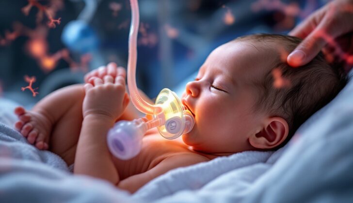
Ventilator-associated pneumonia (VAP) is a type of lung infection that happens when a patient has been on a breathing machine (medical term: mechanical ventilation) for more than 48 hours. VAP is the second most common infection that people catch in the hospital, specifically among children and newborns in intensive care. It contributes to 7% to 32% of infections related to healthcare and 10% of all infections linked to medical devices in children, according to the National Healthcare Safety Network (NHSN). Generally, such infections are less common in children’s intensive care units as compared to adult ones. In newborn babies, lower birth weight is often associated with a higher risk of VAP. Information about young children and infants with VAP is limited so most of the facts we have are based on studies of adults.
Kids who have artificial breathing tubes, such as a tracheostomy tube for long-term breathing issues or an endotracheal tube for emergency breathing management, are at a higher risk for VAP. It’s challenging to diagnose VAP in anyone, especially in young children and in the newborn ICU population. To help with this, the Centers for Disease Control (CDC) and NHSN provided guidelines in 2008 for recognizing VAP. They divided VAP into three categories: clinically defined (based on symptoms), confirmed by laboratory results, and found in patients with weakened immune systems. Infants and children usually fall into the first category.
VAP is a type of infection that can occur in the upper airway after bacteria have gotten into an artificial breathing tube. For simplicity, doctors usually diagnose either tracheitis or VAP in these situations.
Tracheitis describes situations where there are signs of infected tracheal discharge (a fancy term for mucus or other substance from the throat), fevers, difficulty in breathing, and the presence of bacteria and white blood cells in the tracheal tissue, but without signs of pneumonia on an X-ray.
VAP, on the other hand, involves all the symptoms of tracheitis, but with added X-ray evidence of pneumonia. This means there are signs of disease in the lungs along with the presence of harmful microorganisms in the white blood cells in the tracheal tissue.
What Causes Ventilator-Associated Pneumonia?
Ventilator-associated pneumonia is usually caused by a single type of bacteria. However, infections caused by multiple types of bacteria are becoming more common. A large study done in intensive care units (ICUs) at three hospitals found that the types of bacteria causing the infections were the same in both adult and pediatric hospitals. The most common bacteria causing these infections were Staphylococcus aureus (28.4%), Pseudomonas aeruginosa (25.2%), and other types of bacteria (26.6%).
Artificial airways, such as those used for intubation or tracheostomy, often get colonized by disease-causing bacteria shortly after they are put in. The main types of bacteria causing these infections include both gram-positive and gram-negative bacteria: S. aureus (including MRSA), P. aeruginosa, and Klebsiella and Enterobacter species. These types of bacteria can also put patients in a NICU at risk of getting infected by Enterococcus species and group B Streptococcus. Other disease-causing bacteria include Streptococcus, Enterobacteriaceae, and Acinetobacter species. Anaerobic bacteria, which don’t require oxygen to live, don’t often cause Ventilator-associated pneumonia, but they can contribute to infections caused by multiple types of bacteria, especially in cases of pneumonia caused by swallowing something in the wrong way. Bacteria or fungi acquired in the hospital are rarely the cause of pneumonia in hosts with normal immune systems.
Pneumonia that develops early (less than 4 days after being admitted to the hospital) is usually caused by bacteria commonly found in the community that are sensitive to antibiotics, while late pneumonia (more than 4 days) is usually caused by bacteria resistant to antibiotics. However, this difference isn’t useful for children who are frequently admitted to the hospital.
Risk Factors and Frequency for Ventilator-Associated Pneumonia
Pediatric patients, or children, seem to be less likely to get ventilator-associated pneumonia than adults. This could be because children typically have fewer additional health conditions. According to a report by the NHSN, out of 304 participating hospitals, the rates of ventilator-associated pneumonia were between 2.36 and 2.08 for every 1000 days a device was used. These rates were for newborns weighing less than 750 grams and between 750 and 1,000 grams, respectively.
In pediatric ICUs, the estimated rates of infections associated with ventilators range from 1.8 to 8.3 for every 1000 days a ventilator is used.
Signs and Symptoms of Ventilator-Associated Pneumonia
Diagnosing ventilator-associated pneumonia can be challenging. To do so, doctors usually look at a mix of clinical signs, x-ray images, and microbiological tests. The longer someone is on a ventilator, the higher their chances of getting this type of pneumonia. If the patient has a fever, changes in their lung sounds, alterations in their chest x-ray, or a growing need for breathing support, doctors may suspect ventilator-associated pneumonia. However, it’s important to note that the sounds of the ventilator system itself can make listening to the lungs tricky, particularly with high-frequency ventilation. Additionally, many newborns have anomalies on their chest x-rays that can make recognising new infections difficult.
Testing for Ventilator-Associated Pneumonia
If doctors suspect a patient has ventilator-associated pneumonia, they’ll look for signs like fever, a change in white blood cell counts, pus-like secretions, and a worsening ability to exchange gases, like oxygen and carbon dioxide. A chest X-ray is an important tool for diagnosis. It can reveal new or worsening areas of inflammation in the lungs, holes or cavities, indicators of pneumonia such as air bronchograms, or air-filled cysts called pneumatoceles.
Doctors also consider microbiological data, basically an analysis of the bacteria or other germs present. They often collect samples of secretions from the lower respiratory tract for culture, as well as blood samples for culture, in patients suspected of having ventilator-associated pneumonia. However, because most individuals who have been intubated (put on a ventilator) show colonization of bacteria after 48 hours, some experts do not recommend cultural testing of intubated patients.
In adults, these samples may be obtained through a variety of procedures such as bronchoscopy, lung biopsy, lung aspiration, protected specimen brushing, or transtracheal aspiration. But in children, especially newborns in the neonatal intensive care unit (NICU), these procedures can be challenging and potentially risky.
Therefore, for infants less than one year, diagnosing ventilator-associated pneumonia relies more on clinical signs. These include worsening ability to exchange gases, higher oxygen needs, increasing demands on the ventilator, and at least three of the following: unstable body temperature, changes in white blood cell counts, new or changed sputum (mucus coughed up), increased respiratory secretions or need for suctioning, paused breathing, fast breathing, flaring nostrils with retraction of the chest wall or grunting sounds, wheezing, rattling breath sounds or other noise during breathing, coughing, and changes in heart rate such as slower (bradycardia) or faster (tachycardia) beats.
Treatment Options for Ventilator-Associated Pneumonia
When there’s a suspicion of ventilator-associated pneumonia, doctors will collect culture samples to identify the bacteria causing the infection. Treatment will then be decided based on how long the patient has been intubated and hospitalized, whether they’ve had antibiotics before and how severe their condition is. Doctors will also consider the susceptibility of local bacteria to certain antibiotics.
To begin with, doctors would typically provide broad-spectrum therapy, which is a type of treatment that can fight a wide variety of bacteria, including gram-negative bacilli, P. aeruginosa, and possibly methicillin-resistant S. aureus.
The treatment is then tailored to the patient’s needs based on their culture data, which shows what bacteria are present, and what clinical and radiographic findings show about the patient’s condition.
We don’t yet know how long treatment should be in children. However, for adults, simple cases may be treated for 7 to 10 days. For more severe cases, or those suffering from a specific type of pneumonia called necrotizing pneumonia, treatment of at least 14 days is necessary. For children who have ventilator-associated tracheitis, which is inflammation in the trachea, a course of treatment lasting less than 7 days may be enough if there’s no evidence of pneumonia.
What else can Ventilator-Associated Pneumonia be?
When it comes to lung-related issues, doctors look into several health problems that present similar symptoms. These include:
- Aspiration pneumonia
- Virus or bacterial pneumonia
- Pneumocystis jirovecii pneumonia
- Heart failure
It’s crucial for healthcare providers to run appropriate tests to determine the root cause of lung troubles.
Possible Complications When Diagnosed with Ventilator-Associated Pneumonia
Common Complications:
- Stress ulcers
- Deep vein thrombosis
- Failure of multiple organs


