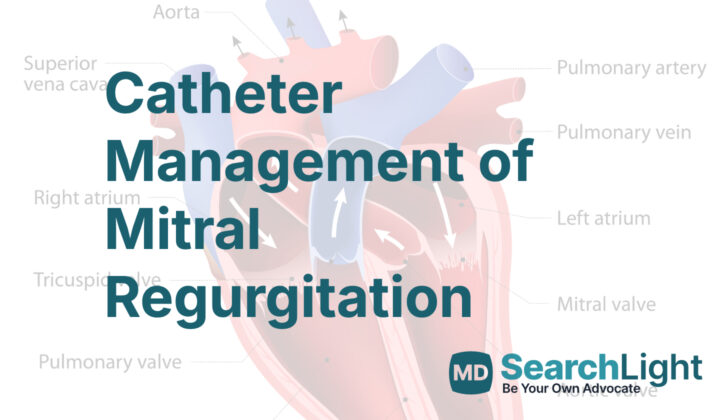
Overview of Catheter Management of Mitral Regurgitation
Mitral regurgitation (MR) is a heart condition where blood flows backward into your heart because the mitral valve isn’t closing properly. It’s one of the most frequent issues with heart valves and is second only to aortic valve stenosis, a condition where the heart valve becomes narrow. The treatment for MR depends on how severe it is and how long you’ve had it.
If you have severe MR, which can happen suddenly because of things like a torn muscle in your heart or an infection, it can lead to serious issues that make your heart unstable. This requires immediate surgery.
Mitral regurgitation can be chronic (long-term) in two forms: primary or secondary. Primary MR is caused by a problem with one or more parts of the valve in your heart, while secondary MR occurs when the shape and function of the left side of your heart changes. If you have mild chronic MR without any symptoms, it can be managed with medication and regular check-ups. However, if you’re showing symptoms, you’ll need to consider surgical treatment. Even if you’re not showing symptoms, surgery might be necessary if other problems are present, including irregular heartbeat, high blood pressure in the lungs, or a larger than normal left side of the heart.
One way doctors check your heart is by using an imaging test called transthoracic echocardiography (TTE). This gives us a clear picture of your heart and its workings. It also helps us measure the severity of the MR and the functioning of your left ventricle (main pumping chamber of the heart). If the images from TTE are not clear enough, they might recommend other types of imaging tests – transesophageal echocardiography (TEE) or cardiac magnetic resonance imaging.
Recently, less invasive treatments, like repairing the mitral valve using a catheter, have been found to be a good alternative for patients at high risk from surgery. Studies have shown that this method has low risk and is effective. Based on a surgical repair method pioneered by Dr. Ottavio Alfieri, the procedure brings the two leaflets of the mitral valve together, reducing or getting rid of the backflow of blood. Typically, this repair results in a double exit from the valve.
There are several less invasive options available for patients with MR who are at a higher risk from surgery due to other health conditions. They can be grouped based on the specific section of the valve they target. These options include the edge-to-edge repair, annuloplasty (repair of the valve opening), chord implantation, or left ventricle remodeling.
This article has discussed these methods for managing MR, including when they’re suitable and unsuitable, how they’re done, and any complications that might arise. The main feature of our discussion has been the edge-to-edge repair devices approved by the United States Food and Drug Administration.
Anatomy and Physiology of Catheter Management of Mitral Regurgitation
The mitral valve, named so because it resembles a bishop’s hat, is a complex feature in our hearts. It includes several parts, such as the front and back flaps of the valve, a supporting ring known as the mitral annulus, and some connected structures that help the valve work properly.
This valve’s job is to make sure that blood can only flow in one direction. It does this by closing off after blood has passed through, so the blood can’t go back the way it came.
There are two main types of problems that can happen with the mitral valve. The first is primary Mitral Regurgitation, or MR, which is when something goes wrong with the valve itself, and it can’t close properly, allowing some of the blood to flow backward. This might happen because the valve has become weak or misshapen over time.
The second type of problem is secondary MR, which is when other issues in the heart cause the valve to stop working properly. This could be because the heart has become enlarged or weak, which means it can’t give the valve the support it needs to work well.
Both primary and secondary MR can be mild, moderate, or severe, and the type of treatment your doctor will recommend can range from medication to surgery, depending on how severe your case is. Treatment is very important because if left untreated, severe MR can lead to conditions like heart failure, which can be very dangerous.
Here’s a bit more detail about the different stages of MR:
– Stage A is the earliest stage, where you might be at risk for developing MR. At this stage, the changes to the valve are very mild and often don’t cause any symptoms.
– Stage B is when MR starts to get more serious. Yyou might see moderate changes, but still, you likely won’t feel anything different.
– Stage C is where the valve’s changes are severe but still might not causing any noticeable symptoms.
– Stage D is the most serious stage. At this point, the mitral valve has severe changes, and you’ll likely notice symptoms like feeling short of breath, especially when you’re being physically active.
Please remember, these stages help doctors determine the best course of treatment, and while it might sound scary, doctors have many different options to help manage any issues with the mitral valve. They only aim to help you keep your heart as healthy as possible.
Why do People Need Catheter Management of Mitral Regurgitation
The main treatment suggested for Mitral Regurgitation (MR), a heart valve disorder, is a technology known as the edge-to-edge leaflet repair device. However, several new technologies are now being explored for treating MR. These include the neo-cords procedure, transcatheter mitral valve repair, and rings.
The edge-to-edge leaflet repair device is recommended for specific situations:
- If the MR is moderate to severe, whether it’s primary (due to problems with the structure of the heart valve itself) or secondary (due to other heart diseases affecting the valve).
- If the person is experiencing symptoms of heart failure.
- If the person is at high or very high risk for surgery.
- If the heart valve’s anatomy is suitable for this treatment.
- If the person’s life expectancy is more than a year.
When a Person Should Avoid Catheter Management of Mitral Regurgitation
There are certain situations where a specific type of heart operation, involving a catheter device used to repair a part of the heart known as the leaflet, may not be suitable. These include:
- If a person can’t tolerate anticoagulation. This means they can’t take medication to help prevent blood clots.
- Having a condition called active endocarditis that affects a heart valve (the mitral valve).
- Another disease called rheumatic mitral valve disease which also involves the mitral valve.
- If a person has a thrombus, which is a blood clot inside the heart, or in particular veins (the inferior vena cava or femoral veins).
- Severe mitral annular calcification involving the leaflets. This is a condition where calcium builds up in the heart valve making it very hard.
- If there’s a significant cleft or hole in the mitral valve leaflets. The leaflets are the parts of the valve that open and close to control blood flow.
- Another condition called mitral valve stenosis, which is when the mitral valve is narrower than it should be.
Equipment used for Catheter Management of Mitral Regurgitation
To perform a procedure called a transcatheter edge-to-edge leaflet repair, certain medical equipment and supplies are needed. These include the leaflet repair device, a kit with catheters, needles, and a wire that’s used for making a hole in the wall between the heart’s upper chambers (a transeptal puncture), an X-ray machine that’s used for imaging (fluoroscopy), a cart with life-saving equipment including a defibrillator (used to deliver an electric shock to the heart), a sterile gown and drape, anesthesia, advanced ultrasound imaging of the heart (transesophageal echocardiography), heart monitoring equipment, and personnel who can operate a heart-lung machine if needed.
The MitraClip is a device that’s been approved for treating both primary and secondary mitral regurgitation (MR), which are conditions where the heart’s mitral valve doesn’t close properly, causing blood to flow backward in the heart. The MitraClip device comes in various sizes, including a traditional width of 4 mm and a newer 6 mm size. The arms of the clip also come in two lengths – 9 mm (called NT clip) and 12 mm (XT clip).
The MitraClip is made up of two sturdy arms made from a cobalt-chromium alloy, and it has flexible grippers made from a metal called nitinol. These grippers are designed to hold onto the heart valve leaflets (flaps) and have either 4 or 6 small hooks, depending on the size of the clip. Some have raised questions about the use of the longer XT devices, especially in patients with complex heart conditions. There are concerns that the increased tension due to more tissue being gripped by the device could potentially harm the leaflet. However, research hasn’t shown a higher rate of adverse events with the longer arm devices compared to the smaller ones.
The PASCAL transcatheter mitral valve repair technology is another device used for this procedure. Introduced in 2016, the device has been updated to include three catheters, allowing for better movement within the left atrium of the heart. Some versions of the PASCAL implant feature a central spacer designed to fit into specific gaps in the heart, reducing stress on the mitral valve leaflets. These implants also offer capabilities that allow for individual leaflet control, which can be beneficial during the procedure.
Who is needed to perform Catheter Management of Mitral Regurgitation?
To safely carry out the procedure involving the TEER device (a tool used in heart treatments), a team of healthcare professionals is needed. In this group, the main doctor is the interventional cardiologist, who specializes in treatments that use tiny, flexible tubes called catheters to approach the heart.
An echocardiographer’s job is essential, too. This could be either a cardiac anesthesiologist or a cardiologist. Their role is to perform echocardiograms, which use sound waves to produce images of your heart.
The cardiac anesthesiologist is a doctor who specializes in giving medicine that makes you comfortable, relaxed, and pain-free during heart procedures.
A first-assist is also required. They assist the main doctor during the procedure. In addition, nurses and technical staff are needed to complete the procedure efficiently.
In case of an unexpected complication, a cardiac surgeon and an operating room staff are on standby. Their job is to step in and take over if things don’t go as planned.
Finally, there might be a perfusionist if there is a need for cardiopulmonary bypass. A perfusionist is a trained healthcare professional who operates the heart-lung machine, which takes over the functions of the heart and lungs during surgery.
Preparing for Catheter Management of Mitral Regurgitation
The TEER procedure, a type of heart procedure, is typically done in a specialized room with advanced imaging technology. This procedure also uses a type of echocardiography called TEE (a technique for creating images of the heart and its blood vessels) to confirm the heart problem, guide the procedure, and check the results of the repair. Because TEE plays a critical role, the patient is usually under general anesthesia to prevent sudden movement that could lead to serious problems.
Before starting the TEER procedure, the doctor performs a pre-procedure TEE to assess the heart valve damage and see if it can be repaired. Sometimes, other types of heart imaging may be required, which are carried out by a heart specialist.
Importantly, before undergoing general anesthesia, the patient’s condition must be evaluated to make sure they can handle it safely and comfortably. For the success of the procedure, it’s crucial for the heart team—including the heart specialist, anesthesiologist, operating room staff, and nurses—to work closely together. It’s also vital to have all necessary equipment readily available. To ensure everyone is on the same page, the team takes a “time-out” to establish effective communication and to confirm all elements are properly set up.
In any medical procedure, cleanliness is extremely important to prevent infection. Therefore, before the procedure, a sterile area is created around the patient. The area where the catheter (a thin tube) is to be inserted is carefully cleaned according to standard protocols. All medical staff working near this sterile space follow strict cleanliness rules, including thoroughly washing their hands and wearing complete protective clothing such as gowns, hats, masks, and gloves. The procedure area is also cleaned and suitably draped to maintain this sterility for patient safety.
Device Selection
Before deciding on the right device for the procedure, the doctors carefully analyze several things including the cause of the heart condition, size of the heart valve, how much the valve is obstructed by blood flow, and the complexity of the valve’s structure. The features of the device have to match the patient’s specific heart condition. For instance, more advanced devices may be used in patients with complex valve abnormalities.
Important factors in choosing a device for this procedure also include the treatment strategy and location of the problematic area of the heart valve. Patients with particular heart conditions that need a certain type of repair may require a larger heart valve area. In situations where there are major valve defects, especially when multiple devices are needed, certain types of advanced devices can be more effective in reducing problems with the heart valve. However, in some cases where multiple devices are considered, certain types like the PASCAL P10 are not recommended due to their unique design. For certain specific lesions, smaller and more controllable implants are preferred.
These decisions are based on detailed assessment of the heart’s condition and structure, ensuring the most suitable treatment is given to each patient.
How is Catheter Management of Mitral Regurgitation performed
The structural heart team is made up of a heart doctor (interventional cardiologist), heart surgeon, a specialist doctor to take care of you during the operation (cardiac anesthesiologist), and a nurse. This team takes care of a heart procedure called transcatheter mitral valve repair using an edge-to-edge leaflet repair device. The room where the procedure is done has special x-ray like capabilities (fluoroscopic capability).
The procedure involves using live x-ray images (fluoroscopic) and special ultrasound pictures (TEE) to see the heart clearly and accurately from different angles while the procedure is being done. The patient is put to sleep (general anesthesia) to ensure comfort and avoid any movement during the procedure.
In the procedure, images are captured from four different views – 2-chamber, 4-chamber, the middle part of the esophagus, and a specialized view for the irregular rhythmic movement of the heart. These images help the surgical team confirm the problem with the heart valve and rule out the presence of any blood clot inside the heart before starting the procedure. This also helps them evaluate the overall function of the heart.
A vein in the groin area is accessed using ultrasound guidance. A very thin wire is guided through this vein under live x-ray monitoring to ensure the correct position and to avoid any problems.
The tissue that separates the two upper chambers of the heart – right and left atrium (the interatrial septum) is punctured. The location chosen for this puncture is at the back top end of the septum. A needle is used to make this puncture and this is carefully monitored on the live images to avoid any injury to the heart.
A particular device is then inserted through this vein from the puncture in the septum. This device is delivered towards the problematic heart valve guided by the live images. The distance from the septum where the puncture was made to the heart valve being treated is important. If it’s too low or too high, it can cause complications. Finally, the device is deployed and checked to make sure it is in the right place.
Risks of this procedure include injury to the heart, formation of a blood clot, air bubble entering the blood vessels, and bleeding from the site where the vein was accessed.
Possible Complications of Catheter Management of Mitral Regurgitation
Although exchanging the patient’s valve through the TEER procedure comes with existing health conditions, it’s generally seen as a safe surgery with a low risk of severe outcomes. Below are common complications that potentially could happen during or after this procedure and the likelihood they will occur.
Some possible complications include issues with the device such as improper attachment or the device moving away from its intended position, ranging from 1.5% to 5.1% and 0.05%-0.7% chances respectively. There’s a 0%-2% chance of injury to the heart’s valve leaflet which controls the blood flow in your heart. Higher blood pressure in the valve than desired could occur up to 15% of patients. Residual heart valve leakage could happen in 3.4%-17.0%. In some rare cases, patients may suffer major vascular complications, severe bleeding requiring blood transfusion, stroke, or even a heart attack, ranging from 0% to 17% chance of occurrence.
There’s a small concern for people with a long-standing secondary heart valve leakage and calcified leaflets as they face a higher risk of leaflet perforation, tear, or issues with device attachment. In some cases, if the medical device moves from its intended position, removing it can be a challenge.
An unusual event called afterload mismatch could happen especially in people with reduced heart function but it’s generally managed with specific medications and doesn’t usually require intensive support. In some cases, patients with severe heart conditions might experience clot formation in the left side of their heart which might need early and heavy treatment with blood thinners.
If there are remaining or recurring leaks in the heart valve, it is important for the team of doctors to reevaluate the need for heart valve surgery or additional interventions. Usually, a repeated heart ultrasound is performed to understand the disease and evaluate the risks.
According to large-scale data from multiple medical centers, failure of the device due to tearing or loss affects about 3.5% of all patients and can potentially increase the chances of severe in-hospital and long-term consequences. In some situations, repeating the TEER procedure may be a better choice than surgery, especially in suitable patients with primary or secondary heart valve leakage, and when the outcomes from surgery are not optimal.
What Else Should I Know About Catheter Management of Mitral Regurgitation?
Using a catheter to treat mitral regurgitation, a condition where the heart’s mitral valve doesn’t close tightly and allows blood to flow backward in the heart, represents an important breakthrough in heart health care. This method offers a possible solution for severely affected patients who can’t safely undergo surgery. Recent research even suggests that in some cases, this catheter-based treatment might be more effective than surgical procedures.
After fixing the mitral valve with a specific device via a catheter, the heart’s ability to contract and pump out blood stays stable. However, the overall volume of blood pumped out by the heart and the stretching and contraction of the heart muscle might decrease. This decrease is probably because the repaired valve stops the backward flow of blood, which reduces the amount of blood in the heart when it is relaxed (end-diastolic volume). This lessens the heart’s demand for oxygen, which can lead to the patient feeling better and more able to participate in physical activities, three months after the procedure according to the New York Heart Association’s classification.


