
What is Aneurysmal Bone Cysts?
Aneurysmal bone cysts are non-cancerous growths that look like tumors and are filled with blood. They can show up in any bone, but are most often found in the thigh bone, shin bone, or backbone. These cysts can cause the bone to swell, leading to pain, inflammation, joint disruption, and damage to areas of the bone responsible for growth. Sometimes, they can grow swiftly, damage the surrounding bone tissue, and make the bone so weak that it fractures.
What Causes Aneurysmal Bone Cysts?
The exact cause of aneurysmal bone cysts, a type of bone tumor, is currently unknown. However, one possible reason is abnormal blood vessels in the bone.
There are three main theories about how these cysts occur:
* Aneurysmal bone cysts may form because of another bone tumor. Since about one third of the cases have a separate bone lesion or abnormal area of tissue, this theory makes a lot of sense. Some of the bone lesions that are often found alongside these cysts include chondromyxoid fibromas, chondrosarcomas, fibrous dysplasia, giant cell tumors of the bone, osteoblastomas, osteosarcomas and others.
* Aneurysmal bone cysts may themselves be the primary or original tumor.
* They may form at a site where the bone was previously injured.
Recent science has found that the cysts could also be formed by abnormal growth of cells. Genetic studies of the tumors have found that up to 69% of these cysts contain a specific genetic change, t(16,17). This genetic change activates a gene called TRE17/USP6 which in turn activates other molecules called matrix metalloproteinases (MMPs). MMPs help break down tissues outside cells, allowing lesions or abnormal areas of tissue to grow quickly. This genetic change isn’t found in secondary aneurysmal bone cysts, which are those that occur because of another tumor or disease.
Risk Factors and Frequency for Aneurysmal Bone Cysts
Aneurysmal bone cysts are a rare type of tumor found in bones, accounting for 1% to 6% of all primary bone tumors. Some studies have shown that for every 10 people, roughly 0.14 will develop this type of bone cyst. Eight out of ten people diagnosed with an aneurysmal bone cyst are children and teenagers under 20 years old. A slightly larger number of females have this condition than males. It’s important to take special care of this as these bone cysts mainly show up in children and can potentially impact growth and cause permanent deformities in limb length.
- Aneurysmal bone cysts are most commonly found in long bones (67%)
- They can appear in the spine (15%), especially in the back parts
- The pelvis is another common area (9%)
- They can also occur, though less frequently, in the bones of the skull and face, as well as in the ends of the long bones
Signs and Symptoms of Aneurysmal Bone Cysts
People with an aneurysmal bone cyst often experience a slow onset of pain that develops over weeks or even months. This pain might be coupled with swelling or a lump that can be felt. However, some people first become aware of the problem when they suddenly experience pain due to the cyst causing a pathological fracture. If the cyst presses against a nerve or involves the spine, neurological symptoms may occur.
Testing for Aneurysmal Bone Cysts
To evaluate an aneurysmal bone cyst, your doctor would heavily rely on imaging tests as they offer the most critical information for diagnosis.
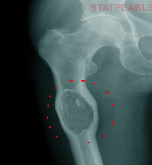
During a simple X-ray, the cyst might show up as a pocket within the bone with thin borders resembling an “eggshell”. These cysts can push the surrounding bone out of shape, illustrating how aggressive they are.
A CT scan, another type of imaging, reveals similar characteristics as the X-ray, but provides a more detailed look at the walls of the cyst, hence, enhancing the “eggshell” appearance of its rim. This test can also identify fluid layers in the cavities, most likely caused by the settling of cellular debris from the serum.
Then there’s an MRI, which again gives results like a CT scan. Here, T1 and T2 image sequences can highlight the dividing walls within the cyst. Blood within these cysts, which can be different ages, shows up as bright spots on both these sequences. Sometimes, a MRI can also show signs of a pathologic fracture, which would appear as bone and tissue swelling.
The role of lab tests in diagnosing these bone cysts is very limited. However, it’s worth noting that the level of an enzyme called alkaline phosphatase might be high in some patients because their osteoblasts (the cells that help in bone formation) are working overtime.
Treatment Options for Aneurysmal Bone Cysts
If diagnosed with an aneurysmal bone cyst, which is a rather uncommon type of bone tumor generally filled with fluid, you would need to see an orthopedic oncologist, a specialized doctor who treats bone tumors. More often than not, the doctor would recommend surgery to avoid the risk of the bone breaking, which can happen when a bone is weakened by a tumor.
The type of surgery you have depends on factors such as the size of the tumor and which bone it has affected. Here are three possible surgical procedures:
1. The first technique, called “intralesional curettage”, involves scooping out the contents of the cyst, similar to how an egg is scooped out of its shell. The empty space is then filled with a bone graft or cement that would lend strength to the bone.
2. The second is intralesional excision. Like curettage, the cyst is opened up and its contents are removed. The difference is the surgeon uses various additional treatments (like high-speed drills, beam coagulations, phenol, and cryotherapy) to lower the chance of the cyst coming back. This procedure allows more of the healthy bone to remain intact. The reason intralesional excision is preferred is that the cyst is filled with a substitute to restore strength to the bone. An intralesional excision is particularly beneficial when the cyst is near joints and structures where maintaining the normal anatomy is key for functionality.
3. The third procedure, known as an “en bloc excision”, involves removing the entire tumor along with a portion of the affected bone. This procedure is more invasive and can result in more significant loss of functionality, so it’s only considered when the other two procedures are insufficient, such as in cases when the bone cyst keeps recurring.
Selective arterial embolization (SAE), a procedure where the blood supply to the tumor is blocked, can either be performed before surgery to reduce the size of the tumor, or as the main treatment in cases where surgical procedures carry significant risks. In close to 40% of the patients treated with SAE alone, the procedure may have to be repeated once or twice.
In cases of recurring bone cysts, radiotherapy (treatment with radiation) can also be used. However, your doctor will need to weigh the potential benefits against the significant risks associated with radiation therapy. There’s a risk of it causing another type of cancer, as reported in a patient who received radiation for a vertebral aneurysmal bone cyst.
The usage of medication with monoclonal antibodies (lab-made proteins that can bind to specific parts of cancer cells) is currently being explored for patients who aren’t candidates for surgery. While this is a promising direction, research in this area is still ongoing.
What else can Aneurysmal Bone Cysts be?
Let’s talk about different types of bone growths that can occur:
- Chondroblastoma: These are non-cancerous tumors typically found in the ends of long bones. They are usually detected when a child experiences constant pain and swelling in a limb that isn’t related to physical activity. They show up as a well-defined lesion in medical images that may cross the growth plate of the bone. They often exhibit a high signal in MR images and are associated with surrounding areas of inflammation.
- Fibrous dysplasia: This is a benign, tumor-like abnormality commonly found in children and young adults. It happens when normal bone is replaced with fibrous connective tissue and immature bone. It can either affect one bone or multiple bones and usually doesn’t cause any symptoms or necessitate intervention. However, issues can arise if the growth presses on neighboring structures. On imaging scans, these lesions appear to have thinning borders and a “ground-glass” appearance. CT and MRI scans are usually not needed unless there’s a suspicion of a fracture or stress injury.
- Giant cell tumor (GCT): GCT is a non-cancerous tumor that typically appears in adults with mature skeletons. Despite being non-malignant, they’re aggressive and can spread to the lungs. They often cause chronic pain and swelling, along with movement restriction of the affected joint. In cases where weight-bearing bones are involved, fractures may occur. Medical images typically reveal a lesion in the bone end, with CT scans showing significant thinning of the bone.
- Telangiectactic osteosarcoma (TOS): TOS resembles another type of bone growth called an aneurysmal bone cyst. MRI scans can help differentiate between the two by showing a nodular appearance in TOS, which corresponds to a cancerous tissue. TOS often reveals destruction of the bone and a related soft-tissue mass. However, a biopsy is the only definitive way to differentiate TOS from other lesions.
- Unicameral bone cyst (UBCs): Appearing in the first two decades of life, UBCs are fluid-filled lesions encased by a fibrous lining. Patients often feel pain due to a fracture located in the long bone. Imaging scans reveal an expanding lesion with well-defined borders that doesn’t exceed the bone’s diameter. A unique feature of UBCs is the “fallen fragment’ sign where fractured fragments can be seen inside the cystic cavity.
Understanding these conditions can aid in early detection and appropriate treatment planning.
What to expect with Aneurysmal Bone Cysts
When a patient has a bone cyst that bulges outwards, known as an aneurysmal bone cyst, it is typically removed through a surgical procedure. This usually solves the problem. However, there’s a chance that these cysts might reappear. Historically, there has been a 19% chance of such a case happening. These recurrences tend to occur within the first year after the surgery.
This is why it is important for patients to have regular check-ups up to five years after the surgery to ensure the cyst hasn’t returned. For younger patients who are still growing, this is particularly important, as a recurrence could affect how the bones in their body grow and could cause them to become deformed.
Possible Complications When Diagnosed with Aneurysmal Bone Cysts
The side effects from the treatment of bone lesions (abnormal areas in the bone) can vary, depending on where the abnormal area is located in your skeleton. Like all surgeries, there’s a risk of bleeding and infection; this can be either a surface wound infection or a deeper bone infection called osteomyelitis. If the lesion is near the growth plate (an area where new bone growth happens), it may get damaged. Potential risks also include a type of blood clot in the lungs called a pulmonary embolism. This can result from other types of blood clots (venous thromboembolism) or fat particles traveling to the lungs, which are common risks in all kinds of orthopedic surgeries.
Possible Risks:
- Bleeding
- Surface wound infection
- Bone infection (Osteomyelitis)
- Damage to the growth plate in the bone
- Blood clot in the lungs (Pulmonary embolism)
Recovery from Aneurysmal Bone Cysts
When a patient can go back to their normal activities after treatment depends on several factors. These include where the affected area is located, how much was removed during the operation (if surgery was performed), and the type of reconstruction done (like replacing the removed part with a bone graft or cement). Physical therapy or occupational therapy are very important to help keep the patient’s health conditions from becoming worse after the treatment.
Preventing Aneurysmal Bone Cysts
The exact cause of aneurysmal bone cysts, a condition where fluid-filled sacs form inside the bone, is still not known. Therefore, we don’t know the measures to prevent them from occurring. It’s very important for young people, especially kids and teenagers whose bones are still growing, and their parents, to understand that they need to get medical help if they start to feel sharp or persistent bone pain.


