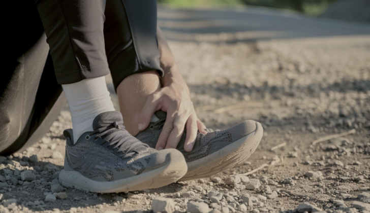
What is Calcaneofibular Ligament Injury (Ankle Sprain)?
Ankle sprains are very common and often the reason why people go to the emergency room. They account for about 7% to 10% of all visits and are the cause of up to 40% of sports-related injuries. Most ankle injuries happen during sports activities and mainly affect the outer part of the ankle. This part of the ankle has three ligaments, or bands of tissue that connect bones to each other, known as the anterior talofibular ligament (ATFL), the calcaneofibular ligament (CFL), and the posterior talofibular ligament (PTFL).
It can be difficult to tell the difference between an injury that involves both the ATFL and CFL and one that only involves the CFL since physical examinations aren’t always precise. However, it is generally agreed that the ATFL is mostly involved in most ankle sprains, making up two-thirds of outer ankle injuries. While there isn’t much information available about injuries that only involve the CFL, injuries that involve both the ATFL and CFL are the second most common type of outer ankle injury.
Because of this, when people talk about CFL injuries in medical research, they usually include them as part of the broader category of outer ankle injuries. In discussing this topic, this article will highlight the common aspects of outer ankle injuries and point out the unique characteristics of CFL injuries.
What Causes Calcaneofibular Ligament Injury (Ankle Sprain)?
Many ankle injuries, specifically those on the side of the ankle, happen because of sports activities. Indoor and court sports, in particular, have the highest risk of causing an ankle injury.
Risk Factors and Frequency for Calcaneofibular Ligament Injury (Ankle Sprain)
Injuries to the CFL (calcaneofibular ligament) are quite uncommon and are usually grouped under larger categories like lateral ligament injuries. Every day, there are about 30,000 instances of twisted or sprained ankles. These contribute to 25% to 40% of all sports injuries. Ligaments on the outer side of the ankle are involved in 85% of these injuries. The daily rate of such injuries is one in 10,000 people.
Signs and Symptoms of Calcaneofibular Ligament Injury (Ankle Sprain)
When dealing with injuries, it’s crucial to take a good look at the injury history and perform a physical examination to decide the next steps. Patients may report hearing a crack, and observe swelling, redness, pain, and an inability to continue their activity. The physical exam should involve steps like:
- Looking at the affected area (inspection)
- Feeling the affected area (palpation)
- Carrying out special tests like the anterior drawer test and the talar tilt test
The anterior drawer test is where the patient’s foot is held in a neutral position and the doctor applies a forward force onto the ankle. If the injured ankle feels looser compared to the uninjured one, this indicates a positive result.
Similarly, during the talar tilt test, the foot is held neutrally and a force is applied to tilt the ankle. The looseness of the injured ankle is compared to the other side. Both these tests however may not always give precise results.
After this initial assessment, if the examination shows bruising and pain when a specific area 4 to 5 days after the injury is touched, there’s a 90% chance of a torn side ligament. Patients with tenderness over the CFL (calcaneofibular ligament) have a 72% risk of ligament damage.
The Ottawa ankle rule is a useful tool for deciding if an x-ray should be done. It involves checking pain at specific points (rear edge or tip of the ankle bone, navicular bone, or base of the fifth metatarsal bone) and seeing if the patient can bear weight. This examination tool is very sensitive (96.4% to 99.6%) and valuable for ruling out ankle fractures.
Testing for Calcaneofibular Ligament Injury (Ankle Sprain)
If your doctor suspects that you may have hurt your ankle, they might use a test known as the “Ottawa ankle test” to check for any issues. If the results are positive, this could suggest an ankle injury and your doctor might ask for an X-ray of your ankle to get a more detailed look. However, it’s worth noting that actual ankle fractures (broken bones) are relatively rare, happening in less than 15% of cases.
Other imaging methods like ultrasound and MRI may be used if your doctor suspects specific types of injuries. For example, ultrasound is a good option for viewing motion in the ankle, but the results can vary based on the skills of the person performing the ultrasound. In general, ultrasound is good at detecting injuries to the ligaments (the tough bands of tissue that hold bones together) with a success rate of 92%, but it’s only able to correctly identify normal ligaments 64% of the time.
If your doctor is really concerned about the possibility of a fracture, they might decide to use an MRI. This imaging method is excellent at spotting fractures (93-96% success rate) and is usually able to accurately rule them out (100%).
Once your doctor has examined your ankle, they will usually classify any injury you have into one of three categories, or “grades”. Grade I injuries happen when a ligament is stretched, grade II injuries involve a more moderate sprain, and grade III injuries are severe sprains that involve full ligament lesions (or tears). The higher the grade, the more severe the injury, which can help doctors decide on a suitable treatment plan, understand how the injury might progress, and anticipate any possible complications.
Treatment Options for Calcaneofibular Ligament Injury (Ankle Sprain)
In simpler terms, often, injuries to the CFL (Calcaneofibular Ligament, located in the ankle) can be treated through non-surgical methods. Healing from these injuries goes through three phases: inflammation (1 to 10 days), repair (4 to 8 weeks), and restructuring (up to one year). Each of these phases gives a specific time window for treatment.
In the initial few days of injury, when inflammation is high, the RICE method (rest, ice, compression, and elevation) is recommended. In the first week, a cast or a boot can help reduce swelling and pain. After that, a brace or medical taping can be used for support when returning to normal activity. It’s not advisable to keep the ankle immobile during the repair phase, as some stress on the ankle helps with its restructuring. For minor to moderate injuries, ankle support with a semi-rigid brace is beneficial, whereas severe injuries might need a cast in the initial stages followed by an orthosis, a device that supports the ankle. Over-the-counter painkillers, like NSAIDs (Non-Steroidal Anti-Inflammatory Drugs), can help manage pain.
Most of the time, these ankle injuries can be managed without surgery. That said, surgery may be considered for patients with long-standing instability in the ankle. Two patients with isolated CFL injuries were studied, with one receiving surgery and the other receiving non-surgical treatment involving immobilization and physical therapy. Both had good results. It’s important to remember that the decision between surgical and non-surgical treatment should be tailored to each patient’s individual situation. In some cases, surgery might help reduce the chance of future ankle instability. However, the overall results, whether through surgery or non-surgical methods, are typically similar.
What else can Calcaneofibular Ligament Injury (Ankle Sprain) be?
When dealing with a CFL (calcaneofibular ligament) injury, it’s common for the ATFL (anterior talofibular ligament) to be involved as well. Therefore, an injury to the ATFL alone should be in the list of possible diagnoses. Other conditions that need to be considered include:
- Injuries to the bone and cartilage
- Fibularis tendon injury
- Ankle fractures
- Rupturing of the Achilles tendon
- Dislocation of the tendons
- Subtalar joint injuries, as CFL injuries can involve this joint.
What to expect with Calcaneofibular Ligament Injury (Ankle Sprain)
When diagnosed with a certain illness, getting back to work or school is an important point to consider. It’s been found that about 25% of patients may take time off from work or school due to their condition.
In some cases, patients may have long-term pain or instability. Roughly 74% of patients can still experience chronic symptoms like pain, swelling, weakness, or instability four years after the initial injury. In fact, 32% of patients reported that they still experienced these symptoms up to seven years after the original injury.
A comprehensive study conducted by Thompson et al. showed that aspects like the intensity of the pain, the ability to bear weight, and range of motion can’t always reliably predict recovery prospects, because individual studies may introduce certain biases. However, understanding these factors can be helpful and provide a rough guide for patients to anticipate their recovery process.
Possible Complications When Diagnosed with Calcaneofibular Ligament Injury (Ankle Sprain)
Injuries to the side of the ankle are recurring and can have long-term consequences. The outside part of your ankle can get injured again, especially if you’ve had a less severe sprain before. This can lead to problems with balance and pain, making it harder for you to move around. Over time, continuous instability in the joint can contribute to damaging the ankle joint, leading to a condition known as post-traumatic osteoarthritis.
- Repeated injuries to the side of the ankle
- Instability and pain
- Difficulty with moving around
- Long-term joint instability
- Potential post-traumatic osteoarthritis
Recovery from Calcaneofibular Ligament Injury (Ankle Sprain)
Starting rehabilitation early, which involves daily exercise routines, is great for helping recovery and movement after an injury. A study by Doherty and his team showed that exercise therapy for a freshly sprained ankle helped patients report an improvement in their functioning. The time spent on rehab exercises varied among patients, with some doing as little as 3.5 hours and others doing up to 21 hours.
There is no one-size-fits-all exercise therapy method for everyone. However, patients are encouraged to focus on activities that improve their strength and balance. Starting these exercises within the first week of the injury can help reduce muscle inhibition caused by swelling and pain. This means that patients can regain the use of their ankle muscles and movement quicker.
Ankle braces or taping can be a big help for patients during the rehabilitation phase. They can relieve pain and prevent further injuries.
Preventing Calcaneofibular Ligament Injury (Ankle Sprain)
Injuries to the ligament located between your heel and outer ankle bone, also known as the calcaneofibular ligament, are not often discussed separately in medical studies. This means much of what we know about these injuries actually comes from information on all types of outer ankle injuries. Medical examinations can help doctors determine whether patients need additional evaluations and imaging tests like X-rays or MRIs. They will explain how long the recovery process might take and what the expected outcome of the injury is.
Patients should be informed that it’s crucial for them to participate in rehabilitation exercises for their ankles. Going back to their usual activities too soon might cause more damage to the calcaneofibular ligament. The decision on when to start putting weight on the injured ankle depends on the severity of the injury, and this varies from patient to patient.


