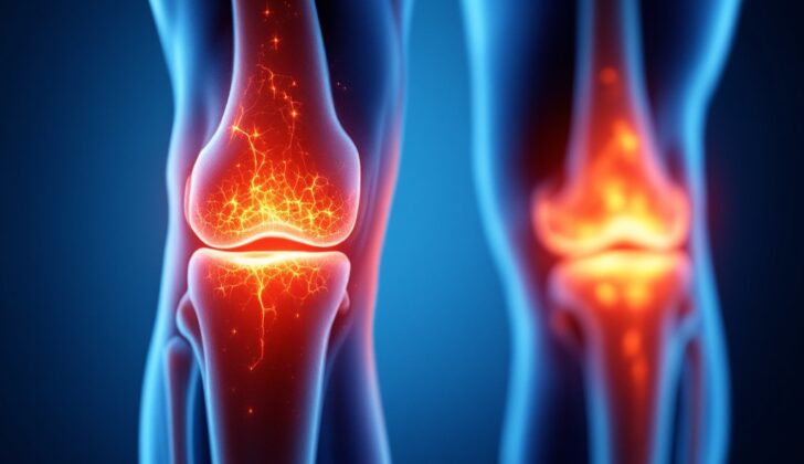
What is Calcium Pyrophosphate Deposition Disease?
Calcium pyrophosphate deposition disease (CPPD) is a condition where specific crystals build up in your joint tissues. How this disease affects people can vary a lot. Some might not even notice any signs, while others might experience sudden or ongoing joint inflammation, also known as arthritis.
There are different names for the various ways this disease may show itself. Acute calcium pyrophosphate (CPP) deposition arthritis is commonly called “pseudogout”. This happens when there’s a sudden increase of inflammation in the lining of the joints, causing symptoms similar to another type of arthritis known as “gout”. On the other hand, chronic CPPD arthritis, often referred to as pseudo-rheumatoid arthritis, shows signs that come and go over several months, much like the symptoms of rheumatoid arthritis that affect the wrists and the joints in the fingers. Chondrocalcinosis is a term used to describe the unique X-Ray finding of joint soft tissue getting hard due to crystal buildup.
The crystals in this disease are made of calcium pyrophosphate dihydrate, and they commonly form in larger, weight-bearing joints like the hips, knees, or shoulders. Many patients suffering from this also have certain other related joint diseases or metabolic abnormalities such as osteoarthritis, trauma, surgery, or rheumatoid arthritis.
What Causes Calcium Pyrophosphate Deposition Disease?
Calcium pyrophosphate deposition disease (CPPD) is thought to happen when there’s an imbalance between the production of pyrophosphate, a kind of salt, and the levels of pyrophosphatases, enzymes that break down this salt in the cartilage of our joints. When too much pyrophosphate accumulates in the synovium (joint lining) and nearby tissues, it combines with calcium to form the substance known as CPP.
There are several health conditions linked to CPPD. Various studies have reported a strong connection between CPPD and hyperparathyroidism, a condition that affects the parathyroid glands in our neck. Following this, the illnesses most often associated with CPPD include gout, osteoarthritis, rheumatoid arthritis, and hemochromatosis, a condition that leads to too much iron in our body. Other related conditions include osteoporosis, a condition that weakens bones; low levels of magnesium in the blood; chronic kidney disease; and the use of calcium supplements.
The deposits of calcium pyrophosphate in the body are thought to trigger the immune system, leading to inflammation and additional damage to the soft tissues.
Risk Factors and Frequency for Calcium Pyrophosphate Deposition Disease
Acute calcium pyrophosphate deposition arthritis, or CPPD, is most often found in people who are 65 years old or older. In fact, between 30 to 50 percent of all patients with this condition are over 85 years old. Based on a study involving over 2,000 cases among US veterans, the condition affects around 5.2 in every 1,000 people, with an average age of 68 and occurring predominantly in men.
This disease is quite rare in people under the age of 60. Moreover, a large study revealed that signs of this condition on X-rays are present in about 4 percent of the general population.
Signs and Symptoms of Calcium Pyrophosphate Deposition Disease
Acute calcium pyrophosphate arthritis often presents similarly to acute urate arthritis, with swelling, redness, and pain in the joints. Up to half of these patients may also have a low-grade fever. The knee is the most commonly affected joint, but the condition can also affect other joints that carry the body’s weight, like the hips and shoulders.
Some patients may have a chronic form of calcium pyrophosphate arthritis. These patients often experience episodes of arthritis that come and go, affecting multiple joints not typically carrying body weight, such as the wrists and the knuckle joints of the fingers. This form of arthritis is similar to rheumatoid arthritis.
Elderly patients who come in with sudden degenerative arthritis in weight-bearing joints may be suspected to have crystal deposit disease. These older patients usually experience a milder course of the disease. Some may have sudden flare-ups after experiencing a traumatic injury.
Testing for Calcium Pyrophosphate Deposition Disease
If your doctor suspects you might have a disease linked with calcium deposits in your joints, known as calcium pyrophosphate deposition disease, they will perform a physical examination. After this, they would likely use a procedure called arthrocentesis, which helps them extract and analyze the fluid from the involved joints. They will also take X-rays of the affected areas.
The definitive confirmation of this disease can be achieved by detecting certain crystals, called ‘rhomboid crystals’, in the extracted joint fluid. These are typically observed under a special microscope that can identify crystals based on how they bend light, a characteristic known as ‘positive birefringence’.
Radiographic images, or X-rays, can reveal evidence of this disease in the form of chondrocalcinosis – calcification, or hardening, of cartilage in the joints. However, not seeing this on an X-ray does not necessarily mean you don’t have the disease. In fact, earlier signs of the disease may be detected via an ultrasound, which can identify abnormalities in your joint cartilage.
Magnetic Resonance Imaging (MRI), especially a type of MRI called gradient-echo sequences, has also proven helpful in assessing the extent of calcium pyrophosphate crystal build-up in your joint cartilage.
Treatment Options for Calcium Pyrophosphate Deposition Disease
The treatment for calcium pyrophosphate deposition disease (CPPD) is aimed at reducing inflammation and managing any underlying conditions that might encourage the development of calcium pyrophosphate crystals in the body.
Patients who are experiencing an acute flare-up involving one or two joints are commonly treated through joint aspiration (a procedure in which fluid is removed from the inflamed joint using a needle) and the administration of glucocorticoids (medicines that reduce inflammation) directly into the joint, unless there’s a medical reason not to do so.
If the acute inflammation impacts three or more joints, the typical treatment involves systemic medication, often with nonsteroidal anti-inflammatory drugs (NSAIDs). If patients have any medical contraindications (reasons not to use certain treatments) for NSAIDs, they might be treated using colchicine (a drug often used to treat gout) or systemic glucocorticoids which work to reduce inflammation throughout the body.
A corticosteroid injection (a medicine usually administered to help reduce inflammation) should only be given when it has been ruled out or considered unlikely that the patient has septic arthritis, an infection in the joint. If the patient displays signs and symptoms that suggest septic arthritis, healthcare providers will typically postpone administering glucocorticoids until they can confidently rule out the infection via negative synovial fluid cultures (laboratory tests on fluid from around the joint).
Patients may also find relief through using ice packs and resting the affected joints as much as possible to reduce inflammation. Those who experience repeated episodes of acute CPP arthritis might find help from daily low-dose colchicine.
While several medications can lower levels of uric acid in the blood and prevent the formation of uric acid crystals, there’s currently no therapy that directly targets the deposition of CPP crystals. That’s why the treatment of CPPD aims at treating any contributing metabolic diseases, controlling inflammation in soft tissues, and relieving symptoms.
What else can Calcium Pyrophosphate Deposition Disease be?
Calcium pyrophosphate deposition disease, or CPPD, can look different in different people, and this can make diagnosing the condition tricky. Doctors often have to consider whether other health issues might be causing the symptoms. These might include conditions such as gout, rheumatoid arthritis, ankylosing spondylitis, and erosive osteoarthritis.
When a person with CPPD gets an x-ray of their knee, the doctor will typically see a certain kind of calcification or hardening within the joint. This might present as a thin, dense cloud that lies parallel to and separate from the bone, or it might show up as a sort of hardening within the flexible tissues called menisci in the knee joint.
Doctors will often suspect CPPD in patients who have joint pain or other related symptoms and who also have these characteristic calcifications on their x-rays. Other imaging methods, such as ultrasound (US) and magnetic resonance imaging (MRI), can also reveal signs of CPPD. If there’s still doubt about the diagnosis, the doctor can look at synovial fluid—the fluid from within the joint—under a special type of microscope to see if there are CPP crystals present, which would confirm the diagnosis.
What to expect with Calcium Pyrophosphate Deposition Disease
Acute calcium pyrophosphate arthritis, also known as acute CPP arthritis, typically resolves itself and the inflammation usually subsides within days to weeks with treatment.
Some patients with chronic CPP inflammatory arthritis may show symptoms similar to those of rheumatoid arthritis. These symptoms can include stiffness in the morning, localized inflammation causing swelling in a particular area, and restricted joint movement. Some may also suffer from tenosynovitis, a condition that causes pain and swelling along the tendon, and can manifest as carpal or cubital tunnel syndrome. It’s common for multiple joints to be affected, and symptoms might come and go or change over time, spanning several months.
Patients with pre-existing joint conditions, like osteoarthritis, are more prone to developing acute CPP arthritis. This is because the calcium pyrophosphate dehydrate (CPP) crystals that form in the joint can trigger an immune response, promoting inflammation and causing injury to fibrocartilage, the tough, resilient type of cartilage It acts as a cushion in the joints to absorb shock from activities like walking and running.
Possible Complications When Diagnosed with Calcium Pyrophosphate Deposition Disease
Calcium pyrophosphate has a specific molecular structure that can cause inflammation. Furthermore, the presence of chondrocalcinosis, a form of calcium buildup in the body, is associated with the breakdown of certain knee tissues (menisci) and tissue that lines the joints (synovial tissue).
In rare instances, after several bouts of acute calcium pyrophosphate crystal arthritis, patients might develop solid, palpable lumps that are similar to the nodules seen in gout. These lumps are found around the joint areas and are essentially an accumulation of calcium pyrophosphate crystals. They tend to reside within the tissues lining the joints (synovium) and the surrounding soft tissues. These crystal accumulations can potentially exacerbate the degradation of the affected joint.
Some patients may have involvement of the spine, which can occasionally cause symptoms like stiffness and bone fusion that appears similar to a condition called ankylosing spondylitis. In certain cases, these symptoms are associated with a calcification of a crucial ligament in the spine (the posterior longitudinal ligament). When this happens, it can compress the spinal cord and lead to symptoms like those seen in conditions such as diffuse idiopathic skeletal hyperostosis and spinal stenosis.
- Calcium pyrophosphate’s molecular structure can trigger inflammation.
- Chondrocalcinosis can lead to the breakdown of knee and joint lining tissues.
- Occasional formation of palpable lumps around joints, resembling gout nodules.
- These lumps can lead to further degradation of the affected joint.
- In some instances, spine involvement leading to stiffness and bone fusion similar to ankylosing spondylitis.
- Certain cases can lead to calcification of a spine ligament, possibly causing symptoms of spinal cord compression.
Preventing Calcium Pyrophosphate Deposition Disease
People with chondrocalcinosis or who have had a previous bout of acute calcium pyrophosphate arthritis should be aware that their condition could be aggravated by certain situations, such as surgery or injuries. In simpler terms, ‘chondrocalcinosis’ refers to the calcification (or hardening by calcium) of the cartilage in joints and ‘acute calcium pyrophosphate arthritis’ is a sudden, painful type of arthritis caused by calcium pyrophosphate crystals in the joints.
If a person is diagnosed with calcium pyrophosphate arthritis at a young age, they might be checked for metabolic abnormalities like hyperparathyroidism or hemochromatosis. To clarify, ‘hyperparathyroidism’ involves the overactivity of the parathyroid glands causing higher levels of calcium in the blood, and ‘hemochromatosis’ is a condition whereby the body absorbs too much iron from the food taken in, causing iron to build up in the body. A detailed family medical history is also important in order to understand and treat the disease better.


