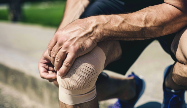
What is Chondromalacia Patella?
In 1906, Budinger and his team were the first to report changes to the patellar cartilage, a form of damage to the tissue covering our bones. This kind of damage was later named “chondromalacia patella” by Kelly and his colleagues. Chondromalacia stems from Greek, where ‘Chrondros’ means cartilage and ‘malakia’ means softening.
In simple terms, chondromalacia is when the protective cartilage layer of our bones gets sick and begins to deteriorate. Specifically, chondromalacia patella happens when the cartilage on the back surface of the patella or kneecap begins to lose its regular density. It becomes softer, which can cause damage and wear, such as tearing or splitting, and then erosion of the cartilage. This condition is often associated with the part of the knee that extends or straightens the leg and is sometimes called runner’s knee.
The back of the kneecap is shielded by this cartilage layer, which connects with another layer of cartilage on a part of the thigh bone known as the femoral groove. Certain injuries, continual minor damage from use over time, or certain medication injections can cause chondromalacia. Though it can occur in any joint, it’s especially common in joints that have experienced trauma or have deformities.
What Causes Chondromalacia Patella?
The chondromalacia patellae, a condition that causes knee pain, develops due to various pathways. These causes are usually a mix of several factors, including:
Misalignment in the Lower Limb and Abnormal Patella Movement. The Q angle, which measures the tug of the thigh muscle against the pull of the knee cap, plays a key role. Usually, this angle is around 14 degrees for men and 17 for women due to wider hips in women. If this angle exceeds 20 to 25 degrees, it indicates excessive sideways pull which causes wear and tear on the knee cap’s cartilage. Additionally, abnormal placement of the patella, either too high or too low, can contribute to chondromalacia.
Foot and ankle variations, like flat feet, can upset the knee’s alignment, causing additional stress on the knee joint. High heels, for instance, can add extra stress leading to chondromalacia.
Chondromalacia patellae might be related to poor alignment syndrome, a condition marked by certain anatomical features that increase the Q angle and lead to abnormal patella formation. This could involve the forward twist of the thigh bone, a knock-kneed position, or feet that turn excessively outward or are flat.
Muscle Weakness. If the thigh muscles and general core muscles are weak, this could play a role.
Direct injuries to the knee cap, prolonged periods of not using the leg, and surgery can all contribute to chondromalacia due to the mini-injuries caused by less pull from the thigh muscle on the knee cap.
In many cases, chondromalacia is linked with abnormal wear and tear of the knee cap’s cartilage. Excessive sideways positioning of the knee cap in the knee joint is a common cause. A tight outer knee band or an inflamed fold of synovial membrane may cause this, but often it’s due to an abnormal Q angle.
While increased vulnerability of cartilage from birth can’t be changed, avoiding chondrotoxic medications can help. Injecting medicines like bupivacaine into the joint or using high doses of corticosteroids can lead to cartilage softening or dysfunction.
The most frequent cause is knee cap dislocation. It often goes unnoticed as it doesn’t cause a notable dislocation.
Risk Factors and Frequency for Chondromalacia Patella
Chondromalacia, a condition affecting the knees, is more common in women than in men due to the increased Q angles that women have. There is no evidence of a hormonal cause for this condition. People who put a lot of stress on their knees, such as active young adults who run a lot and workers who have to climb stairs or kneel repeatedly, are at a higher risk of developing chondromalacia.
Signs and Symptoms of Chondromalacia Patella
Chondromalacia patellar is a condition often associated with pain at the front of the knee. Many patients describe a gradual onset of a vague pain either behind or in front of the kneecap. The pain typically worsens with activities that put pressure on the knee joint (for example, going up or down stairs, squatting, kneeling, running, or even sitting for a long duration). Other symptoms patients might experience include swelling, muscle wasting, and a grating or cracking sound when the knee moves. However, these are not specific to this condition. Therefore, the diagnosis usually involves ruling out other possible causes of the knee pain.
To diagnose this condition accurately and prevent incorrect treatment due to misdiagnosis, a detailed history and physical exam needs to be carried out. The history would check for things like previous injuries, other health conditions, pain or dysfunction in the feet and ankles, instability of joints, and the person’s physical activities. In the physical exam, the doctor would look at the quadriceps to check for muscle wasting, inspect the alignment of the feet and ankles, and specifically assess the knee joint.
Assessing the knee joint involves checking for pain, swelling, muscle strength, kneecap mobility, and a grating sound when the knee moves. The doctor would also look for signs of the kneecap moving incorrectly, which could include: an abnormal forward rotation of the thigh bone, abnormal outward rotation of the shinbone, a subluxated patella (dislocated kneecap), limited movement of the inside part of the kneecap, and a positive patella apprehension test (which indicates the kneecap is likely to dislocate).
The doctor might also perform Clark’s test, a physical examination method specifically used to check for chondromalacia patellae. In this test, the doctor applies pressure on the kneecap while the patient contracts their quadriceps muscle, causing the kneecap to move along the groove in the thigh bone. This can help identify any grinding sensations or pain in the knee joint.
Testing for Chondromalacia Patella
Regular X-rays can help in diagnosing chondromalacia patellae, especially in its advanced stages. These images may show signs like advanced cartilage loss or joint space loss. Though they’re less sensitive in early stages, they can still hint at the underlying cause. For example, they may reveal specific knee conditions like an unusually high or low kneecap position, or a sideways tilt of the kneecap.
A CT scan can provide more detailed information, especially regarding the alignment of the kneecap and thighbone. It can also measure the difference between these two, and detect any unusual twisting in the lower limb.
Although it is less sensitive, an arthrogram (a procedure where a special dye is injected into the joint) can show areas of softened cartilage when combined with regular X-rays or a CT scan. This technique may help to identify issues with the cartilage, but usually only in advanced stages of the disease.
An MRI scan is the preferred method for evaluating the cartilage in the knee. It’s non-invasive, reliable, and tends to detect issues more accurately than an arthroscopy (surgical examination). Abnormal cartilage shows up more clearly with the MRI T2 sequences.
Patellofemoral congruency, or the alignment and conformity of the kneecap and thighbone, can also be evaluated using various measurements from MRI scans. Studies have found correlations between the severity of chondromalacia patellae and measurements like the lateral patellar tilt angle and trochlear depth.
One study using MRI scans even showed that people with chondromalacia patellae had more fat around their knee compared to the general population. This was especially true in women, who had thicker knee fat and more severe chondromalacia patellae compared to men.
Finally, arthroscopy is also used to diagnose chondromalacia patellae. It’s the most effective way of locating and measuring cartilage lesions and assessing kneecap position. However, since it’s an invasive procedure, non-invasive methods are usually used first to diagnose the condition.
Treatment Options for Chondromalacia Patella
The first approach for treating chondromalacia patellae, a condition where the cartilage under the kneecap softens and deteriorates, is a conservative one. That means for at least a year, patients should rest, limit physical activities, and take non-steroidal anti-inflammatory drugs, which are more effective than steroids. Going to a physiotherapist for strength training exercises targeting specific knee and hip muscles could also help.
However, treating this knee problem can be challenging and there’s no single universally-approved treatment. Based on the results of a physical exam, the doctor might recommend wearing a knee brace, going for more physiotherapy, using shoe inserts to correct the foot’s mechanics, and continuing with anti-inflammatory medication. Some might suggest using therapies involving platelet-rich plasma or prolotherapy, but these are not often considered standard treatments as they haven’t shown consistent improvements in patients.
If these methods don’t work, then surgery might be an option, with the choice of procedure depending on the patient’s age and how serious the knee problem is. Surgeries could range from removing or reshaping the kneecap cartilage, making alignment adjustments to the soft tissues and bone of the knee. The aim of these procedures is to avoid causing problems in the quadriceps muscles – the large muscles at the front of the thigh.
For example, arthroscopic (keyhole) surgery could be performed to fix issues with the cartilage or to treat worn or damaged parts of the cartilage. Other surgeries might involve releasing the stiff outer side of the kneecap or capsular (surrounding tissue) release, as well as doing procedures that realign the kneecap. However, such processes might end up worsening the wear and tear of the kneecap joint to a certain extent.
There are several techniques that aim to adjust the bone where the kneecap attaches (tibial tuberosity), but these techniques have their own risks and should be done cautiously. For example, lifting the bone no more than 1 cm to avoid causing the skin to die. Other procedures aim to move the bone forward and inward, but they are not suitable for everyone.
Completing removing the kneecap can be done, but generally only if the patient’s thigh muscles are still strong and the patient can commit to a regular exercise schedule after the operation. This is a drastic procedure and can lead to other complications, and potentially damage to the muscles and ligaments around the knee.
Other outdated procedures include resurfacing the kneecap with an artificial one, but this often resulted in further knee problems and is no longer used.
Another treatment option is cell therapy. This therapy involves injecting the patient’s own cartilage cells or mesenchymal stem cells (which have the ability to develop into many types of cells) into the knee. This has been found safe and effective in treating chondromalacia patellae, can provide relief from symptoms, reduce inflammation, and is less invasive compared to surgery. Research is still being done to understand exactly how this treatment works, but early results are promising.
What else can Chondromalacia Patella be?
When a doctor is diagnosing problems related to the knee, especially pain, they may consider a range of conditions, including:
- Chondromalacia patellae or osteochondral defect, which indicate damage to the cartilage under the kneecap.
- Patellofemoral osteoarthritis, a condition where the cartilage wears away and causes swelling in the kneecap joints.
- Patellofemoral pain syndrome, a condition that causes front knee pain.
- Quadriceps tendonitis/tendinopathy, a condition causing swelling and pain in the tendon attaching the kneecap to the thigh muscles.
- Patellar tendonitis or tendinopathy, another tendon injury causing swelling and pain.
- Saphenous neuroma, a nerve condition that can lead to pain and numbness.
- Hoffa disease, a rare syndrome that causes knee pain.
- Patella Alta, a high-riding kneecap due to abnormal positioning of the kneecap.
- Patella Baja, a low-riding kneecap due to improper positioning.
- Patella instability, where the kneecap does not stay stable within its groove.
- Bi-partite patella, a condition where the kneecap is made up of two parts rather than one.
These potential diagnoses and more should be considered and ruled out through specific tests to correctly identify the source of the problem.
What to expect with Chondromalacia Patella
Chondromalacia patellae, a condition that affects the knee, can either be reversed or develop into a type of arthritis known as patellofemoral osteoarthritis. People suffering from knee pain due to chondromalacia patellae usually have the opportunity to fully recover.
The recovery period varies greatly from one case to another; it could be as short as one month or stretch out over several years. Teenagers generally have a better chance at long-term recovery, since their bones are still developing. Typically, their symptoms lessen once they become adults.
Possible Complications When Diagnosed with Chondromalacia Patella
Patients with knee cartilage softening, also known as chondromalacia patella, may experience complications. This can be due to the side effects of certain anti-inflammatory medications, such as gastrointestinal problems. Sometimes, their skin may react to the material of a knee brace and cause skin issues. Rarely, therapeutic exercises may make the symptoms worse.
If a particular physical activity aggravates their symptoms, the patient and the medical professional should work together to adjust this activity. This could mean changing how often the activity is done, how long it lasts, how intense it is, or even temporarily stopping the activity if it’s necessary. Common issues include:
- Side effects of anti-inflammatory medications
- Skin reactions to brace materials
- Symptoms becoming worse due to therapeutic exercises
- Aggravation of symptoms from specific activities
Preventing Chondromalacia Patella
Teaching patients is focused on making sure they take their medicines correctly, do their healing exercises, follow through with their recovery plans after surgery, and stop doing any activities or movements that could worsen their condition when they can.


