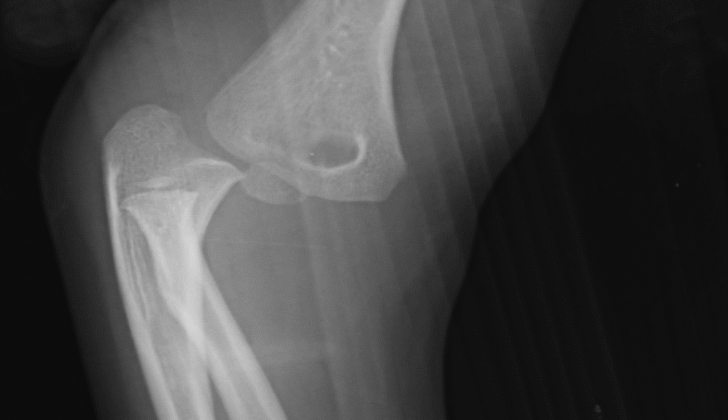
What is Elbow Trauma ?
Injuries to the elbow are pretty common in emergency care. These injuries can range from simple bruising and soft tissue injuries to very severe, complex injuries involving bones and ligaments, such as the “terrible triad” injuries. For adults, such serious injuries often result from high-energy impacts, like car accidents or falls from a significant height. However, older people can also suffer elbow injuries or fractures from simple falls. This happens due to a number of factors including poor physical condition, lack of balance and finesse, poor vision, loss of muscle mass, and fragile bones due to conditions like osteoporosis.
The treatment approach to elbow injuries can vary significantly and depends upon the patient’s medical history and physical examination. That’s why, understanding the structure of the elbow is crucial for healthcare professionals treating such injuries. You see, the elbow joint is one of the most complicated joints in our body. It involves three separate connections between the bones in your arm: the link between the ulna and humerus bones, between the radius and humerus, and between the radius and ulna near the elbow.
On one side of the elbow joint, a part of the ulna bone connects with the humerus. On the other side, a separate part of the humerus connects with the top of the radius bone. Plus, the part of the radius bone near the elbow, called the radial tuberosity, is where the lower bicep tendon attaches to the bone.
The joint also relies on the ulnar collateral ligament (UCL) and lateral collateral ligament (LCL) for stability as they help in keeping the elbow stable during movements to side-to-side. They also help maintain elbow stability during rotational movements.
Furthermore, the transition area between the upper arm and the forearm is a significant area called the antecubital fossa. This area contains important structures such as the radial nerve, brachial artery, and median nerve.
What Causes Elbow Trauma ?
Traumatic injuries can vary in severity, from simple bruises to complicated fractures and dislocations. The more complex injuries often occur after a fall, where someone tries to break their fall using an outstretched hand. Falling directly onto the elbow can also result in different types of fractures or even joint dislocations. There are also isolated soft tissue injuries, which can vary from light bruising, sprains, and muscle strains to deep cuts or bullet wounds that could cause the joint to open up, and requiring medical treatment as a result. While simple elbow dislocations can often be treated without surgery, some people may experience recurring dislocations or partial dislocations, commonly accompanied by pain, clicking sounds, and weakness.
Soft tissue injuries can range from minor ones such as simple bruises, strains, or sprains, to severe ones following bullet wounds or deep cuts. Fractures, fractures-dislocations, ligament injuries, and simple or complex dislocation patterns all belong to the sphere of bone and ligament injuries. ‘Simple’ injuries refer to a dislocation without fracture while ‘Complex’ refers to those with an accompanying fracture. One particularly severe type of elbow injury includes dislocation, a fracture of the radial head/neck, and a fracture of the Coronoid.
Besides traumatic injuries, elbow injuries can also occur over time due to repetitive motions, leading to conditions like lateral epicondylitis (tennis elbow) and chronic partial UCL injuries or strains, especially in athletes or individuals with demanding occupational requirements. These types of injuries are often seen in sports that involve repetitive movements of the upper body, such as baseball and tennis, or in jobs with regular repetitive movements. Patients complaining of elbow pain should have an assessment based on the specific area of the elbow that’s affected.
In children, elbow trauma usually happens through sports activities or falls. Diagnosing these injuries can be tricky because of the unique sequence of bone development and fusion in children. Some common elbow fractures in children include Supracondylar fractures, Lateral condyle fractures, Medial epicondyle fractures, and Radial head and neck fractures. ‘Nursemaid elbow,’ another common injury in children, accounts for 20% of all upper extremity injuries. It usually happens in children aged 1 to 4 and occurs more often in girls than boys. The injury is typically caused by a sudden pull on the forearm while it’s turned inward.
Risk Factors and Frequency for Elbow Trauma
Elbow injuries in young adults often happen during sports activities. They can result from sudden accidents, damage to the ligaments, or from ongoing repeated strain. This is common among weight lifters and accounts for 2.6% of all strains. High impact sports like skateboarding, inline-skating, and skiing often lead to elbow injuries. Overarm-throwing sports like cricket, baseball, and tennis frequently cause a specific type of weakness in a ligament in the elbow.
In children, the pattern of elbow injuries is different. Girls tend to get these injuries earlier than boys due to their elbow joints developing sooner. For girls, the majority of fractures happen between 9 to 12 years, and for boys, they occur between 12 to 15 years. In younger children, the rate of fractures between boys and girls is equal. But in children older than 11, boys are seven times more likely to fracture than girls. Around 15% of all fractures in children are at the elbow, mainly around the age of 6. Elbow dislocation is not as common in children as fractures are. They make up between 3% to 5% of all elbow injuries in children and are most common in early teens, around 12 to 13 years old.
- Elbow injuries in young adults often happen during sports activities.
- They can range from sudden accidents, ligament damage, or from repetitive strain.
- Weight lifters often suffer from elbow sprains and strains, which account for 2.6% of all such injuries.
- High impact sports, like skateboarding and skiing, and overarm-throwing sports, like cricket and tennis, are often associated with elbow injuries.
- In children, elbow injury patterns differ. Girls usually get these injuries earlier than boys due to faster joint development.
- Most fractures in girls occur between 9 to 12 years, and for boys, between 12 to 15 years.
- In children younger than 11, the rate of fractures is equal between boys and girls. In children older than 11, boys are seven times more likely to fracture.
- About 15% of all child fractures are in the elbow, primarily occurring around age 6.
- Elbow dislocation is less common in children, accounting for 3% to 5% of all child elbow injuries, and traditionally occurs in early teens, around 12 to 13 years old.
Signs and Symptoms of Elbow Trauma
Patients with acute injuries typically experience varying levels of swelling and deformation. Accompanying symptoms include pain and restricted movement range. A thorough physical evaluation is needed, focusing on the arm affected, from the shoulder to the fingertips. Some patients might present a combination of fractures in the forearm, elbow, and upper arm. In rare cases, a high-energy trauma could result in a dislocated elbow, fractured upper arm, and dislocated shoulder at the same time.
The examiner should consider the following:
- Skin integrity: it’s important to check if there are any open wounds or injuries.
- If there’s any swelling or fluid accumulation.
- A comprehensive nerve and vascular examination.
How the patient positions their arm can also give some insights into the diagnosis. Additional factors such as patient’s age, existing medical conditions, medications, and previous injury history can affect the assessment of injury severity.
Common bone injuries include supracondylar fractures, which can be of two types – flexion and extension. With the flexion type, a patient would hold the injured forearm with their other arm and elbow flexed at 90 degrees. There might be noticeable loss of the elbow’s bony prominence. In the extension type, the patient’s arm would naturally hold a S-shaped configuration.
Elbow dislocations can be either posterior or anterior. A posterior dislocation might show an unusual prominence of the elbow’s bony part. In contrast, anterior dislocations might have a loss of this prominence. If a radial head subluxation – a dislocation of the forearm’s upper bone – is suspected, patients have difficulty moving their elbow while keeping it slightly bent and the forearm turned inward.
Motor and sensory nerve testing also constitute a part of the examination. Median nerve function, responsible for sensation and movement in parts of the hand, can be tested using a two-point discrimination test over the tip of the index finger. Ulnar nerve function, which affects sensation and movement in the arm and hand, can be checked through resistance tests. Other nerve and vascular injuries could involve the anterior interosseous nerve, radial nerve, and brachial artery.
Compartment syndrome, a condition that involves increased pressure in an arm or leg that causes serious muscle and nerve problems, can develop acutely or chronically. Acute symptoms start soon after an injury and include persistent deep pain, exaggerated pain for the injury’s severity, limb numbness and tingling, and swelling, tightness, and bruising. Chronic compartment syndrome may develop over time and is characterized by pain or cramping in the affected muscle during exercise, relieved by rest, without loss of muscle function. If symptoms are experienced more in the hand and wrist area, it might indicate compartment syndrome of the forearm, also known as Volkmann ischemic contracture.
Acute compartment syndrome (ACS) is mostly diagnosed based on clinical features. Classical symptoms include pain, pale skin, loss of pulse, pressure, abnormal sensations, changes in temperature, and in severe cases, paralysis. Regular examinations are needed if ACS is suspected and especially if paralysis occurs, as it suggests late-stage ACS, possibly causing irreversible damage.
When ACS is suspected, the pressure inside the compartment is measured for a definitive diagnosis. This requires invasive monitoring. Some procedural details are prevalent, such as securing the catheter within 5 cm of the fracture level and taking note of some pitfalls which could give false reading. Pediatric patients and infants should be assessed with extra caution, and child abuse should be ruled out if applicable.
Testing for Elbow Trauma
In a situation of suspected elbow injury or fracture, there are several imaging methods to figure out what’s going on.
Firstly, a kind of X-Ray test known as ‘anteroposterior’ (AP) can show a head-on view of the elbow, while a ‘lateral’ view displays the elbow from the side. ‘Oblique’ views (if needed) capture the elbow from an angle, and a ‘traction view’ can help better understand complex fracture patterns.
In more serious cases, such as high-energy trauma injuries, the doctor might recommend ‘orthogonal’ views. These are X-rays taken from the shoulder to the wrist to provide comprehensive images of the entire arm.
One important sign that doctors look for on these images is the ‘fat pad sign’. This is where the areas of fat around the elbow joint become visible due to joint injuries. If you can see the ‘posterior’ (rear) fat pad on an adult’s X-ray and there are no other obvious fractures, this usually means the ‘radial head’ (part of the arm bone near the elbow) is fractured. In children, this sign could indicate a fracture above the elbow joint or ‘supracondylar fracture’.
An ultrasound is less commonly used for trauma injuries. However, it does have its uses in quick assessments or in trauma cases combined with an infection.
In more complex cases, like fractures with multiple breaks, doctors might use a Computerized Tomography (CT) scan. This can help them plan surgeries and procedures. Magnetic Resonance Imaging (MRI) is another option if there’s the need to look at soft tissues, ligaments, or suspected hidden fractures.
In children, understanding elbow injuries can be trickier because fractures often occur in areas not yet fully hardened into bone. Doctors may use several techniques to clarify such situations, such as drawing a line on the X-ray images or comparing the injured elbow with the healthy one. They also consider the child’s age and growth stage, as there are certain areas, or ‘ossification centers’, that are prone to injuries at specific ages.
Finally, doctors might need to consider both elbows in children for full understanding. Occasionally, a nurse may be able to adjust the elbow back into place by twisting the forearm.
Treatment Options for Elbow Trauma
Mild injuries of soft tissue can usually be treated with rest, ice, and over-the-counter anti-inflammatory medications. It’s important to keep the elbow moving so the joint doesn’t become stiff. The individual might need to see a physical therapist to ensure the best possible recovery. If the injury is severe, for example, if bones have been displaced, surgery might be needed. If the fracture isn’t displaced, a splint at first might suffice, but generally, any fracture displacement greater than 2mm needs to be considered for surgery.
If the elbow is dislocated but there’s no fracture, doctors can put the elbow back in place and immobilize it with a sling for 10 to 14 days. The initial examination after the elbow has been put back in place will guide how long the elbow needs to be immobilized, and whether the person might be at risk for long-term elbow instability during physical exertion. In this scenario, surgery might be necessary to mitigate the risk of poor outcomes.
For emergency treatment, orthopedic consultations are standard. Stable fractures with no displacement can be stabilized with a splint until a follow-up with an orthopedist can take place within 24 to 48 hours. Some fractures, especially ones involving the surface of the joints or complex fractures, will generally require an orthopedic consultation. Elbow dislocation should be addressed immediately if blood vessels are compromised. In these cases, the elbow is repositioned, and then the arm is bent to 90º and placed in a splint.
If a medical check suggests a fracture but the radiograph doesn’t confirm it, the patient should wear a splint and return for re-evaluation in 24 to 48 hours. Pain relief and sedative medications are often required during the reductions due to the close involvement of blood vessels and nerves in the joint.
Continual injuries, like tennis elbow or inflammation of tendons, usually respond well to non-surgical treatments. These can include rest, ice, NSAIDs, physical therapy when suitable, corticosteroid injections, and possibly treatments with platelet-rich plasma.
Whether a hospital stay is required depends on factors like vascular injuries, open fractures needing surgical intervention, or considerable swelling or bruising. Stable fractures or successful reset dislocations might mean discharge unless they deteriorate. Orthopedic follow-up appointments should be scheduled within 24 to 48 hours. Simple soft tissue injuries can continue being managed supportively at home.
Long-term immobilization of the elbow can cause it to become stiff. So, the primary goal is to restore movement as soon as possible. A score, known as the Mayo Elbow Performance Score, has been found to be good for measuring the results of treatment after surgery.
What else can Elbow Trauma be?
When diagnosing a patient’s injury, doctors might consider several possibilities:
- Fractures (broken bones)
- Dislocations (bones forced out of their normal position)
- Sprains (stretching or tearing of ligaments)
- Strains (stretching or tearing of muscle or tendon)
- Ligament weaknesses (for example, UCL)
- Bursitis (inflammation in the cushioning disk near a joint)
- Conditions related to tendons, either sudden (acute) or long-term (chronic)
For diagnosing children’s injuries, doctors may need to consider:
- Child abuse
- Injuries to the growth plate (distal humeral physeal injuries)
- Nursemaid’s elbow (dislocated elbow common in toddlers)
- Fractures (broken bones)
- Avulsions (severe injury where a body structure is forcibly detached)
- Monteggia fracture-dislocations (an uncommon injury affecting the arm)
- Growth plate injuries or reactions
What to expect with Elbow Trauma
Generally, patients with treated fractures recover well. Some might experience a loss of final extension by about 10° to 15°, but this is usually not a cause for concern. Dislocations can press on nerves or blood vessels, but immediate treatment can help avoid complications.
Patients with bursitis also usually have a positive outcome. Although infectious bursitis can potentially lead to a widespread infection, this risk is relatively low.
Possible Complications When Diagnosed with Elbow Trauma
The main complications after a fracture and dislocation usually involve the nerves and blood vessels. Around 10% of people may experience a condition called ulnar neuropathy, that affects a nerve in the arm. A less common condition is median nerve entrapment, where a nerve in the forearm gets compressed. Other issues may involve the blood vessels, such as a decreased or lost radial pulse. Keeping the elbow immobilized for a long time can also cause stiffness and a loss of fully straightening the arm, which can be particularly problematic for children and athletes.
Common Complications:
- Neurovascular complications
- Transient ulnar neuropathy
- Median nerve entrapment
- Vascular complications (e.g., reduced or lost radial pulse)
- Elbow stiffness due to prolonged immobilization
- Loss of full arm extension due to prolonged immobilization
Preventing Elbow Trauma
Patients should follow the after-care guidelines and rehabilitation plans given by the medical professional treating their injuries or surgeries. This is especially important for intricate elbow injuries and severe fracture-dislocations, like terrible triad elbow injuries. It’s important for patients to understand what to expect during recovery. Almost all patients experience loss in Range of Motion (ROM) after recovery is complete. Therefore, ROM is a crucial factor in predicting the outcome of recovery.


