What is Fingertip Injuries?
The fingertip is the part of the finger that’s furthest away from the hand. It helps us touch, feel, and understand the world around us, then sends that information back to the brain It’s technically also described as the portion of the finger that is farther away from the flexing and extending tendons linked to the last bone of the finger, or in the thumb’s case, from the joint in the thumb.
The fingertip gets its blood supply and nerve supply from the digital arteries and nerves, which branch out near the last joint in the finger. The pad of the finger is served by specialized nerve cells – Pacinian and Meissner corpuscles, and Merkel cells – that allow us to discern touch and feel sensations in detail. The fingerprints on the fingertip are unique to each individual and consist of grooves and ridges.
The back of the fingertip includes the fold of skin overlapping the nail, the nail bed itself, and the nail plate. Some specific parts of the back of the fingertip include the eponychium (the soft tissue just before the nail), the paronychium (the folds of skin on the sides of the nail), and the hyponychium (a seal of hardened skin where the nail bed meets the skin at the edge of the nail). The white part of the nail near its origin, called the lunula, marks the separation between the sterile area and the germinal matrix below, which is responsible for producing new cells. The nail bed itself is split into the sterile matrix, where the nail sticks on to, and the germinal matrix, which is further away, and is responsible for 90% of nail growth.
What Causes Fingertip Injuries?
Injuries to the fingertip can be sorted by how the injury happened or how severe it is according to a commonly used system called the Allen classification.[4][5] The most usual causes of these injuries are:
* Crushing injuries, which happen when pressure is put on the finger. This could be from a door closing on the finger, getting hit by a hammer, or objects being dropped onto fingers. These injuries might not break the skin or could break the skin and even cause fractures in the fingertip bone.
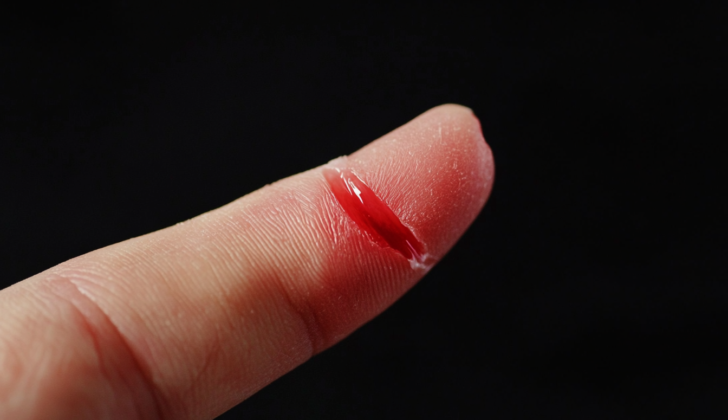
* Cuts, which usually occur due to household items (like knives, scissors, and cans) or workplace tools (such as rotary saws). These cuts could affect the fleshy part of the fingertip or the nail and the nail bed.
* Amputations, which involve losing some or all of the soft tissue and part or all of the fingertip bone. These injuries can lead to changes in how the finger looks and works.
Less common injuries happen when the finger is suddenly bent or straightened, causing the tendon in the fingertip to pull away.
The Allen classification sorts these injuries into four types:
* Type 1: affects only the fleshy part of the fingertip
* Type 2: involves the fleshy part of the fingertip and the nail bed
* Type 3: includes partial loss of the fingertip bone
* Type 4: injury is closer to the palm than the crescent-shaped area at base of the nail (lunula)
Type 1 injuries usually heal well naturally over time. Type 3 and type 4 often need some skin grafts.
Risk Factors and Frequency for Fingertip Injuries
Fingertip injuries are the most frequent types of hand injuries. There are several million estimated check-ups with primary care doctors and emergency room visits every year due to these injuries. Almost half of these injuries that happen outside work are cuts to the fingers.
Signs and Symptoms of Fingertip Injuries
People suffering from a digital injury typically show signs like pain, bleeding, or an inability to use the affected finger or toe. Key factors to consider include the person’s age, gender, job, use of drugs, tobacco or alcohol, which hand they use the most, which finger or toe is injured, how the injury happened, and any past medical or surgical history. It’s important to examine the injured digit carefully in a well-lit setting so that the extent of the injury can be properly assessed. This will often reveal any cuts, fractures (which may either be closed or open), and even instances where the tip of the digit has been amputated.
Testing for Fingertip Injuries
The evaluation of a potential hand injury should include several steps: checking for sensation or feeling, testing the finger’s motion, and observing how quickly blood returns to the nail after being pressed (capillary refill). Your doctor will also need to take X-ray images of your injured finger and hand, from two to three different angles.
There are certain situations when it becomes necessary to consult with a hand surgeon. These include if there is a possibility of injury to a tendon (the tissue that connects muscle to bone), if a bone is fractured in a way that it isn’t in its normal position or the break extends into a joint, if a bone is dislocated to the point where it is visible, if there is a significant loss of finger skin, if a deep cut is present at the base of the nail (which is called the eponychium), or if there is an amputation that leads to significant exposure of the bone.
Treatment Options for Fingertip Injuries
The goal of treating finger injuries centers around reducing pain and trying to limit blood loss. The specific approach depends on the type and how severe the injury is.
A subungual hematoma is a blood clot under the fingernail, typically caused by a strong crushing injury. This type of injury often happens at work and is identified by severe pain and a discolored nail. It occurs when the blood vessels in the nail bed get damaged. Over half of such injuries need a process called trephination, which relieves pressure and drains the blood clot. If a bone in the finger is also broken, a careful examination of the nail bed is recommended, followed by using an aluminum splint to immobilize the finger until the pain goes away.
Injuries to the nail and nail bed can range from simple to complex cuts, nail being ripped off, or amputations. These injuries are often associated with partial or complete fingertip avulsion — where the fingertip is forcibly detached from the rest of the finger. The best management for simple and complex cuts is to join the skin together without damaging it further for a pleasing cosmetic result. In children, absorbable stitches should be used so they don’t have to be removed. If the nail is partially or completely ripped off or the area around the nail is significantly damaged, then the nail must be removed. If the nail bed is ripped off, it should always be repositioned. This helps the new fingernail grow properly. After the nail is removed and the nail bed is stitched up, the nail should be put back in place. Around half of nail bed injuries may also have a broken bone in the fingertip. Nail bed injuries resulting in the nail being ripped off often have a poor outlook.
Closed fractures, which are minimally displaced, can be managed with a splint. If the broken bone is angled or displaced, the bone needs to be moved back into place. Unstable and breaks in the joint need to be looked at by a hand or orthopedic surgeon. Open fractures need to be managed with a digital nerve block, deep cleaning of the wound, and soft tissue repair. The patient should take antibiotics and they will need close follow-up care either by an orthopedic or a hand surgeon.
Seymour fractures often happen through the growth plate. These fractures are usually open and have a high chance of infection and growth problems. Seymour fractures look similar to mallet fingers, but the bone is moved through the fracture not the joint in the finger.
A mallet finger occurs when there is a tear in the extensor mechanism leading to an inability to extend the DIP joint. It is the most common tendon injury among athletes.
Swan-neck deformity results in the chronic untreated mallet finger where the bending of the main knuckle and straightening of the joint closest to the fingernail tip.
A Boutonnière deformity is when the middle joint of the fingers bends and the farthest joint straightens. The treatment includes keeping the joint continuously straightened for about 5-6 weeks and a follow-up with a hand surgery service.
Amputations present a challenge in preserving function and good look. Several techniques have been described for finger and thumb amputations including V-Y plasty, homodigital neurovascular island flap, and first dorsal metacarpal flap.
Infections can happen due to small injuries such as a splinter, thorn, or biting one’s nails. When the nail structures are affected, a paronychia occurs while a felon abscess affects the fingertip. Pain, redness, and a decreased movement of the affected finger are common signs. These cases need an early evaluation, antibiotics, warm water or Betadine soaks, and a cut to let out pus when severe.
What else can Fingertip Injuries be?
Here are some conditions related to hand injuries that your doctor may consider during the diagnosis:
- Base of thumb fracture
- Digital collateral ligament injury
- Hook of hamate fracture
- MCP dislocations
- Metacarpal fracture
- Pisiform fracture
- Phalanx dislocations
- Phalanx fracture
- Scaphoid fracture
- Thumb collateral ligament injury
Possible Complications When Diagnosed with Fingertip Injuries
- Nail loss
- Finger deformity
- Loss of the fingertip
- Having abnormal sensations like tingling or numbness


