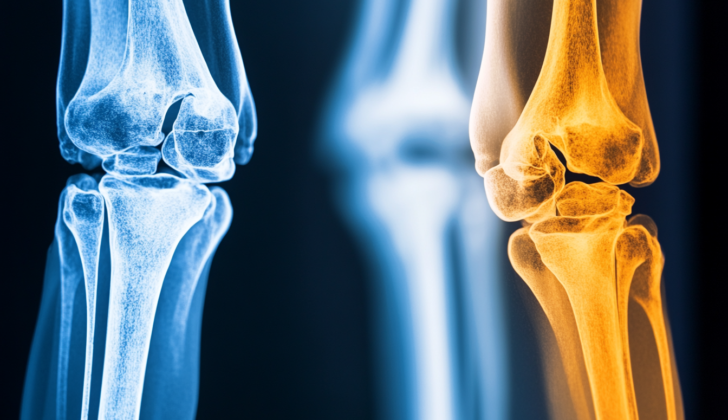
What is Floating Knee?
The term “floating knee” was first used by Blake and McBryde in 1975 to explain a situation where both the femur (thigh bone) and the tibia (shin bone) on the same side are broken. These fractures can happen at any place on either bone, but for it to be a “floating knee” injury, both bones must be fractured. This term refers to the knee joint, but isn’t specifically about the connection to either long bone. Although it’s not rare for fractures to occur in the femur and tibia, it’s pretty uncommon for both bones on the same side to be injured at the same time. These types of injuries, known as floating knee injuries, are usually complex and require meticulous treatment processes. They might be caused by various types of impacts or accidents.
Another term for “floating knee” injuries is “flail knee”. These injuries can be categorised into many different types:
Blake and McBryde’s classifications are based on the location of the injury.
- Type I: Fractures occur in both shafts of the two long bones.
- Type II-A: The injury affects the knee joint.
- Type II-B: Requires involvement of the hip or ankle joints.
Letts-Vincent’s classifications are specifically for children and firstly identify the area of the fracture and whether it is an open (skin is broken) or closed (skin is not broken) fracture.
- Type A: Two fractures occur in the middle section of the bones, both closed.
- Type B: Two fractures, one in the middle and the other towards the ends of the bones, both closed.
- Type C: Two fractures, one in the middle and the other at the end connections of the bones, both closed.
- Type D: At least one of the fractures is open.
- Type E: Both fractures are open.
Bohn-Durbin’s classifications are also for children and, like Letts-Vincent, first identify the area of the fracture and if it’s open or closed.
- Type I: Double fractures in the middle section of the bones.
- Type II: Injuries are close to the joints.
- Type III: Injuries involve the end connections of the bones.
Fraser et al.’s classifications are:
- Type I: Fractures are in both bone’s midsections without involving the knee.
- Type II: Fractures extend into the knee and are further subdivided into:
- Type IIa: The injury involves the top of the shinbone.
- Type IIb: The injury includes the bottom of the thigh bone into the knee.
- Type IIc: Both areas of the shinbone and the thigh are involved within the knee joint.
What Causes Floating Knee?
Floating knee injuries are complex fractures often caused by powerful forces, such as car accidents, falls from great heights, or accidents involving pedestrians and cyclists. These injuries usually involve strong blows to the knee and often come with other injuries to various parts of the body, including severe damage to soft tissues. These other injuries can be quite serious, with an average injury severity score (essentially a measure of how serious an injury is) of over 16.
These injuries can come with a variety of severe complications:
– Serious head injuries occur in 14% of cases.
– Injuries to the chest, abdomen, and other soft tissues are also connected to these kinds of injuries.
– Injuries to the ligaments in the knee, which make the knee loose and unstable, happen in 19% of cases.
– Additional fractures happen in up to 44% of cases.
Some of these complications can be life-threatening or harmful to the limb:
– Vascular injuries, mainly to the popliteal artery (the main artery in the knee), happen in 7% of cases. Among these, up to 69% are open fractures, where the broken bone pierces the skin.
– Injuries to the popliteal artery and severe open fractures can lead to the need for amputation within the first 24 hours after the injury, this occurs in up to 9% of cases.
– Conditions like compartment syndrome, where pressure within a muscle compartment increases, and fat embolism syndrome, where fat particles enter the bloodstream and block blood vessels, are often reported as well.
– Up to 10% of patients are reported to die upon admission due to the severity of these injuries.
Risk Factors and Frequency for Floating Knee
Floating knee injuries are uncommon and we don’t have enough information to accurately say how often they occur in the general population. The biggest study mentioned involved 222 patients over 11 years. It appears that these injuries are more common in men than women. The reason for this might be because men engage in high-speed and physically demanding activities more often than women. Men could also have a higher prevalence because they tend to take more risks.
Signs and Symptoms of Floating Knee
A “floating knee” injury refers to a type of injury where both the bone of the thigh (the femur) and the shin bone (the tibia) have been broken. This usually happens due to multiple traumas in the same area, often from serious accidents such as falls, car crashes, or being hit by a car as a pedestrian.
People with this kind of injury will usually report intense pain in the leg, difficulty bearing weight on the injured leg, and possibly feeling like the knee is unstable. This instability could be due to damaged ligaments, which often come with this sort of injury.
When examined by a medical professional, the person will usually feel pain when the doctor touches both the broken tibia and femur. The injured leg may appear deformed or shortened in comparison to the uninjured leg. More complicated fractures may also become apparent during the examination. Furthermore, it’s crucial to check the nerves and blood flow in the injured leg, as the injury may have caused damage in those areas as well.
Testing for Floating Knee
If you have a “floating knee” injury, where both the knee joint and one of the bones in your lower leg are broken, your doctor will follow standard procedures to assess the extent of your trauma. This means they’ll look at your general health and any other injuries you might have. If you have multiple serious injuries, the doctor will follow a protocol called the Advanced Trauma Life Support® (ATLS®) which identifies and treats the most life-threatening issues first. The ATLS® approach includes checking your Airway, Breathing, Circulation, Disability, and Exposure (ABCDE).
Once it’s confirmed that your condition is stable and other life-threatening injuries have been ruled out or treated, the doctor can then focus on your floating knee and other less urgent injuries. One of the key checks will be to examine the sensory and blood supply to your lower limb. This involves testing the sensation in all the areas of skin (dermatomes) in your lower extremity and checking for pulses in the arteries of your foot. If the back part of your thigh bone (distal femur) is displaced, it can harm the nearby artery (popliteal). If this is suspected, a Doppler ultrasound test will be conducted to check for damage to the inner lining of the artery. In addition, due to the significant bone trauma usually involved in these injuries, you will be closely monitored for signs of fat embolism. Fat embolism happens when fat tissue enters your bloodstream and blocks blood flow. If this is detected, surgery to manage the fractures will be postponed until the condition stabilizes.
For diagnosis, the doctor will initially use plain film radiography, better known as X-ray imaging. This provides a general view of the affected limb and can identify any clear fractures. If the fractures are complex, a CT scan might be conducted next. This can provide a more detailed view of the fractures, including the number of fragments (comminution) and bone loss. It can help with planning treatment strategies and predicting potential complications. Because “floating knee” injuries often involve damage to the ligaments (the tissue that connects bones), an MRI might also be conducted. This test can provide detailed images of these soft tissues.
Treatment Options for Floating Knee
When someone has a “floating knee” injury, the first step in their care is to stabilize any life-threatening injuries, like those to the head, chest, or abdomen. If the patient is stable, attention then turns to stabilize any fractures in the femur and tibia bones, which can be done temporarily. For stable patients, an immediate effort is made to fix the fractures as surgery typically leads to better recovery outcomes.
The specifics of treatment rely on many details about the fractures and the patient, such as whether the fracture is open or closed, the fracture pattern, its location, and the patient’s age and overall health. Because floating knee injuries are rather rare, treatment plans often draw more from the experience of the doctors than from large scientific studies.
For younger patients whose bones are still growing, treatment often avoids surgery. Instead, doctors may use a long leg cast to stabilize the injury, allowing the body to heal. In some cases, a special kind of fracture known as a physeal fracture may be treated with a combination of surgery and casting.
There are several surgical options available. One common approach is intramedullary nailing, a procedure in which metal rods are inserted into the marrow cavity of the bone. This allows the fracture to remain stable while the bone heals. This method is usually used in cases where the fracture involves the long, narrow part of the bone (the diaphysis). The femur is usually fixed first, followed by the tibia.
In other cases, like those where the fracture is in the joint itself (type II fractures), a technique known as plating may be preferred. Plating involves affixing metal plates to the bone to secure the fracture and allow healing. This approach has the added benefit of enabling doctors to address related soft tissue injuries at the same time.
An external fixator, a stabilizing metal frame attached to the bones outside the body, can be used in cases of open fractures or where there is extensive soft tissue damage.
There are also specific circumstances where other methods might be used, like a knee replacement, or the use of plates if the fracture is near a previous artificial joint. In treating these types of fractures, it’s essential to ensure the prosthesis remains stable.
In addition to bone injuries, patients with floating knee injuries may also have damage to the ligaments and menisci (a type of cartilage within the knee), which may be found through techniques like MRI or arthroscopy and treated appropriately.
What else can Floating Knee be?
Patients who have ‘floating knee’ injuries usually have other severe injuries as well. Because of this, doctors always have to check for other serious injuries that could threaten their life. There can also be complications from this type of trauma. It’s important to keep an eye out for deep vein thrombosis and fat emboli, which can occur secondary to injuries to the skeleton. Patients might also have injuries to their ligaments (like the ACL, PCL, or meniscus). These injuries aren’t life-threatening and can be treated once the patient is stable and any related fractures have been reduced.
What to expect with Floating Knee
There are several factors that can lead to worse outcomes in cases of “floating knee” injuries, a condition where both the thighbone (femur) and shinbone (tibia) of the same leg are broken. These factors include having an open tibial fracture (broken shinbone where the bone pierces through the skin), fractures involving multiple segments or affecting the knee joint’s surface, and the need for initial external support or additional surgeries to treat the injuries. It’s been found that patients who undergo surgery typically have shorter hospital stays compared to those receiving non-surgical treatment.
Other things that could affect how well a person recovers include their age and whether they are a smoker. Older people and those who have smoked for a long period of time at the point of injury are more likely to experience delayed bone healing and a delay in their ability to fully bear weight on the injured limb. The same can be said for those with open fractures. Furthermore, the severity of the injury (the injury severity score or ISS), knee joint involvement, and serious pretibial soft tissue injuries (injuries to the soft tissues in front of the lower leg) are also noted to affect the recovery outcomes of floating knee injuries.
Possible Complications When Diagnosed with Floating Knee
There are different complications that may occur with injuries to the knee. Broken bones in the knee might not heal directly or fast enough. This is called a “malunion” or “delayed union”. In a study with 89 patients who had floating knee injuries, which are quite severe, the average time it took for the bone in the thigh (femur) or shin (tibia) to heal was around 9.5 to 10.5 months. The healing process for fractures involving multiple parts of the femur took an extra 6 months. On the other hand, similar fractures on the tibia didn’t experience this delay. More than half of these patients ended up with good or excellent outcomes.
- Malunion (incorrect healing of fracture)
- Delayed union (slow healing of fracture)
Another complication could be a difference in leg length. This is possible whether the damaged limb becomes shorter or longer than its healthy counterpart. Kids whose bones are still growing may also experience this if their growth plate is disrupted causing premature closing.
- Limb length discrepancy
Infections are more common when fractures are open and expose the bone surface, and if several surgeries are needed. Specialists mainly attribute the elevated infection risk in floating knee injuries with an open femur break to the fact that these injuries often come with severe tissue injury as well.
- Increased risk of infection, especially with open fractures
There are other complications related to floating knee injuries. These include compartment syndrome (a painful condition due to pressure buildup from internal bleeding or swelling of tissues), loss of joint movement, and in severe cases, the need for limb amputation. Another finding was that the use of a specific method to treat these injuries, known as retrograde intramedullary nail, resulted in a higher risk of abnormal bone growth around the knee compared to another method known as anterograde femoral nails, which doesn’t involve entering the knee joint.
- Compartment syndrome
- Loss of joint motion
- Potential requirement of limb amputation
- Increased risk of abnormal bone growth around the knee with certain treatment methods
Preventing Floating Knee
Patients need to adhere to the directions given by their doctor. In most cases, surgery tends to provide better outcomes for patients, often allowing them to heal around 8 weeks sooner than if they had chosen a non-surgical route. After surgery, patients will need to avoid putting weight on their leg and their leg will be enclosed in a full-length cast.
Patients who do not undergo a specific type of surgery involving ‘intramedullary nails’ (a type of rod inserted into the bone to stabilize it), or those who have more complex fractures, may need to avoid bearing weight on their leg for a longer period of time. Additionally, if patients fail to follow weight-bearing instructions, there can be some serious issues. These can include breaking of the surgical devices used during the operation and even a re-fracture, which could lead to other complications, including the bone not healing properly (a condition known as ‘nonunion’). So, it’s extremely important to follow your doctor’s instructions for recovery to avoid these issues.


