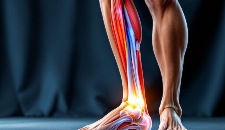
What is Gastrocnemius Strain?
The gastrocnemius muscle, known commonly as your calf muscle, has two parts. One part of the muscle starts from the inside back area of your thigh bone (medial femoral condyle), and the other form the outside part (lateral femoral condyle). This calf muscle is more prone to injuries because it extends over three joints: the knee, ankle, and a joint near your ankle called the subtalar joint.
The two parts of the calf muscle start from two different points on the back of your thigh bone. After reaching a point near the junction of muscle and tendon, this muscle turns flat and joins with another muscle in your calf, known as the soleus muscle. This joint area together forms the Achilles tendon. Though Achilles tendon injury is common, injuries to the back of lower leg, including areas such as the gastrocnemius, soleus, and a muscle called the flexor hallucis longus, are less common.
It is vital for a proper and timely diagnosis to effectively treat patients with injuries in the back of the lower leg. With the right diagnosis and treatment, patients usually recover well.
What Causes Gastrocnemius Strain?
A strain in the gastrocnemius muscle, which is in the calf, typically occurs when the knee is fully straightened out and the ankle is flexed upwards. This position stretches the muscle to its maximum, creating a lot of tension. This tension makes it easier for the muscle to tear, particularly when the muscle is contracting while it lengthens – a motion known as ‘eccentric contraction’.
Risk Factors and Frequency for Gastrocnemius Strain
Gastrocnemius strain, or strain of the calf muscle, is often found in middle-aged or older individuals, as well as younger athletes. This issue is sometimes called “tennis leg” because the steps of serving in tennis can lead to this strain. The posture used in tennis – fully extending the knee and flexing the ankle – can cause strain, particularly in the medial head, or inner section, of the gastrocnemius muscle.
You might also see this type of injury in young athletes who play various sports, including racquet sports, running, basketball, football, and skiing. In an interesting case, it was reported that this type of injury occurred after praying Namaz. The act of praying includes kneeling and touching the ground with the head. When standing up from this position, the calf muscle is stretched significantly, which could lead to injury.
Signs and Symptoms of Gastrocnemius Strain
When a person injures their gastrocnemius muscle, which is found in the calf, they might describe it as feeling as if something hit their calf. Some people even hear a snapping or popping sound that is similar to a twig breaking. At the time of injury, they might not feel any pain, but after taking a few steps, they usually start to experience pain on the back and inner part of the calf. The pain can get so severe that it becomes hard to walk.
Before the injury, some people might experience warning signs or prodromal symptoms. They might feel a dull ache in the calf that will later be injured. In fact, studies show that about 20% of patients recall experiencing such symptoms before the injury.
Different signs can be noticed during a physical examination of a patient with a gastrocnemius muscle injury. These can include:
- Swelling (edema)
- Bruising (ecchymosis)
- Tenderness along the muscle
- Pain when the muscle is touched
- A noticeable gap under the skin where the muscle has retracted
Additionally, the doctor will check the Achilles tendon to make sure it’s not injured. If the tendon’s condition isn’t clear, the doctor might perform a Thompson test. They will also check the patient’s pulses. They might ask the patient to move their foot and ankle to see if these movements cause pain, indicating a more serious injury. Lastly, a neurological examination will be performed to test movement and feeling. In severe instances when the muscle is completely torn, a large bruise (hematoma) can form, pressing on the sural nerve and causing a loss of feeling in the outer calf and ankle.
Testing for Gastrocnemius Strain
A gastrocnemius strain, or a strain of the calf muscle, is usually diagnosed through a physical examination by a doctor. However, imaging studies can help determine whether the muscle tear is partial or complete. Simple x-rays or CT scans are not very useful in this case. Ultrasound, however, can provide useful information while avoiding any radiation exposure, and it’s relatively inexpensive.
An ultrasound can reveal certain signs, like the disruption of the muscle fibers at the junction where the muscle becomes tendon, a blood clot or hematoma, and fluid accumulation between the calf muscle and the muscle beneath it. It can also show whether a muscle tear is partial or complete, and reveal the size of the hematoma. Larger hematomas usually indicate a complete tear of the calf muscle, rather than a partial tear. Ultrasound can also guide doctors in draining the hematoma if needed, and check for a deep vein thrombosis, a blood clot in the leg which can sometimes occur with a calf muscle strain.
One study looked at 141 patients with a clinically diagnosed condition referred to as “tennis leg”, which is usually associated with a calf muscle strain. Out of these patients, 67% had a partial calf muscle tear, 1.4% had an associated rupture of the plantaris tendon, and 21% had fluid build-up without a calf muscle tear. Ten percent of the group had deep vein thrombosis, but no visible calf muscle injury.
The healing process can be monitored using ultrasound as the signs of recovery include a decrease in the size of the hematoma, the appearance of healing tissue, and rearrangement of muscle fibers.
If necessary, Magnetic Resonance Imaging (MRI) may be used because it offers extremely detailed images of soft tissues. These images can reveal a rupture or discontinuity of muscle fibers and pull back of the torn muscle fibers. MRI can also differentiate between an injury to the calf muscle and the Achilles tendon. This information can help doctors determine the best course of treatment. In some cases, an MRI scan can provide additional information about the injury to the surrounding tissues. Connective tissue injury plays an essential role in deciding when a person can return to sports after a muscle injury.
Treatment Options for Gastrocnemius Strain
Early and correct diagnosis and treatment can make a big difference to a patient’s recovery. The first steps of treatment aim to alleviate symptoms. This includes reducing further bleeding, decreasing pain, and preventing the muscles from becoming too tight or shortened – also known as ‘contracture’. To minimize swelling and pain of the injured muscle, resting the area, applying ice, using a compressive bandage, and elevating the limb are common strategies. Using moist heat and giving massages are not suggested early in the treatment as they are believed to increase the risk of further bleeding.
Medications may be used in order to lessen pain and reduce muscle contractions. Getting moving again early is also important to avoid ‘contracture’. Nonsteroidal anti-inflammatory drugs are not recommended within the first one to three days of injury due to them increasing the risk of bleeding because of their effects on platelets, the elements of blood that help in clotting. During this period, medications such as Celebrex and possibly other COX-inhibitors that have a lesser effect on platelets may be options. Pain relief can also be managed with acetaminophen or even stronger pain medications. If a patient’s condition does not improve with these methods, further assessment and imaging studies need to be performed to check for complications or surgical requirements.
Once the patient has successfully surpassed the initial phase of treatment, the next focus is rehabilitation. Physical therapy can help improve functional recovery. Initially, gentle stretching can help reduce the scar within the muscle caused by the injury. Gradually, strengthening exercises, heel raises, and balance training should be added, along with exercises for the central muscles and general conditioning. The patient should aim to move as comfortably as possible and gradually increase their activity level after the pain subsides. In severe injuries, the patient may need to limit the weight put on the affected lower extremity and use a cast or orthosis for walking. Exercises that involve weight-bearing or stretching the foot upwards, known as dorsiflexion stretching, should be delayed until the pain subsides.
Most patients recover well without surgery. Surgical repair of a torn muscle may be needed in certain situations. However, this can be a difficult procedure due to the challenges of sewing through muscle tissue. Scarring and contraction at the operation site are other potential concerns. The exact cases for which surgery is essential are not yet fully identified.
What else can Gastrocnemius Strain be?
When trying to diagnose what’s wrong with your calf muscle, doctors must consider and rule out several possibilities before they arrive at the correct diagnosis. These potential conditions include:
- Achilles tendon problems
- Soleus muscle injury (a muscle in your calf)
- Plantaris muscle injury (another muscle in your calf)
- Deep vein thrombosis (a blood clot in a deep vein)
- Compartment syndrome (a painful and potentially serious condition caused by pressure buildup from internal bleeding or swelling of tissues)
Of these, the most common injury is to the medial head of the gastrocnemius muscle, a key muscle in the calf. The plantaris muscle, also in the calf, is injured less frequently. The soleus muscle, which is mostly made of slow-contracting muscle fibers and only stretches over the ankle joint, also has a lower risk of injury compared to the gastrocnemius muscle. When the soleus muscle does get injured, the injury tends to be less severe than an injury to the gastrocnemius muscle.
What to expect with Gastrocnemius Strain
Most research shows that strains to the gastrocnemius muscle, which is a large muscle located in the calf, typically have a good outcome. With proper care and treatment, patients often experience significant pain reduction and can return to physical activities like exercise.
Possible Complications When Diagnosed with Gastrocnemius Strain
Potential complications after a surgical procedure can include the following:
- Formation of scar tissue
- Chronic pain or dysfunction
- Chance of reinjury
- Formation of a deep vein thrombosis (DVT), a dangerous blood clot in a deep vein
- Development of compartment syndrome, a painful condition that occurs when pressure within the muscles builds to dangerous levels
Preventing Gastrocnemius Strain
It’s important for patients to understand the value of keeping the large calf muscle, known as the gastrocnemius muscle, flexible. Doing regular, gentle stretches for the lower body could help prevent the calf muscle from becoming over-stretched or injured again. Patients should also make sure they attend all their physical therapy appointments as suggested, and keep doing the exercises they’ve been given to do at home during their recovery period. For people involved in sports, it’s crucial to know that they should only return to their sport once they are completely free from pain and have fully regained their mobility.


