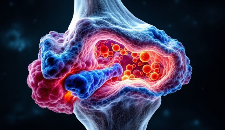
What is Giant Cell Tumor (Osteoclastoma)?
A giant cell tumor (GCT) is a common non-cancerous bone tumor that mostly affects young adults between the ages of 20 and 40. GCTs often return and can sometimes behave aggressively even though they are mostly non-cancerous. This type of tumor is typically found near the ends of the tibia (shinbone) or femur (thighbone), where the bone is either growing or has stopped growing.
These tumors can cause varying symptoms depending on their aggressiveness. They may cause mild symptoms, such as bone damage or the expansion of tissues surrounding the bone. In rare instances, they can spread to other parts of the body. When a GCT forms in areas of the skeleton closer to the center of the body, it can pose a higher risk of severe complications and is often considered too risky to remove with surgery.
When the tissue from a GCT is examined under the microscope, it shows three distinct types of giant cells:
1. Giant cell tumor stromal cells: These come from cells that help form bone (osteoblasts).
2. Mononuclear histiocytic cells: These are a type of immune cell.
3. Multinucleated giant cells: These belong to a group of cells involved in both bone formation and breakdown (osteoclast-monocyte lineage).
Within the tumor, it’s primarily the giant cells that break down (resorb) the bone. The spindle-like stromal cells play a crucial role in attracting immune cells, helping them form giant cells, and enhancing their ability to break down bone. This contributes to how the tumor overall breaks down bone.
What Causes Giant Cell Tumor (Osteoclastoma)?
The exact cause of GCT (Giant Cell Tumor) is still not completely known, and experts are debating whether it’s a real tumor or just a body’s reaction to a certain condition. Notably, a significant change (called 20q11 amplification) is discovered in 54% of GCT cases, and 20% of those cases show over-production of p53, a protein connected to cell growth.
Also, increased centrosome numbers (a part of a cell that helps with cell division) and heightened telomerase activity (which helps protect the ends of chromosomes from deteriorating) prevent shrinkage of telomeres (the ends of chromosomes), suggesting that GCT may have its roots in the abnormal growth of cells, which is what typically characterizes a tumor.
Risk Factors and Frequency for Giant Cell Tumor (Osteoclastoma)
Giant Cell Tumors (GCTs) account for about 4% to 10% of all primary bone tumors and 15% to 20% of benign bone tumors. They commonly affect young adults, with half of these tumors found in individuals in their thirties and forties. People above 50 rarely have these tumors. Females are slightly more likely to have GCTs than males, and they’re more common in Asian populations compared to Western populations.
- Knee joint: 44% of cases
- Distal radius: 10% of cases
- Proximal humerus: 6% of cases
- Hands and feet: 13% of cases
While the spine and skull are rarely affected, when they are, the ala of the sacrum is the most common location in the spine and the vertebral body in the axial skeleton. In the head, the mandible and maxilla are often affected. In the hand, GCTs typically occur in the phalanges.
Even though GCTs are generally benign, they can be locally aggressive and can spread to other parts of the body. Metastasis, or the spread of cancer, occurs in about 1% to 5% of cases. The spread correlates positively with the local aggressiveness and recurrence of the tumor. The lungs are the most common location for these metastases. GCTs may also break through the bone cortex, extend to surrounding tissues and joint structures, causing serious local complications. The recurrence risk is about 35%.
The tumor often occurs suddenly in people under 20. However, it’s quite rare in youngsters with less than 5% of cases being in skeletally immature patients. These patients may have a higher incidence of vertebral GCT and multicentricity, with multifocal lesions tending to be more aggressive than solitary ones. Patients with Paget disease experience a higher occurrence of GCT, often in flat bones such as the skull and pelvis.
Signs and Symptoms of Giant Cell Tumor (Osteoclastoma)
Giant Cell Tumors (GCTs) can show up in different ways. Most commonly, people might experience some of the following symptoms:
- Pain, typically resulting from the tumor destroying a part of the bone.
- Swelling or change in shape of the afflicted area. This generally happens with larger tumors.
- A lump or mass that can be felt. This happens when the tumor breaks through the bone and starts to spread outside. It’s usually found close to a joint and may restrict movement in that area.
- Joint fluid buildup and inflammation, which can also occur.
- Fractures: About 12% of people with GCTs have fractures when they are first diagnosed. The rate of fractures in such cases ranges from 11% to 37%, which suggests that the disease could be more severe with a higher likelihood of returning and spreading.
- Location within the bone: In 90% of cases, the tumors are located in the ends of the bones and can extend towards the joint. This type of tumor rarely invades the joint or its covering. In younger patients whose bones are still developing, tumors are usually found in the metaphysis, the wider part at the end of a long bone. Only around 1.2% of GCTs occur in the metaphysis or diaphysis (the shaft of the bone) without involving the ends of the bone.
GCTs most commonly affect the lower end of the femur, the upper end of the tibia, the lower end of the radius, and the sacrum. Approximately half of all GCTs are found around the knee region. Other locations can include the upper femur, fibular head, and the upper humerus. It is relatively rare for the pelvic bone to be involved.
In a few cases, GCTs can be found in multiple locations at the same time, although these cases are extremely rare. Generally, GCT is a solitary lesion. Multicentric involvement, happening in less than 1% of cases, can be more severe and generally affects the small bones of the hands and feet. These tumors exhibit different characteristics compared to single ones. Patients with multicentric lesions are generally younger than those with lesions in other locations.
Testing for Giant Cell Tumor (Osteoclastoma)
When checking for giant cell tumors (GCT), a mix of lab tests, imaging, and biopsies are used to reach a final diagnosis. Let’s break this down.
Blood Tests
As part of the preliminary check-up before any operation, standard blood tests are carried out. These tests include checking the level of a protein known as acid phosphatase in the blood, as this is usually higher in patients with GCT and can help track how well treatment is working. This is particularly helpful for cases where the tumor comes back. For instance, a study found that a specific type of this protein (tartrate-resistant acid phosphatase 5b) tended to be higher in younger patients and those with fewer changes to their cells resulting from disease. Interestingly, the level of this protein does not always match with the size of the tumor.
Imaging Techniques
X-rays typically show a distinct, radiolucent geographic appearance with a narrow transition zone at the lesion margin, which is the edge of the tumor. Unlike many benign (non-cancerous) lesions, GCT lacks a prominent hardened rim at the lesion margin. The tumor is usually found in the epiphyseal portion (end of the bone) and often extends about a centimeter into the bone directly under the cartilage.
Other imaging methods, like computed tomography (CT) scans and magnetic resonance imaging (MRI), help confirm the location of the GCT within the bone and assess if there is any tissue mass either outside the bone cortex or within the nearby joint. Functional positron emission tomography (PET) scans and bone scans can also help determine how far the disease has spread.
CT scans offer a more accurate view of the thinning and penetration of the cortex and the mineralization (build-up of minerals) in the bone than plain X-rays. The formation of a new cortex and the density of the tumor can be seen, which helps distinguish between a primary osteosarcoma (a type of bone cancer) and other tumors. A chest CT scan may be requested in patients whose disease has returned, to check for the spread of the disease.
MRI is crucial for evaluating the surrounding soft tissues, neurovascular structures, and the extent of subchondral extension into adjacent joints. On MRI, GCT lesions typically show uniform signal intensity, indicating a well-defined lesion. MRI also helps assess soft tissue mass and cystic components (fluid-filled sacs) within the tumor. For the evaluation of residual or recurrent GCT, fat-suppression fluid-sensitive sequences in MRI are useful.
Bone scans help stage multicentric disease (disease occurring in several locations at once), although the findings are not specific to GCTs. GCTs show an accumulation of the FDG tracer, distinguishing it from many benign bone tumors, probably due to the active metabolism of osteoclast-like giant cells. However, the advantages of FDG PET evaluation compared to conventional imaging with CT, MRI, or a bone scan remain unclear.
Biopsy and Immunohistochemistry
Biopsy samples are tested using immunohistochemistry, which helps identify the H3.3-G34W mutation. A mutation is a change in DNA, the building blocks of our cells. This specific mutation is found in patients with GCT, and it helps to differentiate this condition from other tumors that also contain giant cells. Identifying this mutation helps doctors to better understand the molecular characteristics of GCT which can guide therapy decisions and predict the outcome of the condition for patients.
Treatment Options for Giant Cell Tumor (Osteoclastoma)
The treatment of Giant Cell Tumors (GCT), a type of bone tumor, involves a team of healthcare professionals, using a combination of surgery, medication, and sometimes radiation. What the team chooses to do can depend on where the tumor is in the body, how big it is, how aggressive it is, and whether it is the first occurrence or a recurrence.
Surgery is a common treatment for GCT. Often, the type of surgery is tailored to individual patients because most GCTs are non-cancerous, occur near joints in young adults, and the aim is to preserve the body’s bone structure. Generally, an “intralesional” approach, scraping out the tumor from within, is advocated over complete tumor removal to maintain bone integrity.
Although comprehensive tumor removal has been linked to a lower risk of the tumor coming back, it carries with it higher rates of surgical complications. Hence, the decision between scraping the tumor out or removing the tumor whole depends on where the tumor is, its size, how aggressive it is, and the patient’s overall health and preferences.
In some cases, particularly when the tumor involves the end of a long bone and affects joint function, reconstructive surgery may be necessary. Many options are available for these cases, including joint replacement and several types of bone reconstruction procedures.
In the past, GCTs were often treated with amputation, wide resections, or reconstructions. Yet, since GCTs are usually benign but locally aggressive, removing the tumor from within (intralesional approach) is often chosen. Radiation treatment is recommended when complete removal isn’t possible due to surgical risks or patient health. Using liquid nitrogen, phenol, or HO with argon beam coagulation, especially with intralesional curettage, has shown good success in preventing recurrence.
Emerging treatments include the use of bisphosphonates, medications that slow down or prevent bone loss. Another is the monoclonal antibody denosumab, which specifically targets a substance called RANKL, involved in bone breakdown. Denosumab is widely used for GCTs that can’t be surgically removed. However, the optimal dose and treatment duration are still under debate, as extended use could lead to complications.
Finally, there are potential new treatments on the horizon that are being explored, such as Sunitinib, a drug that targets some of the molecular pathways involved in tumor growth, and cyclolinopeptide, a molecule extracted from linseed shown to inhibit some of the biochemical pathways involved in GCTs.
What else can Giant Cell Tumor (Osteoclastoma) be?
When we talk about Giant Cell Tumor (GCT) of the bones, the diagnosis can sometimes be confusing, because there are quite a few other medical conditions that can show similar symptoms or may have similar findings in X-rays. These conditions include, but aren’t limited to:
- Cancer that has spread to the bones (like from thyroid or kidney)
- Other types of bone tumors
- A condition called hyperparathyroidism which can cause ‘Brown Tumors’
- A bone defect called Nonossifying fibroma
- An abnormal, blood-filled bone cyst called an Aneurysmal bone cyst
- Bone defects common in children called Fibrous metaphyseal defects
- Another type of benign bone tumor called Osteoblastoma
- A bone tumor called Chondroblastoma that often affects children and young adults
- A type of cancer called Malignant fibrous histiocytoma
- A rare form of bone cancer called Telangiectatic osteosarcoma.
Looking for mutation in a gene named H3F3A can help doctors differentiate between GCT and the other conditions mentioned above, as up to 96% of GCT cases show this mutation. However, other bone tumors rich in cells that break down bone (the osteoclasts) like Chondroblastoma, Aneurysmal bone cyst, or Nonossifying fibroma may also show mutations in H3F3A. So, it’s important to note that the presence of H3F3A mutation doesn’t rule out all types of malignancy.
Chondroblastoma, in particular, often has mutations in genes associated with histone 3.3, a protein involved in DNA packaging in the nucleus of the cell.
What to expect with Giant Cell Tumor (Osteoclastoma)
GCT, or Giant Cell Tumor, recurrence after 3 years is a rarity, observed only in exceptional cases. The possibility of a locally recurring GCT ranges from 20% to 50%, with an average of 33%. With the help of new curettage techniques – methods to remove tumors – we’ve been able to get better control over local GCT.
A substance called ‘total serum acid phosphatase’ is often used to track a GCT’s response to treatment. Increasing tumor grades, which are used to describe the severity of a tumor, do not necessarily mean the tumor is more biologically aggressive. However, grade III lesions – which are the most severe – have been observed to recur more often.
On the other hand, malignant, or cancerous, transformation of a GCT has been documented, but only in very few instances.
GCT’s spreading into the lungs (also known as ‘pulmonary metastases’) occurs in 16% to 25% of recurrent cases, but only in 1% to 6% of initial cases. Treatments generally include a combination of surgery and interferon alfa – a protein used in cancer treatments -, along with chemotherapy, and radiation. When surgery isn’t possible, the usual alternatives are radiation and chemotherapy.
Bone metastases – or when these giant cell tumors spread to the bones – is a rare event, not seen in more than 3% of cases. But when it comes to those spread to the lungs, prediction becomes trickier. A younger patient, those with reoccurrence, advanced stages of the disease, or involvement of central parts of the body, have higher risk. These lesions when spreading to other parts mostly resemble their original forms.
We’ve learned that the average time from when the tumor starts to when it’s detected in the lungs is about 1.5 to 2 years. Full removal of the tumors has shown success with good chances of long-term survival, although patients with other serious illnesses may still end up succumbing to the disease.
It’s also important to think about an initial detection of a specific kind of bone cancer, bone sarcoma if even a newly discovered GCT shows large areas of giant cell reaction and bleeding. Malignant transformations in GCT can lead to bone sarcomas, malignant histiocytoma, or fibrosarcoma, all of which are very serious. This malignant transformation might only be detected several years after first surgery, sometimes decades later.
However, GCT has a reasonably good outlook overall. Deaths due to the tumors spreading to the lungs range from 16% to 25%. If a GCT becomes malignant, the prognosis becomes grimmer. However, it is still usually somewhat better than for other highly dangerous sarcomas.
Possible Complications When Diagnosed with Giant Cell Tumor (Osteoclastoma)
Giant cell tumors of the bone can lead to several complications such as:
- Reappearance of the tumor
- Swelling and stiffness in the knee joint, akin to osteoarthritis
- Development of stress fractures
- Limited mobility or movement
- Spread of the tumor to the lungs, known as pulmonary metastasis
- Local or deep-seated infections
- Osteomyelitis, which is an infection in the bone
- Joint deterioration
- Failures related to surgical implants or hardware
Preventing Giant Cell Tumor (Osteoclastoma)
The exact reason why GCTs, a type of tumor, occur is still unknown, just like most other tumors. This means we don’t have known ways to prevent them. However, it’s very important that patients know about the possible signs of the tumor growing into nearby areas or spreading to other parts of the body. This knowledge can encourage patients to seek medical help early on, and catching the tumor at an early stage often leads to better treatment outcomes.
Another important aspect of patient education is knowing about the different surgical treatments available. This information can really empower patients, as it allows them to make informed choices about their own health care, which can greatly improve the results of their treatment. Patients’ awareness and active participation in their own care are key factors in managing GCTs and making the treatment process as effective as possible.


