What is Jumpers Knee?
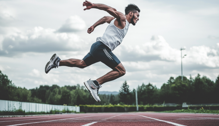
“Jumper’s knee,” also known as patellar tendinopathy, is a painful knee condition caused by small tears in the patellar tendon, the tissue that connects the kneecap to the shinbone. It’s mainly related to activities and is commonly seen in sports that require a lot of jumping, leading to knee tendon damage. This includes sports like volleyball, track, basketball, long-distance running, and skiing, which place high demands on leg muscles.
Jumper’s knee is more common in males, and it often occurs in adolescents and young adults. Despite the name, “jumper’s knee” does not involve any inflammation in the knee tendons. In fact, research over the past 40 years shows that jumper’s knee is a degenerative condition, meaning it stems from wear-and-tear over time.
Diagnosing jumper’s knee involves taking a detailed medical history and performing a physical exam. An ultrasound, which is widely available and affordable, may also be used to aid diagnosis.
Treatment largely involves reducing activities that put heavy strain on the knee. When the pain lessens, physical and exercise therapy can help restore function. Surgery is typically considered as a last resort for cases that don’t improve with these other treatments.
What Causes Jumpers Knee?
Jumper’s knee is a common injury among athletes, often caused by excessive stress on the knee due to activities like jumping, landing, and changing direction quickly. This injury usually results from small tears developing in the knee extensor tendons, which are the bands that connect your knee muscles to your bones. These micro-tears can occur when these movements are repeated too often in one workout, or if you don’t allow enough rest time between workouts.
The part of the knee most commonly affected is the lower part of the kneecap, or patella, where the patellar tendon attaches. Sometimes, the injury can also occur at the top part of the kneecap, where the quadriceps tendon (the tendon connecting the muscles at the front of your thigh to the kneecap), attaches, or at the point where the patellar tendon connects to the shinbone.
For convenience, we often refer to this condition as patellar tendinopathy, which is a term that more accurately describes the damage to the tendon rather than inflammation. This label is more accurate because this condition is primarily about damage to the tendon, not inflammation.
There are several factors that could make someone more likely to develop jumper’s knee. This could be due to loose ligaments, tightness in the muscles on the front or back of the thigh, an unusual angle of the knee, abnormal knee height, ongoing inflammation, and putting too much force on the knee. Workout habits, like training too much, how well the athlete performs, and the hardness of the surface where the sport is played, could also lead to jumper’s knee. Other risk factors include the athlete’s weight, body mass index, ratio of waist to hip, difference in leg length, the height of the arch in the foot, strength of the thigh muscles, and how well they can jump vertically. These factors can increase the strain on the patellar tendon.
Risk Factors and Frequency for Jumpers Knee
It’s challenging to find the exact number of knee tendon issues, officially called patellar tendinopathy, as sporting injuries are often under-reported. Despite this, we know that the condition is common in sports that involve a lot of jumping, such as volleyball, basketball, and jumping sports.
Research has shown us that professional athletes experience this issue more widely than those who play sports recreationally. The age range that this condition affects extensively runs from teenagers to people who are in their thirties. It’s also noticed that males suffer from this problem more frequently.
- About 45% of professional athletes in jumping sports might experience symptoms of jumper’s knee at some point in their careers.
- Similarly, up to 14% of recreational athletes may also face the same situation.
Signs and Symptoms of Jumpers Knee
Patellar tendinopathy is a condition typically diagnosed through a careful medical history and physical examination. The patient may be involved in sports and can describe their training schedule, competition frequency, their role in the sport, and performance level. Commonly, they will describe a pinpoint pain and sensitivity at the bottom of the kneecap.
This condition can share symptoms with other knee-related problems, such as pain when sitting for extended periods, squatting, or climbing stairs. Known familiarly as the “Movie Theatre sign,” patients may also experience discomfort during activities involving prolonged bending of the knee. Quick, sharp bouts of tendon pain that occur under strain and generally stop as soon as the strain is removed can also be a sign. However, pain at rest is rare.
An examination could reveal any swelling located above the patellar tendon, which may be tender to the touch. Two signs could help identify this condition:
- “Passive extension-flexion sign”: The doctor will feel the front of the fully extended knee to find the most sensitive point, typically found at the bottom of the kneecap or the upper part of the patellar tendon. The examination will be repeated with the knee bent at 90 degrees. If there’s a sharp decrease in sensitivity when the knee is bent, then it shows this sign.
- “Standing active quadriceps sign”: The medical professional will feel all of the patellar tendon while the patient stands. The examination is repeated while the patient stands on the involved leg with the knee slightly bent at 30 degrees. A significant reduction in tenderness when the thigh muscle is contracted can be seen as this sign.
Other important factors can include alignment issues involving the foot, heel, or shin bone as these can put more strain on the knee extensor tendons, increasing the risk of developing tendinopathy.
The recovery and severity of tendon overuse can be assessed using certain scales, with the Victorian Institute Sports tendon Assessment (VISA) score being one of the more reliable ones. This questionnaire evaluates symptoms, function tests, and the ability to play sports, offering a useful insight into patellar tendinopathy.
Testing for Jumpers Knee
At present, there is no universally agreed-upon best method to diagnose certain issues affecting the knee. Ultrasound is often preferred because it’s quick, affordable, non-invasive, accurate, and provides a live picture of the knee structures. Both ultrasound and the more advanced Magnetic Resonance Imaging (MRI) can be used to identify abnormalities in the patellar tendon, which is a key structure in your knee. These imaging tools also help doctors understand how serious the issue is.
Simple X-ray images, taken from different angles, are often the first step to rule out any immediate bone injuries or diseases. You might see changes on the X-ray images of the tendon, like elongation, calcification, which is a build-up of calcium salts, an inferior traction spur (also known as an enthesophyte) in long-term cases, or higher density within the tendon itself. However, these changes are rare in the first six months of symptoms.
If the condition becomes chronic or if surgery is considered, an MRI scan may be required. MRI can reveal a thickening of the patellar tendon, which is a more clear sign than swelling. The MRI could also show intense signals both in T1 and T2 images. For long-term cases, MRI scans may show that the back of the fat pad, a soft structure behind the tendon, is missing. Generally, it is the upper part of the tendon which is affected and thickened.
Although it’s not usually necessary, a bone scan can be used in the early stages of the disease. This test shows increased blood flow and specific tracer activity, which means the scanner specifically detects changes, in the affected region.
Treatment Options for Jumpers Knee
There’s not a one-size-fits-all treatment for “jumper’s knee”, also known as patellar tendinopathy. This condition is commonly stubborn and often has patients and healthcare providers exploring different treatment options.
Early diagnosis and treatment of jumper’s knee is extremely important as it can worsen over time. Traditionally, doctors prescribed medications to reduce inflammation, like non-steroidal anti-inflammatory drugs (NSAIDs), but it’s been discovered that the condition might not exactly be due to inflammation, so these drugs might not offer any long-term relief. Also, corticosteroid injections, used to reduce inflammation, are not advisable because they could potentially cause the patellar tendon to rupture or tear.
Instead, non-surgical treatment is usually recommended initially, focusing on the following: A period of ‘relative rest’ where the knee is not immobilized, helping to avoid the weakening of the tendon and muscle. Appylying cold to the area (cryotherapy) provides pain relief and opposes the growth of new blood vessels, which contributes to the problem. Modifying activities and sports training to include sufficient warm-up exercises and physiotherapy also helps to make the quadriceps and hamstring muscles more flexible.
One type of exercise that could help is eccentric training, an exercise strategy that has been found to potentially provide same benefits as surgical treatment, in managing jumper’s knee. It is typically advised to try eccentric training for twelve weeks before considering surgical treatment. As the patient starts experiencing less pain, they can slowly increase the intensity of their physiotherapy and sports-specific training.
Pain and pressure on the patellar tendon can also be minimized using supportive straps (also known as infrapatellar straps). These straps adjust the angle between the kneecap (patella) and patellar tendon, reducing strain.
In response to the stubborn nature of jumper’s knee, recent advancements in treatment have brought new methods, such as dry-needling, injection therapies including platelet-rich plasma and Aprotinin (an inhibitor of matrix metalloproteinases), extracorporeal shock wave treatment, and hyperthermia thermotherapy. There is also a three-stage rehabilitation protocol focusing on pain reduction then progressive loading. The three stages include pain and load management, strenghthening exercises and load progression, and finally functional strengthening and return to sports.
Surgery is typically reserved for severe cases, such as partial tendon tears, or if pain persists even during rest and normal activities. It is also considered when other treatments have not provided relief. Earlier surgical approaches mostly involved the removal of unhealthy tissue from the lower part of the kneecap and from the patellar tendon, and then reattaching the tendon with sutures or anchors. Nowadays, knee arthroscopy (a minimally invasive surgical procedure using a small camera) is gaining popularity for the treatment of jumper’s knee as it allows for the same tissue removal and repair through smaller incisions.
What else can Jumpers Knee be?
Jumper’s knee, also known as patellar tendonitis, can sometimes be confused with other injuries or conditions. These include:
- Osgood-Schlatter disease
- Meniscal injuries
- Patellofemoral syndrome
- Quadriceps injury
- Knee bursitis (a condition affecting the fluid-filled sacs around the knee joint)
- Osteochondritis dissecans (a condition that can cause joint pain and stiffness)
- Patellar subluxation (partial dislocation of the kneecap)
- Problems with the knee fat pad
- Chondromalacia (breakdown of cartilage under the kneecap)
- A patellar tracking problem (where the kneecap moves out of position during activity)
Because these conditions can have similar symptoms to jumper’s knee, it’s important for healthcare professionals to perform correct evaluations and tests for an accurate diagnosis.
What to expect with Jumpers Knee
Most people suffering from patellar tendinopathy, which is an injury or damage to the tendon connecting the kneecap to the shinbone, can recover without surgery. However, even with treatment, mild to moderate knee pain may continue for as long as 15 years in adult athletes who have this condition, though it usually doesn’t limit their physical activities.
According to experts like Rudavsky and Cook, it often takes a long time for athletes with this condition to return to their sport. This recovery time depends on several things, including how severe the pain is, how severe the dysfunction is (how much it impacts the normal function of the knee), what sport they play, how good their rehab is, their performance level, and other internal and external factors.
Research shows that an athlete with a mild injury might need anywhere from 20 days to start playing sports again, but more severe cases might need up to 90 days. In the most severe cases, the recovery period could be 6 to12 months. In a study with patients treated with surgery, it took on average 4 plus or minus 3 months to return to their sport.
In terms of treatment, Joshua and team conducted a research review and found that eccentric squat-based therapy (a type of progressive rehabilitation process which involves strengthening the muscles surrounding the knee), shockwave therapy (a non-surgical technique using sound waves), or Platelet-Rich Plasma (PRP, a method that uses a concentration of the patient’s own platelets to accelerate the healing of injured tendons) can be effective in speeding up recovery. If these treatments don’t work after six months, surgery or shockwave therapy should be considered. Since patellar tendinopathy is not caused by inflammation, corticosteroid injections (a type of anti-inflammatory medication) should not be used.
Unfortunately, patellar tendinopathy can sometimes be so severe that it leads to athletes retiring early from their sports. In a small study, it was found that 53% of athletes with a condition called “jumper’s knee,” a type of patellar tendinopathy, quit their sport due to the symptoms, compared to only 7% of those without symptoms.
Possible Complications When Diagnosed with Jumpers Knee
People involved in sports such as athletes, trainers, coaches and healthcare professionals should be aware that healing from patellar tendinopathy, an injury to the tendon connecting your kneecap to your shinbone, can be a slow and sometimes challenging process. There can be several hitches along the way, particularly with managing pain.
An athlete’s own understanding and perception of their pain and potential damage to their tendons can affect their recovery process for unresponsive tendon issues. For example, if an athlete has been told that their tendons are weakened due to tears and degeneration, and that there’s a high risk of full rupture, this could cause them to start avoiding actions that they fear may cause harm, which can worsen their functional progress once they are affected by lower-limb tendinopathy.
Relying too much on non-invasive treatments, such as shockwave therapy or injections, without incorporating rehabilitative exercises into the treatment plan, can also lead to complications. Ignoring how an athlete lands after a jump during their sport could also cause issues. Once an athlete has undergone adequate rehabilitation, they should be coached on how to relearn the mechanics of jumping and landing.
Important points to remember:
- Healing from patellar tendinopathy can take time
- Managing pain is crucial in the process of recovery
- Athletes’ understanding of their condition and pain can influence the treatment process
- Over-reliance on non-invasive therapies can lead to complications
- Rehabilitative exercises are a crucial part of the treatment plan
- Jump-landing mechanics should be reviewed and corrected after rehabilitation
Recovery from Jumpers Knee
If you have a procedure to clean and fix your patellar tendon (a tendon in your knee), you’ll need to keep your leg fully extended for two weeks. This means you need to keep it straight, without bending it at the knee. After this two-week period, you can begin rehabilitation, which includes a gradual increase in movements and exercises to help your knee recover. You’ll also be allowed to put as much weight on your leg as you comfortably can from the first day post-surgery.
Preventing Jumpers Knee
Currently, there isn’t enough scientific evidence to support specific preventative interventions for a condition called patellar tendinopathy – a type of knee pain often referred to as “jumper’s knee”.
However, some commonly used approaches include static stretching (holding a stretch for a set amount of time), training that focuses on strengthening the body’s core muscles, the use of foot orthotics (devices inserted into the shoes to correct foot alignment), shock-absorbent insoles, and hormone replacement therapies for women.
Additional proposed prevention strategies include proprioception training (exercises designed to improve the awareness of the position of one’s body), eccentric training (types of exercises that focus on strengthening muscles as they lengthen), stretching of the tendon and lower leg muscles, cutting back on the length or frequency of training sessions, and using braces or tape.
Meanwhile, targeted balance training (enhancing stability) tailored specifically for soccer can potentially reduce the occurrence of patellar tendinopathy. The impact of this training seems to be linked with how often it’s done – a more pronounced impact was observed with more frequent training.


