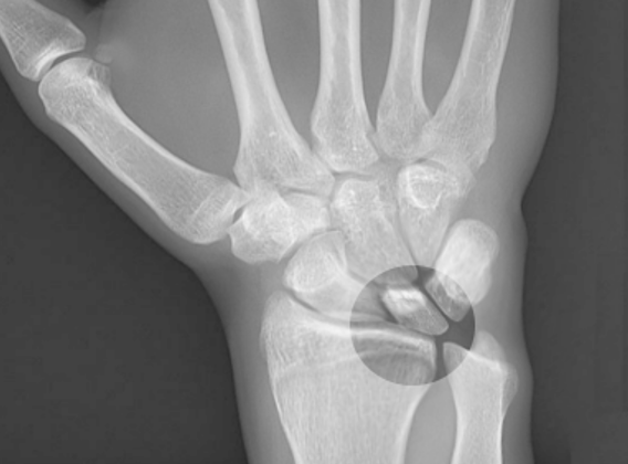
What is Kienbock Disease?
Kienböck disease is a condition that involves the deterioration of a specific bone in the wrist called the lunate carpal bone, due to a lack of blood supply. This is also known as lunatomalacia. It was first identified and named by an Austrian radiologist named Robert Kienböck in the year 1910.
To better understand this, let’s talk about the structure of the wrist: The middle joint of the wrist (or mid-carpal joint) is mainly responsible for wrist movement. This is different from the part of the wrist farthest from the body (the distal carpal row) which is relatively stable and does not move much. The lunate is positioned centrally in the part of the wrist closest to the body (proximal row), where it connects with other bones including the scaphoid, capitate, triquetrum, sometimes the hamate, and the radius (arm) bone via a structure called the triangular fibrocartilage complex (TFCC). Notably, about 10% of the force from the arm is transferred through this structure to another bone called the ulna, and 35% through the connection between the radius and the lunate.
What Causes Kienbock Disease?
Kienböck disease is a complicated health issue with no single agreed cause. It seems to happen due to several different factors:
– Ulnar negative variance or ulna minus: This is a fancy way of saying that the ulna (one of the two bones in your forearm) is shorter than the radius (the other forearm bone). As a result, the lunate bone (a small bone in your wrist) has to deal with more stress and minor damages because of the longer radius. In some studies, up to 78% of people with Kienböck disease had a shorter ulna.
– Blood supply to the lunate bone: This bone in your wrist gets blood from different arteries that come off two loops of blood vessels in the wrist. Sometimes, there aren’t enough connections between these blood vessels inside the lunate bone. If the number of these connections, particularly on the under (or palmar) side of the wrist, is low, it makes it more likely that you can develop this disease.
– Lunate size and shape: Researchers found that people with smaller lunate bones are at higher risk of Kienböck disease because the smaller bone has to support more weight. The bone can be square, rectangular, or triangular. The triangular shape, which doesn’t have a medial joint part, has a weaker inner structure and is more prone to Kienböck disease.
– Radial inclination angle: This is an angle measured in the wrist. If this angle becomes smaller, it increases the chances of Kienböck disease.
Risk Factors and Frequency for Kienbock Disease
Kienböck disease is quite prevalent, being the second leading cause of a medical condition known as avascular necrosis affecting the wrist bones. The only type of avascular necrosis of the wrist more common than Kienböck disease is that which affects the scaphoid bone. This disease typically impacts men between the ages of 20 and 40.
Signs and Symptoms of Kienbock Disease
Patients commonly show up with pain on one side of the back part of the wrist, restricted movement in the wrist, weakness, or a mix of these. Their pain often gets worse when they extend the wrist or put weight on it. The symptoms can vary from mild to severe. It’s rare that both wrists are affected, and often, there’s no history of injury.
- Pain on one side of the back part of the wrist
- Restricted wrist movement
- Weakness
- Pain worsens when extending the wrist or putting weight on it
- Symptoms vary from mild to severe
- Rarely affects both wrists
- Often no history of injury
- Wrist swelling
- Tenderness over the lunate region of the wrist
- Synovitis (inflammation of the synovial membrane)
- Loss of grip strength
Testing for Kienbock Disease
Kienböck’s disease is a condition that can be diagnosed through clinical examination and imaging techniques. Both X-ray/CT scan and MRI can be used for the identification of this disease. However, the MRI is considered the most sensitive and can detect cases that might not be apparent through X-ray.
An MRI involves creating detailed images of the lunate bone, a small bone that is part of the wrist. In patients with Kienböck’s disease, the MRI usually shows a decrease in the bone marrow signal on images called T1-weighted images. This is a characteristic feature of the disease. Other types of images, such as T2-weighted images or short-TI inversion recovery (STIR) images, show changes that vary depending on the severity and extent of bone death (osteonecrosis). MRI can also evaluate the health of the cartilage in the joint.
X-rays may appear normal in the early stages of this disease. But as the disease progresses, X-ray findings may include a variety of changes such as: general thickening of the lunate bone, formation of cyst-like structures, collapse of the joint surface, hand bone (carpal) collapse, and signs of arthritis in the wrist. The lunate bone can also develop fractures.
CT scans are helpful to plan surgery, as they are more sensitive than X-rays for detecting smaller fractures, fragmentation, instability of the hand bones, and the extent of disruptions within the bone’s structure. Patients’ disease stage is often revised after a CT scan due to more detailed findings.
Nuclear scintigraphy, a form of imaging that uses small amounts of radioactive material to diagnose diseases, used to be used as a supportive diagnostic tool for early-stage Kienböck’s disease. However, with the advent of MRI, this method is not commonly used anymore.
Treatment Options for Kienbock Disease
The main aim of treating Kienböck disease, a condition that affects the wrist, is to relieve pain, preserve the movement of the wrist, and maintain grip strength.
The treatment for this disease depends on what stage it’s at and what has caused it. If it’s caught in the early stage (Stage I), treatment usually involves wearing a cast or splint to immobilize the wrist.
Stage II can also use a similar approach, only if the bone is not fully dead (incomplete necrosis). In cases where the bone is fully dead (necrosis is complete), or if the disease is in the later stages (Stage III and IV), more advanced treatments are needed. These treatments include “joint-leveling” surgery, a process to help balance the length of the bones in the wrist. This may be carried out alongside a procedure to supply blood to the dead wrist bone by using grafts or branches from nearby arteries.
If the disease has advanced further to cause the lunate bone (one of the small bones in the wrist) to collapse or for the wrist to degenerate, more complex surgery may be required. For example, surgery might aim to remove part of the wrist bones (proximal row carpectomy) or join them together (intercarpal arthrodesis). If the disease is accompanied by an uncommon condition where the bone of the forearm (ulna) is unusually short, a procedure to reduce the length of the other bone in the forearm (radius) may be performed to ease the pressure on the lunate bone.
While these treatments can help improve symptoms and function of the hand, they may not always alter the changes seen in medical imaging in the later stages of the disease.
What else can Kienbock Disease be?
When diagnosing conditions like Kienböck disease, there are several other diseases that could show similar symptoms. These conditions need to be ruled out:
- Ulnar Impaction Syndrome: often seen in individuals having a relatively long ulna. This condition is similar to Kienböck disease but impacts the ulnar head and triquetrum, which are not typically affected in Kienböck patients.
- Lunate Intraosseous Ganglion: This condition appears on MRI scans as small cysts within the bones. These cysts can be differentiated from Kienböck disease by their sharp, smooth borders in MRI and radiography images.
- Bone Contusion: Early-stage Kienböck disease may be hard to separate from a bone bruise on an MRI scan. A history of recent trauma or associated wrist or hand injuries would help make the correct diagnosis.
- Arthritis: Arthritis can also mimic Kienböck disease, with similar bone marrow changes seen in MRI scans. However, the patient’s age, symptoms, and absence of a particular wrist feature called ‘ulna minus’ can help separate arthritis from Kienböck disease.
- Osteoid Osteoma: This is a rare condition that can affect the wrist bones. It can be differentiated from Kienböck disease by the patient’s symptoms and by an imaging finding of a clear area within a dense outer rim seen in CT scan.
- Enostosis/Bone Island: This condition shows a maintained structure and the area appears to be star-shaped and intertwined with the normal bone tissue in imaging results.
Identifying the correct diagnosis out of these conditions is essential for appropriate treatment. Proper understanding of symptoms and the usage of diagnostic tools like MRI and CT scans are invaluable.
What to expect with Kienbock Disease
Kienbock disease tends to get worse over time, and it can cause damage to the joints within 3-5 years after it starts. The future health of someone with Kienbock disease depends on several things:
– The Functional Stage: This refers to how much of the bone is still healthy. The more healthy bone there is, the better the outlook.
– Negative ulnar variance: This is a term that refers to the length of the ulna (one of the forearm bones) in relation to the radius (the other forearm bone). If the ulna is shorter than the radius (negative variance), the disease tends to be more severe and is more likely to get worse.
– Age at diagnosis: People who get diagnosed later in life tend to have a more advanced stage of the disease and are more likely to see it get worse.
It’s important to remember, though, that the severity of symptoms doesn’t always match up with the stage of the disease based on its physical characteristics.
Possible Complications When Diagnosed with Kienbock Disease
Kienböck disease can result in a number of complications. These include:
- The separation of the scaphoid and lunate bones in the wrist (known as scapholunate dissociation)
- Wearing down of the wrist joints (also called secondary radiocarpal and mid-carpal degenerative arthrosis)
- Misalignment of the triquetrum bone, one of the small wrist bones


