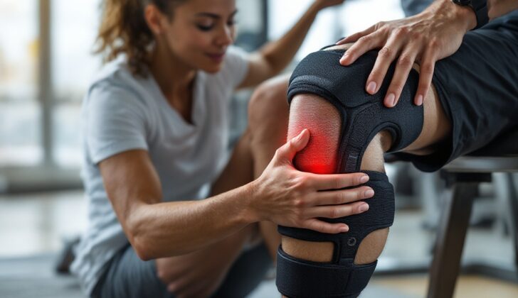
What is Knee Extensor Mechanism Injuries?
Knee injuries, especially those that affect the extensor mechanism, are quite common and can impact a person’s normal daily activities, walking, and sports participation. The extensor mechanism is the part of the knee that helps in straightening or extending the leg. There are two ways these injuries can happen – through trauma (a sudden, severe event like a fall or accident) or non-traumatic causes (slow wear and tear over time).
The knee’s extensor mechanism consists of specific parts – the quadriceps muscles, the patella (knee cap), and the patellar tendon (a strong band of tissue that connects the kneecap to the bones of the lower leg). The quadriceps consist of four muscles named the rectus femoris, vastus lateralis, vastus intermedius, and vastus medialis, which originate from different areas around the hip and thigh bone. These muscles come together to form the quadriceps tendon, which is attached to the knee cap. Contraction of these muscles helps straightening the lower leg. The kneecap rests within a groove of the thigh bone and acts as an anchor point for the tendons of the quadriceps. The patellar tendon starts from the bottom of the kneecap and attaches to the shin bone.
There are certain ligaments known as retinacula that provide stability to the knee. These are fibrous bands from the quadriceps and are divided into medial (inner) and lateral (outer) portions. These include several specific ligaments that connect different parts of the knee.
There are also soft tissues like fat pads and fluid-filled sacs known as bursae around the knee. The bursae help reduce friction and aid smooth movement. Blood reaches the structures of the knee through certain arteries, and nerves in the area help convey messages to and from the brain.
Understanding all these aspects of the knee is crucial to diagnosing and treating both traumatic and non-traumatic injuries that can affect your day-to-day activities and quality of life.
What Causes Knee Extensor Mechanism Injuries?
There are certain things that can trigger a higher chance of developing this disease. These are divided into two categories: factors that weaken collagen, a protein that helps form the structure of our skin, and injuries to the local area.
Certain diseases and conditions can cause weakness in the collagen. These include illnesses like systemic lupus erythematosus (SLE), a condition where the body’s immune system mistakenly attacks healthy tissue, and rheumatoid arthritis (RA), a chronic inflammatory, autoimmune disorder that mainly affects the joints. Other conditions include chronic kidney disease, diabetes mellitus, hyperparathyroidism (an overload of parathyroid hormone), and long-term use of steroids and a type of antibiotics called fluoroquinolones.
Certain genetic disorders that affect connective tissues, like ‘hypermobility syndrome’, Ehlers Danlos syndrome, and Marfan syndrome, can also make people more prone to extensor injuries. Extensors are muscles that help to straighten joints. People who undergo a treatment that removes waste products from the blood, called hemodialysis, are most likely to experience tendon degeneration, which can lead to rupture.
Local injuries that can trigger the disease are due to instability in the kneecap, known as patella, difficulty with the patella, or its degeneration. The instability can be there from birth or result from differences in anatomy. The most common causes of instability are a titled kneecap, a high-lying kneecap, underdevelopment of the groove in the lower part of the knee, or weakness in a thigh muscle called Vastus medialis. Overuse or repeated injuries can also lead to issues with tendons and their eventual breakdown.
Risk Factors and Frequency for Knee Extensor Mechanism Injuries
Quadriceps and patellar tendon ruptures are quite rare occurrences, but they still happen. The former is more common in men over 40, whereas athletes under 40 are more prone to the latter, which requires a strong force. Patellar fractures, which make up 1% of all bone injuries, also occur often due to a heavy blow, like from a car crash or a fall, and are most prevalent in 20 to 50 year-olds, twice more so in men. While closed fractures are more frequent, open fractures can still be found in 7% of these cases.
Patellar dislocations, which account for 3% of all knee injuries, happen most in patients under 20, including dancers, other sportspeople, and military personnel. These dislocations can be due to a direct impact or indirect hit on a knee with unstable patella. It’s also important to know that the risk of dislocation lessens as people age.
- Patellar subluxation is actually a slightly dislocated patella, but it’s more common than a complete dislocation, plus it can fix itself over time.
- Osgood-Schlatter disease affects the tibial tubercle, commonly seen in boys during their teenage years. It often starts showing symptoms alongside an increase in physical activity or a sudden growth spurt. In general, girls will see it emerge between 8 to 13 years of age, whereas boys may experience it between 10 to 15 years due to later maturation of their growth plate.
- Idiopathic chondromalacia patellae (ICP), also known as patellofemoral syndrome or pain, is the most typical cause of knee pain in young athletes. It’s classified as an overuse injury and is seen more often in women (two to ten times the rate in men), runners, and obese patients. Also, nearly 30% of all adolescents suffer from this idiopathic knee pain.
Signs and Symptoms of Knee Extensor Mechanism Injuries
If you suspect you have a knee injury, a doctor will often carry out a physical examination. This includes checking your leg’s movement, feeling for swelling or other changes, and testing your strength and circulation. They’ll also look at the overall shape and condition of your knee, for any signs of bruising or open fractures, and whether the kneecap looks symmetrical compared to your healthy knee.
If your quadriceps or patellar tendon is completely torn, you might not be able to lift your straightened leg against gravity. If it’s only partially torn, extending your knee could be difficult and painful.
In the case of a broken kneecap, or patellar fracture, you may have pain when the kneecap is touched, swelling of the knee, limited movement, and difficulty extending your leg. Your kneecap could also sit too high or low.
If your kneecap is dislocated, it would usually shift to the outside of your knee, limiting the movement and extension of your knee. If you’ve had previous dislocations or injuries to the kneecap, that increases the possibility of this happening. Swelling of the knee could mean the injury is more complicated, such as an injury that involves cartilage. It’s crucial for the doctor to check your lower leg’s pulse to make sure blood flow isn’t affected.
For non-traumatic knee pain conditions like Osgood-Schlatter and idiopathic chondromalacia patellae, patients will often experience prolonged pain in the front of their knee, especially during physical activity and knee extension. The pain usually improves with rest. For idiopathic chondromalacia patellae, symptoms such as pain when standing up from prolonged sitting are common.
Osgood Schlatter patients typically experience tenderness at the point where the kneecap’s tendon attaches to the shinbone, and knee bending or restricting knee extension can trigger symptoms. Usually, the doctor can diagnose this condition by examination alone, without needing imaging tests.
Testing for Knee Extensor Mechanism Injuries
If you are experiencing knee pain, your doctor will likely start with taking X-rays. These images are taken from different angles, including the front-back (anterior-posterior or AP), the side (lateral), and a diagonal angle (oblique). Additionally, a specific X-ray called the “sunrise view” might be taken to get a better look at the kneecap (patella). These images can help your doctor identify things like fractures or certain abnormalities.
Sometimes, X-rays can show “avulsion fractures,” where a piece of bone is torn off due to a torn tendon or ligament. The X-rays can also show a buildup of fluid (effusion), or changes in the knee cap position, known as patella alta (high kneecap) or baja (low kneecap). However, in some cases, X-rays might not reveal any abnormalities, even when there’s a problem.
Another option for initial imaging is the point-of-care ultrasound, which could be especially helpful if a tendon injury is suspected. The ultrasound can provide additional details about the injury. It can show whether a tendon tear is partial or complete, and the ultrasound’s color Doppler feature can even reveal increased blood flow, which is common in injuries.
In certain situations, other imaging techniques, such as a computed tomography (CT) scan or magnetic resonance imaging (MRI), may be needed. A CT scan can be particularly useful when there’s a suspected fracture as it offers a detailed view of the bone structure. It’s often used to plan surgery. Usually, though, a CT scan isn’t necessary. The role of the MRI is to reveal injuries to the extensor mechanism – a group of structures that help extend the knee. It helps in understanding whether a tear is partial or complete and assists the surgical team in planning their approach. An MRI can also detect fluid buildup (effusion) that signifies an injury within the joint.
Treatment Options for Knee Extensor Mechanism Injuries
If you have an injury affecting your ability to straighten your knee, it’s important to consult with an orthopedic specialist, as surgery may be required. Early diagnosis and treatment often lead to a good outcome. Depending on your individual condition and the severity of your injury, your treatment may vary.
For young, active patients with partial injuries to the extensor, which is the group of muscles that help to extend or straighten the leg, early surgical repair can be beneficial to restore function. However, for older patients with other serious health conditions, it may be possible to manage the injury without surgery. This usually involves wearing a knee brace to keep the knee steady, combined with a plan to gradually increase knee movement and weight-bearing over time. Regardless of the treatment approach, full recovery usually takes about six months of extensive rehabilitation.
When it comes to fractures of the kneecap, the treatment plan is influenced by whether the extension function of the knee is still intact and what kind of fracture it is. Stable fractures that have no displacement and retain the knee’s extension ability can usually be treated conservatively. This can be done by wearing a knee brace and gradually increasing knee flexion and weight-bearing in stages. However, some fractures, especially those that are unstable or interfere with the knee’s extension, may require surgical repair.
For cases of patellar dislocation, when the kneecap moves out of its normal position, if it hasn’t relocated naturally, it can sometimes be repositioned based on a physical examination alone. If there are any concerns about associated bone injuries or if the patient is elderly, an X-ray may be needed before repositioning. In some cases where the kneecap can’t be relocated, surgery may be necessary. If the injury is successfully managed initially, the patient will typically be given a knee brace to wear and be advised to see an orthopedic or sports medicine specialist in 1 to 2 weeks. Those with constantly recurring kneecap dislocations may need surgery to regain stability, such as MPFL (Medial Patellofemoral Ligament) reconstruction or graft.
Osgood Schlatter disease and Infrapatellar Contracture Syndrome (ICP) are conditions resulting from overuse of the tendons in the knee, and patients are generally advised to rest and avoid strenuous physical activities. Other recommendations may include applying ice to the affected area, taking NSAIDs (Non-Steroidal Anti-Inflammatory Drugs) for pain and inflammation, and undergoing physical therapy to strengthen the associated muscles. Jumping mechanics may need to be corrected in athletes who demonstrate abnormal bending or bow-leggedness.
What else can Knee Extensor Mechanism Injuries be?
When it comes to knee pain, the causes can be diverse. These causes can be sorted into two categories: injuries that are caused by trauma and those that are not.
Here are some examples of knee issues that can manifest as a result of trauma:
- Fracture
- Tendon rupture
- Patellar subluxation (or knee cap dislocation)
- Patellar dislocation (where the kneecap moves out of its normal position)
There are also non-traumatic causes of knee pain, such as:
- Osteoarthritis (a type of joint disease)
- Osgood-Schlatter Disease (a condition that often occurs in kids experiencing growth spurts)
- Idiopathic chondromalacia patellae (a condition where the cartilage within the knee deteriorates and softens)
What to expect with Knee Extensor Mechanism Injuries
Generally, patients say they recover better and are pleased with the results when injuries to their knee extensor mechanism, which is essentially their kneecap and its supporting structures, are treated promptly. However, delayed treatment, late diagnosis, or mistakes made during surgery can be linked to poorer results.
Possible Complications When Diagnosed with Knee Extensor Mechanism Injuries
The complications from delayed diagnosis or treatment for knee problems vary based on the severity of the damage. These complications include:
- Instability in walking patterns
- Osteoarthritis, a condition that affects your joints
- Difficulty in extending the knee
- Possible infections
- Injuries inside the joint
Preventing Knee Extensor Mechanism Injuries
Patients should be informed that most knee extensor injuries, which are injuries to the muscles at the front of the thigh that help in straightening the leg, usually have a good outcome. It’s important for patients to follow the recommended rehabilitation plan. This includes how much weight they can put on the healing leg and the type of physical activities they can do. By sticking to these guidelines, they can support their recovery process and help avoid any setbacks.


