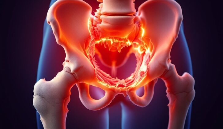
What is Legg-Calve-Perthes Disease (Perthes Disease)?
Legg-Calve-Perthes disease, often simplified as LCPD, is a condition that causes the bone in the hip to break down due to a lack of blood supply. This break down affects the upper part of the thigh bone, called the ‘femoral head’. This condition was first described by three doctors – Arthur Legg, Jacques Calve, and Georg Perthes – in 1910. To explain further, the disease can also be referred to as coxa plana, Legg-Perthes, Legg Calve, or just Perthes disease. It’s important to understand the name is not what matters, rather recognizing the condition itself.
What Causes Legg-Calve-Perthes Disease (Perthes Disease)?
The exact cause of Legg-Calve-Perthes disease, a condition that affects the hip joint in children, is not clearly understood. It might occur on its own or it could be caused by numerous other factors that interrupt the blood flow to the ball-shaped upper part of the thigh bone (femoral epiphysis). Such factors could include major or recurrent minor injuries, an impairment of the body’s ability to form blood clots (coagulopathy), or the use of steroids.
About half of the patients with this disease have blood clotting disorders (thrombophilia), and as many as three quarters show some kind of blood clotting abnormality. Blood clotting disorders can interfere with normal blood flow, which could potentially explain why they are seen so often in this condition.
Risk Factors and Frequency for Legg-Calve-Perthes Disease (Perthes Disease)
Legg-Calve-Perthes disease is typically seen in children aged between 3 to 12 years, with the highest rate seen in kids aged 5 to 7 years. It is quite rare, affecting 1 in 1200 children under the age of 15. This disease is more common in boys, with boys contracting the disease 4 to 5 times more often than girls. In 10% to 20% of cases, it can occur in both hips but the disease usually affects each hip in varying degrees. If it affects both hips equally, other conditions might be at play. This disease occurs more frequently in White and Asian children. It is also seen more often in urban areas among children with lower socioeconomic status.
There are several risk factors for Legg-Calve-Perthes disease, including:
- About 10% of cases have a family history and show a delay in bone development by around 2 years.
- Up to 5% of HIV patients can have this disease.
- The presence of inherited clotting disorders like Factor V Leiden.
- Problems with increased clotting or reduced ability to break down clots.
- Exposure to secondhand smoke (this increases the odds by five times).
- Having a low socioeconomic status.
- Boys weighing less than 2.5 kg at birth.
- Children of short stature.
Signs and Symptoms of Legg-Calve-Perthes Disease (Perthes Disease)
Patients with this condition commonly present with a limp that could appear suddenly or develop subtly over time (1 to 3 months), often without any pain. If there is pain, it’s typically localized to the hip area or could be felt in the knee, thigh, or abdomen. The pain usually gets worse with physical activity as the condition progresses. However, it’s important to note that there should be no systemic symptoms.
- Limp that appears suddenly or develops over 1 to 3 months
- Possibly painless or with localized pain to the hip or referred pain to the knee, thigh, or abdomen
- Pain that gets worse with progression and activity
- Absence of systemic symptoms
Upon physical examination, doctors often observe decreased rotation and abduction of the hip. The pain might be referred to the front inner part of the thigh and/or knee. There may be noticeable muscle loss in the thighs and buttocks due to discomfort leading to lack of use. Patients are usually not feverish and may present a discrepancy in leg lengths.
- Decreased internal rotation and abduction of the hip
- Pain on rotation felt in the front of the thigh and/or knee
- Atrophy or muscle loss in the thighs and buttocks due to disuse
- Absence of fever
- Discrepancy in leg length
The gait, or way of walking, of the patient is also evaluated. In its acute form, the patient may have an antalgic gait, which means they spend less time standing on the affected leg due to pain. In chronic conditions, a patient may exhibit a Trendelenburg gait, characterized by a downward tilt of the pelvis away from the affected hip during the swing phase of walking.
- Antalgic gait in acute condition: Less time spent standing on the affected leg due to pain
- Trendelenburg gait in chronic condition: Downward pelvic tilt away from the affected hip during walking
Testing for Legg-Calve-Perthes Disease (Perthes Disease)
When your doctor thinks you may have a certain condition, they’ll conduct some tests to rule out other possible causes. These tests typically include a complete blood cell count and ESR, which measures inflammation in your body.
Besides these lab tests, there may be a need for imaging tests to get a clearer picture of what’s going on in your body. In the early stages, regular X-rays might not show anything abnormal. But if your doctor still suspects an issue, they might prefer using more detailed imaging tests like a bone scan or an MRI.
In its early stages, the affected joint space might appear wider due to increased growth in the soft, spongy cartilage at the end of long bones (called epiphyseal cartilage). The affected bone (epiphysis) might appear smaller and denser. There’s also a “Crescent sign,” which is a zone near the end of the bone that appears less dense on an X-ray (also known as a subchondral fracture).
During later stages, the top part of your thigh bone that fits into your hip socket (the femoral head) might appear flatter and show signs of breaking down and healing. Bone scans might show less blood flowing to the top part of your thigh bone. An MRI may reveal changes within the bone, suggesting a condition known as Legg-Calve-Perthes.
Treatment Options for Legg-Calve-Perthes Disease (Perthes Disease)
The aim in treating hip problems is to manage pain and other symptoms, improve the range of motion in the hip, and ensure the top of the thigh bone (femoral head) fits properly into the hip socket (acetabulum).
If a child is under the age of six or has a particular type of bone involvement (classified as lateral pillar A), the child might not need surgery. In this case, doctors recommend reducing physical activity and avoiding full weight-bearing activities until the bone has fully developed. That said, the child can still engage in physical therapy. The use of shoe inserts (orthotics), braces, or casts aren’t usually recommended. Nonsteroidal anti-inflammatory drugs (NSAIDs), a type of pain reliever, can be prescribed to help with discomfort. It’s also a good idea for the child to see a pediatric orthopedist — a specialist in children’s bone conditions. With these non-surgical treatments, up to 60% of patients reported good outcomes.
In some cases, a surgical procedure might be necessary, especially for children older than eight years. Two types of surgeries called femoral or pelvic osteotomy may have better outcomes for certain patients (classified as lateral pillar B and B/C). Research has shown that having these surgeries earlier, before the femoral head becomes deformed, can be beneficial.
Another type of surgery, known as Valgus or Shelf osteotomies, can be helpful for children with a condition called hinge abduction, which affects the way their hip moves. This surgery can enhance the functioning of the abductor muscles, which help us move our legs sideways, away from the body.
A newer type of procedure, hip arthroscopy, is now being used more frequently to treat mechanical symptoms or a condition called femoroacetabular impingement, which is where extra bone growth along one or both of the bones of the hip joint gives the bones an irregular shape and causes them to rub together and damage the joint.
A controversial treatment option known as hip arthrodiastasis is being considered as well. This treatment involves attempting to increase the space in the hip joint. The effectiveness of this treatment is still being studied.
What else can Legg-Calve-Perthes Disease (Perthes Disease) be?
When interpreting results from radiographic tests, doctors need to consider several potential conditions that may match the observations. Some of these potential diagnoses include:
- Infections such as septic arthritis, osteomyelitis, or pericapsular pyomyositis
- Transient synovitis – a temporary inflammation of the hip joint
- Multiple epiphyseal dysplasia (MED) – a condition affecting the growth of bones
- Spondyloepiphyseal dysplasia (SED) – a genetic disorder affecting bone growth
- Sickle cell disease – a group of blood disorders
- Gaucher disease – a genetic disorder affecting metabolism
- Hypothyroidism – a condition resulting from insufficient production of thyroid hormones
- Meyers dysplasia – a disorder affecting the development of the hip bones
What to expect with Legg-Calve-Perthes Disease (Perthes Disease)
Your age when diagnosed can greatly influence the results of the treatment. Generally, younger patients have a better chance of recovery. Patients less than 6 years old may develop a normal hip joint, while patients older than 6 years may continue experiencing pain and later develop arthritis.
The seriousness of your condition is also a key determining factor. This is rated from A (least serious) to C (most serious) based on how much the femoral head, a part of the hip joint, is involved. For example, patients who are more than 8 years old and those who are in group B or B/C (border group) benefit more from surgery than from non-surgical treatments. Regardless of the treatment choice, patients younger than 8 years old and those in group B typically do well. Unfortunately, patients in group C typically have poor outcomes for their hip condition, regardless of the treatment chosen.
Generally speaking, half of the patients recover almost completely without long-term negative effects. On the other hand, half of the patients start to develop pain and disability in their 40s and 50s, leading to joint disease that often requires hip replacement in their 60s and 70s.
Gender also plays a role. Female patients tend to have worse outcomes than male patients if they are diagnosed after 8 years of age.
Possible Complications When Diagnosed with Legg-Calve-Perthes Disease (Perthes Disease)
Legg-Calve-Perthes disease can cause different kinds of deformities in the femoral head, which is the ball part of the hip joint. The most frequent deformities are referred to as ‘coxa magna’, which is an enlargement of this ball, and ‘coxa plana’, which is a flattening. If the femoral head gets damaged, it can cause premature stoppage of bone growth, possibly resulting in one leg being shorter than the other. If the femoral head is poorly formed, it can also lead to abnormal development of the hip socket, creating hip misalignment. This can change the way the joint functions and potentially cause tears in the labrum, which is the cartilage that surrounds the socket.
Another associated issue that could result in unfavorable outcomes is lateral hip subluxation or extrusion, meaning the hip joint partially or completely dislocates. This can cause lifelong challenges for the person. Late-stage complication of this disease that originates in childhood is arthritis of the hip.
Common Effects and Complications:
- ‘Coxa magna’ – an enlarged femoral head
- ‘Coxa plana’ – a flattened femoral head
- Premature stoppage of bone growth – which can lead to differing leg lengths
- Disformed femoral head – which can cause hip misalignment
- Labrum tears – caused by misalignment and altered joint function
- Lateral Hip Subluxation or Extrusion – partial or complete hip dislocation
- Hip Arthritis – a late-stage complication


