What is Madelung Deformity?
Madelung deformity is a medical condition affecting the lower part of the arm (the radius), leading to a distinct lack in the inner front corner of the wrist. This commonly results in wrist pain and physical deformity. The typical root cause for this condition is unusual development of the Vickers ligament, which limits growth on the inner front side of the lower forearm, leading to Madelung deformity.
There are several potential treatments for Madelung deformity, varying depending on the severity of the condition, the symptoms, and the stage of bone development at the time of diagnosis. The surgical approaches to this condition may involve lengthening the radius (lower arm bone), shortening the ulnar (inner bone of the forearm), or a combination of both. Surgical intervention is usually considered based on the level of wrist pain the patient is experiencing.
What Causes Madelung Deformity?
Madelung deformity is a condition that can happen because of abnormal development or due to an injury. The exact cause of the inborn type of Madelung deformity isn’t fully understood. However, it is believed to be caused by an unusually short and thick ligament in the wrist (known as the Vickers ligament) which forms abnormally and limits the growth of the radius bone in the wrist.
In a study looking back at patients with this condition, a shortened radiotriquetral ligament (a band of fibrous tissue connecting the radius bone of the forearm to the small carpal bone) was found to be a common feature.
This abnormal growth of the bone in the forearm alters the normal tilt and movement of the wrist bones causing them to sink and also changes how the small bones in the wrist move. A study using advanced imaging on the wrists of people with Madelung deformity found that the movement of some wrist bones was reduced.
There is another condition known as Leri-Weill dyschondrosis, which is a type of dwarfism presenting with the trio of Madelung deformity, short stature, and shortened limbs. This syndrome is linked to a change in the ‘short-stature homeobox-containing gene’ (a type of DNA). Madelung deformity is also common in patients with Turner syndrome, a genetic disorder in girls.
A Madelung-like deformity can occur from damage to the growth plate in the wrist known as the ‘volar ulnar distal radius physis’. An example is “gymnast’s wrist,” which develops from repeated minor injuries to the growth plate in the radius bone. Other injuries, conditions like multiple hereditary exostoses (a disorder causing bony spurs or lumps), and Ollier disease (a rare bone disorder) can also cause Madelung-like deformities. The presence of the Vickers ligament separates true Madelung deformity from these similar conditions.
Risk Factors and Frequency for Madelung Deformity
Madelung deformity, a condition causing abnormal growth in the bones of the wrist, is more often seen in women than men—four times as likely, in fact. This condition is often tied to a certain syndrome called Leri-Weill dyschondrosteosis, which generally affects both wrists in a uniform way.
Typically, doctors identify this deformity in children aged 8 to 14. Throughout their growth, the deformity becomes more pronounced because their growing bones get tethered or restricted. However, once they reach maturity, the deformity tends to stay the same. Despite all this, pediatric hand deformities are actually pretty rare—they only occur in about 1 to 2% of children in general. In specific, Madelung deformity is found in less than 2% of cases of pediatric hand deformities and only has a prevalence of 0.03% in the overall population.
Interestingly, less than 10% of people with a condition called Turner Syndrome present this deformity.
- Madelung deformity is primarily seen in women, four times more than in men.
- It is commonly found in those with Leri-Weill dyschondrosteosis, usually affecting both wrists symmetrically.
- Typical age of diagnosis is between 8 and 14 years old.
- The deformity gets worse as children grow, but stabilizes after reaching maturity.
- Overall, pediatric hand deformities are uncommon, occurring in about 1 to 2% of children.
- Madelung deformity represents less than 2% of pediatric hand deformities and has a prevalence of 0.03% in the general population.
- Less than 10% of individuals with Turner Syndrome have a Madelung deformity.

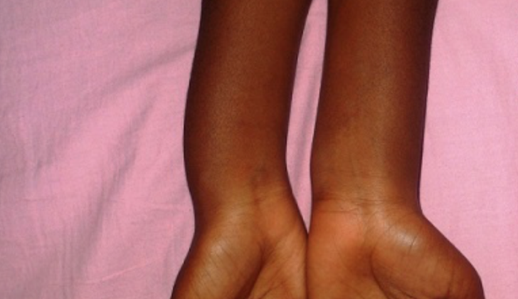
bowing in a patient with Madelung deformity.
Signs and Symptoms of Madelung Deformity
People with the classic form of a condition known as Madelung deformity generally start showing symptoms when they are 8 years old or older. However, Madelung-like deformities caused by injuries can happen at any age. Those with the inherited form of this condition may have family members who also have it. The main concern for these individuals usually revolves around the physical appearance of the deformity, rather than any functional issues it may cause.
However, they can experience symptoms like wrist pain, a decrease in their grip strength, and losing the ability to move their wrist as easily over time. This wrist pain can be felt on either the thumb (radial) or little finger (ulnar) side, and it’s usually more noticeable when putting weight on an extended wrist. A reduction in wrist extension is generally due to a condition where the head of the ulna bone moves backwards, although this doesn’t usually affect rotating the forearm.
People with Madelung deformity have a noticeable bump on their wrist because the ulna has grown more than the radius. There are also variations of Madelung deformity where the entire radius bone is affected, or in rare cases, the radius bone sticks out more than the ulna. In cases where the the entire radius is involved, this is often linked to a condition called Leri-Weill dyschondrosis, which is characterised by a curved radius bone and severe deformity. If the radius is more prominent than the ulna, it can cause what is known as dorsal carpal subluxation, a condition where the wrist bones dislocate towards the back.
Testing for Madelung Deformity
If your doctor suspects Madelung deformity, which is an abnormality of the wrist and forearm, they might begin with X-rays of these areas to better understand the situation. When performed from the front (anteroposterior or AP), X-rays can reveal specific changes like an increased angle of the radius bone, a v-shaped wrist, or a condition where the lunate bone descends into a hollow space called the lunate fossa. Side X-rays might show an increased front (volar) tilt and a related volar displacement of the wrist. Moreover, the disorder may lead to the dorsal part of the ulna bone moving towards the back, which could be visible in X-rays. In severe cases, the lower end of the ulna may appear scattered and irregular.
Typically in Madelung deformity that affects the entire radius bone, the joint between the distal radius and ulna bones remains in its normal position. Because of this, X-rays of the forearm and elbow might show the head of the radius bone separating from another bone, the capitellum, by more than 4 mm. This is also known as radiocapitellar distance. The X-rays may also show a bending or bowing of the radius bone.
To accurately detect Madelung deformity, various standards have been set based on the results of X-rays. For instance, at least 33 degrees of ulna tilt, 4 mm of lunate subsidence, displacement of 20 mm or more of the wrist towards the palm, and a lunate fossa angle of at least 40 degrees are measures that can help diagnose this deformity.
While X-rays are usually the first line of investigation, Magnetic Resonance Imaging (MRI) of the wrist isn’t routinely used for Madelung deformity. However, MRI is the best tool to identify the Vickers ligament, a key finding in this disorder, showing it in around 85% of cases. Despite this, it’s not recommended for routine screening even in patients with a family history of Madelung deformity. However, as the deformity worsens, MRI may reveal certain changes such as a slanting disc of fibrous cartilage that joins two bones in the wrist, or a bone bridge across the hollow space on the volar ulnar side. Occasionally, these changes might be visible on X-rays as well.
X-rays and MRI might also reveal a flame-like notch in the lower end of the radius at the point where the Vickers ligament starts. This sign is strongly associated with Madelung deformity. They may also show a widened part of the radius bone near the wrist. It’s worth noting that an acquired form of Madelung deformity will not show the Vickers ligament in MRI, but swelling around the growth plates of the bone and a bone bridge across it might be seen in cases resulting from injury.
A study has found that the Vickers ligament was seen during surgery in 83% of patients with Madelung deformity, which is close to the 85% detection rate of this structure by MRI. Also, finding changes in the entire radius bone and a notch in the lower end of the radius were important indicators of the existence of Vickers ligament.
Treatment Options for Madelung Deformity
For Madelung deformity, which is an abnormality of the wrists, the kind of treatment that is recommended will hinge on three main factors: how severe the deformity is, the root cause of the deformity, and when the deformity was detected. People experiencing minor to no symptoms might typically find relief simply through non-invasive methods. These may include taking over-the-counter anti-inflammatory medication, changing their daily activities to avoid strain on the wrist, and using a wrist splint for support.
Those undergoing these conservative treatment methods need to have periodic X-rays taken every six months until the bones have stopped growing. This is to check whether the deformity is worsening. In cases where it does continue to worsen while the patient’s bones are still growing, a preventive surgery might be carried out to prevent the condition from deteriorating further. Nonetheless, the decision on when to perform this procedure remains a subject of discussion among medical experts.
If the symptoms of Madelung deformity become more severe, there are several surgical techniques available to help correct the problem. One is the removal of Vickers ligament, a band of fibrous tissue, typically performed in patients who are still growing and have this deformity since birth. Another involves reshaping the growth plate, a part of the bone that facilitates the growth in length, along with introducing fat tissue, if there’s a problematic growth plate with an unfavourable future prospect. Some experts believe it might be better not to rush into any kind of intervention if the deformity isn’t severe, considering that the associated pain might naturally diminish over time.
A procedure commonly carried out on still-growing patients with Madelung deformity is the Langenskiold procedure, which involves halting the growth of a radial bone, removal of Vickers ligament, and insertion of fat tissue onto the treated area.
Treatment could also involve adjusting the joint between the two bones in the forearm over time and inhibiting the lower growth of bones in the forearm. Other methods include a dome-shaped reshaping of bones to correct the deformity from multiple directions in fully-grown patients. Shortening the ulna bone or removing part of it, and correcting a protruding or symptomatic ulnar head, is also considered in some cases.
Options like replacing the joint formed by the two bones in the forearm are not extensively studied and hence not yet widely implemented. A study found that reshaping the lower part of the radial bone that ends at the wrist joint and removing Vickers ligament results in good functionality in patients with Madelung deformity. Isolated or combined surgical reshaping of the two bones in the forearm produced satisfactory results in terms of both clinical improvement and X-ray findings, after an average follow-up period of 8.1 years.
Operating on Madelung deformity remains a complex task since it necessitates changing the deformity safely from multiple directions. However, new tools like tailor-made guides for bone reshaping during surgery could improve the outcomes. A study found that simulating the combined surgical reshaping of radial and ulna bones before the actual operation accurately corrected Madelung deformity. Another study showed that three-dimensional planning substantially helps in managing the deformity during the surgery, although the results might not be consistent across all aspects of the procedure.
What else can Madelung Deformity be?
If you’re experiencing wrist pain and limited mobility, your doctor might consider several potential causes including:
- Fractures
- Arthritis
- Tendinopathy (a condition where the tendons are damaged)
- Tumors
There is a condition in the wrist called Madelung-type deformity which could be caused by either an injury or a tumor. However, it’s crucial to separate it from a true Madelung deformity since both conditions have different management strategies and progress in different ways.
What to expect with Madelung Deformity
The course and progression of Madelung deformity, a certain bone growth disorder, are quite unpredictable and hard to understand. This condition tends to develop throughout the teenage years until the person’s bones are fully grown. Although it varies, most patients experience some level of pain relief after undergoing surgical procedures such as Vickers ligament release and volar dome osteotomy. Some may require an additional procedure called ulnar shortening osteotomy.
Research has indicated that combining Vickers ligament release and dome osteotomy can improve symptoms of Madelung deformity as seen on X-rays. However, because this condition is rare and treatment varies greatly, the results are not well understood. This is due in part to a lack of large, long-term studies on this condition and its treatment.
Possible Complications When Diagnosed with Madelung Deformity
Patients with extreme deformities, especially if not corrected by surgery, are at a higher risk of having their extensor tendons wear out and eventually break — the longer the deformity is present, the higher the risk. This typically affects the extensor tendons in the ulna, one of the bones in the forearm. Patients may continue to experience discomfort in the wrist, regardless of whether or not they have gone under the knife. This discomfort could be due to ulnar impaction, a condition in which the ulna is excessively pressing against the nearby bones. Additionally, people with untreated Madelung deformity can develop wrist arthritis.
Common Risks:
- Increased risk of extensor tendon rupture with severe deformities
- Persistent wrist pain, with or without surgery
- Ulnar Impaction
- Development of wrist arthritis in cases of untreated Madelung deformity
Recovery from Madelung Deformity
After undergoing a Vickers ligament release and removal of physeal bar – part of the bone growth area, patients are advised to wear a short-arm brace for about 3 to 4 weeks. This is then followed by a gradual reintroduction of movements and exercises which involve carrying weights. For patients who have undergone surgical procedures like ulnar shortening (a surgery to shorten the longer bone of the forearm) and dome osteotomy (a surgery to correct bone deformities), they’ll need to have their entire arm immobilized in a long-arm brace for 2 weeks, followed by 4 weeks of short-arm immobilization.
After 6 weeks of having their arm immobilized, patients can now begin doing exercises to regain a full range of motion in their arm. By the third month post-operation, they can start to gradually introduce exercises which involve carrying weights.
Preventing Madelung Deformity
Madelung deformity is an unusual condition that a person is born with. It’s tricky to stop it from happening because we don’t have a lot of established ways to predict and prevent it. However, there are some things that might help lessen the effects of this condition.
The first is genetic counseling. Specialists in this area, known as geneticists and genetic counselors, can provide information about the condition, analyze if the family might be at risk, and offer advice on possible genetic testing or decisions around family planning.
Second is regular check-ups with a children’s doctor, known as a pediatrician. This doctor can watch for changes in the child’s bones and muscles. Detecting abnormalities in bone growth or wrist structure early on can help to get the right medical care and treatment quickly. The third recommendation is ensuring the child eats a balanced diet and stays healthy overall. Lack of certain vitamins can trigger or worsen irregular development of bones and muscles.
In addition, children who might get Madelung-type deformities are generally advised to avoid putting repeated stress or strain on their wrists during their growing years and to properly prepare their bodies for sports activities.
These strategies may help prevent the condition from happening, but keep in mind that we don’t have a lot of evidence proving their effectiveness because this condition is complex and not very common.


