What is Os Odontoideum?
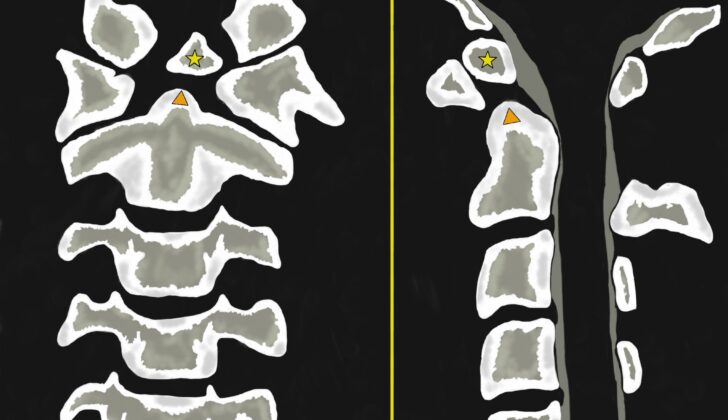
Os odontoideum is an unusual structure of the upper part of the neck or cervical spine, first identified by Giacomini in 1886. This condition is identified in X-ray images as a small, circular or oval-shaped piece of bone with smooth rounded edges. This bone represents an underdeveloped tooth-like structure of the spine known as the odontoid process, which doesn’t connect with the second cervical vertebra, also known as C2.
Usually, the small bone moves upward from the expected position of the tip of the odontoid process. It can occur in two main types— one that remains in the normal position of the odontoid process (orthotopic), and one that moves up near the base of the skull in the area of a large opening called the foramen magnum (dystopic).
Experts have studied the dimensions of the os odontoideum and have found that most are about half the size of a normal odontoid process. Some are exceptionally small and located more towards the head, making them difficult to identify using plain X-rays or a type of detailed imaging test called computed tomography (CT). The small bone is usually found slightly behind and above the front arch of the first cervical vertebra, known as C1. Often, the os odontoideum is attached to the front arch of C1 by a horizontal band of tough, fibrous tissue called a transverse ligament.
A major risk associated with this condition is the forward slipping of the first and second cervical vertebrae, known as anterior atlantoaxial subluxation, while backward slipping is extremely rare. This instability in the joint between the first and second vertebrae can result in cervical spinal stenosis, a condition where the spine narrows and compresses the spinal cord. This can lead to cervical myelopathy, a condition that arises when the spinal cord in the neck is squeezed, resulting in problems with blood supply and could lead to compression or stretching of the spinal cord.
What Causes Os Odontoideum?
Doctors are not entirely sure what causes os odontoideum, a disorder affecting the upper spine, but there are two main theories. The first theory suggests that os odontoideum develops after a trauma or injury that disrupts the blood supply to certain part of the spine called the odontoid. This can happen before or after birth and the patient may not even remember the injury. There are reports of patients developing os odontoideum after trauma where previous spine scans were normal.
The second theory believes that os odontoideum is caused by a birth defect that prevents certain parts of the spine from fusing together correctly. This theory might explain why the condition sometimes occurs in identical twins who have no history of trauma, or even certain families. What’s more, studies showing genetic differences between people with os odontoideum and without it backs up this theory. The theory is also supported by the fact that os odontoideum is often seen with other abnormal conditions that affect the upper spine and neck.
In children, understanding how the spine develops normally is important to avoid falsely diagnosing os odontoideum. The top point of the odontoid (a small upward-pointing part of the second vertebra in the neck) comes from the fourth occipital sclerotome (a block of tissue in a developing embryo), while the odontoid process and anterior arch of C1 (the first vertebra in the neck) come from the first cervical sclerotome, and the body of the axis (second vertebra in the neck) comes from the second sclerotome. While these parts usually begin to harden into bone between the ages of 3 to 6, they don’t typically join with the body of the odontoid until a child is around 10 to 12 years old. Similarly, the sub-dental synchondrosis, a junction located between the odontoid and the body of C2, usually joins together when children are between the ages of 3 and 8.
Risk Factors and Frequency for Os Odontoideum
Os odontoideum is a rare condition that often doesn’t cause any symptoms. This makes it difficult to determine how many people actually have the condition. It is usually discovered by chance in kids or diagnosed in adults who start showing symptoms. A look back at a study, which used MRI scans to examine the upper spine in 133 people aged from 19 to 81, found that only one person had os odontoideum. This suggests that it may affect about 0.7% of the population.
Signs and Symptoms of Os Odontoideum
People diagnosed with this condition can have a wide range of symptoms, from having none at all (only discovered during unrelated medical checks), to experiencing neck pain and severe neurological issues. In extreme cases, individuals may suffer from permanent paralysis.
Often, the first symptoms people notice are quite vague and can include neck and shoulder pain, a twisted neck (torticollis), headaches, and a tingling or numb sensation in the neck and upper limbs. Some may also find they have weaker lower limbs and find it challenging to walk. These symptoms are believed to be caused by repeated minor traumas to the spinal cord or an ongoing pressure on the cord.
Specific movements can sometimes restrict blood flow to the brain, causing symptoms such as seizures, fainting, dizziness, visual disturbances, and even sudden death following minor trauma.
- Neck and shoulder pain
- Twisted neck (torticollis)
- Headaches
- Tingling or numb sensation in neck or upper limbs
- Weaker lower limbs
- Difficulty in walking
- Seizures
- Fainting
- Dizziness
- Visual disturbances
In a physical examination, doctors might discover a loss of motor skills or neurological deficits. Symptoms like decreased sensitivity (hypoesthesia), hyperactive reflexes (such as the patellar (knee-jerk) reflex, Shimizu reflex, Tromner reflex, Hoffmann reflex, Babinski reflex, and clonus), a spastic or irregular walking, and reduced dexterity in hands could be signs of chronic spinal cord compression. However, these signs are not unique to this condition, and doctors should also consider other diseases causing similar symptoms.
Testing for Os Odontoideum
Identifying a spinal condition known as os odontoideum early and with accuracy is necessary to avoid serious complications and a potential worsening of the patient’s health. Even a small accident can cause significant damage to the nerves if this spinal condition is not diagnosed. To spot this condition quickly, your doctor will typically start by taking standard X-rays of the neck, including a special view of a neck bone called the odontoid.
X-rays can reveal several signs of this condition, such as an increase in the gap between your first two neck bones, called the anterior atlantoaxial distance. The same gap may also appear wider in the back, and the line that visually joins the back part of the spine may be disrupted. Another sign is a small bone fragment, round and with well-defined edges, located just above and at the back of the first neck bone. It’s important to note that there won’t be any swelling in the soft tissues in front of the vertebrae. Radiographs taken when the patient is bending and straightening up can help measure how unstable the patient’s neck is.
Computerized tomography (CT) images can also show the same bone fragment with clear edges, depicting a shortened odontoid bone and a smooth bone fragment. Apart from these findings, a CT scan can also show if the back part of the first neck bone has not fused completely. If the front part of the first neck bone looks larger than normal, it often indicates a long-term instability. CT scans can display landmarks inside your body which are useful for planning surgery, including the length and direction of certain bone structures called pedicles, and some blood vessels, especially if their anatomy is unusual.
Magnetic resonance imaging (MRI) is another test your doctor can use. An MRI can show fluids in the widened gap between the anterior atlantoaxial bones, with possible focal narrowing of the spinal canal due to the odontoid bone tilting towards the back and pressing against the spinal cord. In some cases, certain areas may show an increased magnetic resonance signal, indicating an abnormal condition called myelomalacia.
Measurements taken from X-rays or CT scans can help the doctor know the amount of instability in your neck or decide the best surgical approach. For example, the gap between the odontoid process and the back border of the front arch of the first neck bone shouldn’t exceed 3.5 mm in adults. If it exceeds 10 mm, doctors often consider surgery. In children, a measurement of up to 5 mm is considered normal. Another measurement, known as the posterior space available for the cord or for the spinal cord, is the distance between the back surface of the dens and the front surface of the back arch of the first neck bone. If this distance is less than 14 mm, there’s usually an increased risk of nerve damage, often warranting surgical intervention.
Treatment Options for Os Odontoideum
When treating atlantoaxial instability, a condition causing weakness in the neck and leading to severe pain and discomfort, the treatment options depend on the severity of the symptoms and instability of the affected area. This can be determined through medical imaging techniques. If patients do not show symptoms and the neck appears stable under imaging, doctors often suggest watching and waiting. During this time, it is recommended that these patients avoid physical activities that might damage the neck such as contact sports.
However, there is ongoing debate about whether certain protective treatments, such as proactively fusing the spine, might be beneficial for these patients. While this procedure can limit the ability for patients to rotate their necks by up to 50%, the benefits of limiting further nerve damage might outweigh this disadvantage.
Patients who show signs of atlantoaxial instability even if they do not present symptoms should consider surgery. Those who have symptoms are always advised to opt for surgical treatment. Surgery is also suggested especially for patients where imaging of their neck demonstrates instability or signs of potential instability such as increased movement, anyone with persistent neck pain, and individuals with temporary or continuous neurological symptoms.
Overall, the goal of the treatment is to prevent a sudden worsening of the symptoms, improve neurological health, stabilize the neck, and enhance the quality of life for the patients. Typically, treatment follows a certain pattern: identifying the main cause of the symptoms, determining whether the unstable neck can adjust itself, deciding if the removal of the causing lesion is necessary, and then choosing the appropriate surgical operation.
There are several surgical options available, such as atlantoaxial fusion, occiput-C2 fusion, and occiput-C3 fusion, which can be chosen based on the location of the compressed spinal cord, the potential for fusing the bones together (arthrodesis) and the quality of the patient’s bones. If needed, these surgical operations may include an additional procedure to relieve pressure from a patient’s neck. However, this additional procedure can also bring complications such as infection, leakage of spinal fluid, nerve damage, and loosening of hardware.
Successful treatment strategies do exist. One study reported patients experiencing relief from the segmental atlantoaxial fixation procedure, a type of fusion surgery, without the need to remove bone or apply additional treatments. Recommending fusion surgeries is generally more common if there is instability between the head and neck or after unsuccessful atlantoaxial fusion attempts.
In summary, the treatment options for atlantoaxial instability range based on the specific symptoms and instability present in the patient. Treatments can vary from non-invasive management such as monitoring and physical restrictions, to invasive measures such as spine fusions. The most appropriate treatment for patients with atlantoaxial instability is typically made on a case-by-case basis, always ensuring the overall goal to improve a patient’s quality of life and alleviate discomfort.
What else can Os Odontoideum be?
Os odontoideum is a condition that can cause instability and dislocation in the upper part of the spine. There are other conditions that can induce similar instability. These include:
- Acute dens fracture – this is identified by a thinner and more irregular space, as opposed to the wide and smooth one seen in os odontoideum
- Unusual shapes or features of the dens such as an absence or duplication of the dens, a split dens, or a persistent ossiculum terminale
- Down syndrome
- Juvenile rheumatoid arthritis
- Klippel-Feil syndrome which is a rare disorder affecting the spine
- Morquio syndrome, a serious genetic disorder
- Neurofibromatosis, a genetic disorder that affects nerve tissue
- Normal development in children which can sometimes be mistaken for a disorder
- Rheumatoid arthritis
- Injury to the transverse ligament in the spine
Healthcare professionals should be familiar with these other conditions to make an accurate diagnosis.
What to expect with Os Odontoideum
The outlook for a condition called os odontoideum can change based on symptoms and certain key measurements taken from X-rays or other types of medical scans. A factor called C1-C2 instability, tied to certain bones in the neck, plays a big role in this outlook. This instability occurs when there’s more than 3 mm of forward movement of a certain part of the top neck bone in relation to a bone called the dens in adults, or more than 4 mm in kids. If there’s more movement or displacement, the outcome may be worse.
Interestingly, the amount of instability in these neck bones doesn’t determine the prognosis as much as the actual size of the spinal canal, the hollow space in the spine where the spinal cord sits. Also, there’s a specific measure called the PADI, which assesses the width of the spinal canal. If the PADI measure shows the spinal canal is less than 12 mm wide, it predicts that paralysis will occur in all cases of C1-C2 instability.
Possible Complications When Diagnosed with Os Odontoideum
The complexities that arise from os odontoideum, a rare abnormality in the spine, depend greatly on the level of spinal cord compression, instability, and unexpected incidents. Even a minor accident can cause devastating damage to the nervous system and may even lead to unplanned death. This risk comes from possible instability between the first and second vertebrae because of a deformed portion of the spine and dysfunction in the related ligament complex due to the abnormal position of a free bone fragment. It is vital to accurately identify instability on x-rays to avoid complications linked to pressure on the spinal cord.
Common Complications:
- Spinal cord compression
- Instability in the spine
- Nervous system damage
- Unexpected death
- Deformation in a part of the spine
- Abnormal position of a free bone fragment
Preventing Os Odontoideum
If a patient is diagnosed with a condition called ‘os odontoideum’ and they’re experiencing symptoms, doctors typically suggest immediate surgery. ‘Os odontoideum’ refers to a small bone separated from the second vertebra in the neck. Non-surgical treatment options are usually only recommended for those who don’t have any symptoms and there’s no sign of neck instability. Doctors must also warn the patients about the possible severe outcomes linked to this condition.
Regular check-ups and X-ray monitoring are extremely important. Healthcare providers will usually recommend that patients should avoid all contact sports to reduce the risks of potential harm. If patients show symptoms related to ‘os odontoideum’ or if X-rays indicate instability between the first and second vertebrae in the neck (known as C1-C2 instability), then surgery to stabilize these bones is often the best choice of treatment.


