What is Os Peroneum?
The os peroneum is a small, extra bone in the foot, found near the cuboid (one of the seven tarsal bones in the foot) within a tendon known as the peroneus longus. Usually, this extra bone doesn’t cause any problems and is often only noticed when an image of the foot is taken for some other reason. However, because of where it’s located, if there are issues with the peroneus longus tendon, the os peroneum can also be involved.
Painful os peroneum syndrome is a broad term used to describe conditions that cause pain on the outside middle area of the foot, linked to the os peroneum. This could be due to a sudden break, stress fracture caused by ongoing, repetitive trauma, bruise, lack of blood supply leading to tissue death (avascular necrosis), or inflammation, tears, and displacement of the peroneal tendons. Other causes of pain in the same area can also include damage to ligaments (specifically, the talofibular or calcaneofibular), fractures of the bottom of the fourth or fifth long foot bone (metatarsal), the anterior (front part) of the heel bone (calcaneus), or the cuboid.
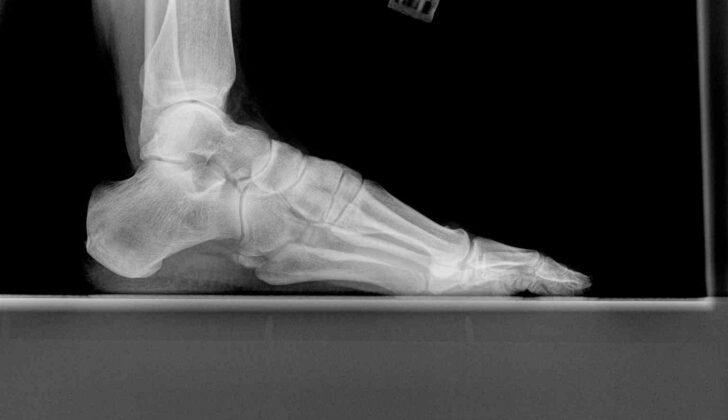
Normal foot x-rays are typically the first step doctors take when trying to figure out why there’s pain on the outside middle area of the foot. Changes to the shape and position of the os peroneum can be easily seen on these x-rays, especially from an angle. This can hint at a possible injury to the peroneus longus tendon.
What Causes Os Peroneum?
Accessory ossicles are small, extra bones that are common and usually cause no symptoms. They are natural variants that can be found throughout the bone-muscle system, especially in the foot and ankle. Knowing about these extra bones is crucial to prevent misdiagnosis, as they can be mistaken for disease conditions. The source of these extra bones can vary, but they might develop from parts of bones not fully combining, or form in response to stresses within tendons.
A recent study conducted on cadavers suggests that some of these accessory bones were present in fetuses. Also, a fibrocartilaginous node is a fibrous, non-bone structure similar to these extra bones, which can be identified during a magnetic resonance imaging (MRI) scan as a distinct oval shape within the peroneus longus tendon (one of the tendons in your leg).
Some of these extra bones, such as the os navicular, os trigonum, and hallux sesamoids, are known to cause painful symptoms. The Os peroneum is one such extra bone that can sometimes cause pain or be linked to disease conditions. Painful Os peroneum syndrome is identified as foot pain on the outer side that could have various causes. It could be due to an acute fracture of the Os peroneum, a tear in the peroneal tendon, or chronic inflammation of the tendons (tendinopathy). Fractures of the Os peroneum can occur along with tearing of the peroneus longus tendon, or they could be linked with chronic disintegration and tendinopathy. An acute fracture of the Os peroneum usually happens due to a strong contraction of the peroneus longus muscle, typically during an inversion or supination (twisting or turning motion).
Risk Factors and Frequency for Os Peroneum
The os peroneum is one of several small bones in the foot and ankle. It’s present in around 5% to 30% of people. In 60% of adults with this bone, it can be found in both feet. In up to 30% of cases, it can have a split or multipart look. There is also an extra muscle called the peroneus quartus in the back part of the foot which appears in 6% to 21% of people and can cause foot and ankle pain.
Twisting injuries to the foot and ankle are a frequent reason for emergency department visits. The ankle joint is the one most often damaged, with side ankle sprains being the joint injury most caused by trauma. Most patients get better with non-surgical treatment when there’s no fracture. However, 10% to 20% continue to experience pain and instability.
If patients don’t get better with non-surgical treatment and there’s no evidence of ligament instability from exam results, problems with the peroneus tendons or the os peroneum bone should be considered. Not thinking about these less common causes can result in a delayed diagnosis.
Signs and Symptoms of Os Peroneum
The Os peroneum is a bone in the foot that usually doesn’t cause any problems. But sometimes, it can break or move out of place if the peroneus longus tendon (a tendon in your foot) tears. This can happen from a direct injury, or from suddenly flexing your foot upwards or twisting it inward. Sometimes, this bone can fracture without any known injury.
The typical signs of this injury include pain on the outer side of your foot or ankle, feeling unstable when you stand or walk, swelling, and discomfort when the area is touched. You could also have swelling, pain, and weakness when you try to move your foot downwards or sideways. Your healthcare provider may also check how your foot and ankle align while you’re standing, as different alignment can be associated with issues in the peroneal tendon.
- Pain on the outer side of foot/ankle
- Instability
- Swelling
- Discomfort when touched
- Swelling, pain and weakness in foot movement
- Possible alignment issues with the foot and ankle
Testing for Os Peroneum
The os peroneum is a small bone that can be seen near the joint of the cuboid bone in the foot. It’s usually round or oval-shaped and can be best seen on angled foot x-rays. If the os peroneum is made up of more than one piece (bipartite or multipartite), it will look fragmented, but smooth edges distinguish it from a fracture, which would have sharp, rough edges. A gap bigger than 6 mm can suggest a fracture in the os peroneum with a tendon tear, while a gap of 2 mm or less may be seen in minor fractures or in a naturally multipartite os peroneum.
When there’s chronic stress injury to the os peroneum, it can become larger and harder, and if it has moved relative to a previous x-ray, that may indicate a tear in the surrounding tendon.
Detecting the difference between a multi-piece os peroneum and a fracture can be difficult with x-rays alone, especially without any other x-rays for comparison. As the os peroneum is usually found in both feet, an x-ray of the other foot may provide valuable insight. In more complex cases, a CT scan can offer a more thorough evaluation of the bone edges to tell the difference between a minor fracture and a multi-piece os peroneum.
Ultrasound imaging, which shows the os peroneum as a bright structure casting a shadow, can also be used. A broken os peroneum can appear lengthened or have irregular outlines on ultrasound. Ultrasound can also pick up signs of any related injuries to the nearby peroneus longus tendon such as fluid in the tendon or enlargements due to injury or a tear.
Magnetic resonance imaging (MRI) is another imaging technique that can be used, which shows particular characteristics of the os peroneum similar to the cuboid bone. However, its use can be limited due to high costs and the need for specialized expertise for correct image interpretation. MRI is beneficial for assessing related peroneus longus tendon injuries and can show visibility of fluid in the tendon, abnormal shaping, and possible continuity issues.
A rating system has been used to describe tendon injuries that also help explain the x-ray appearance of the os peroneum. These tendon injuries are divided into three categories based on their proximity to the os peroneum. Isolated tears of the tendon near the os peroneum usually show as normal os peroneum form and position. Tears occurring at the os peroneum are usually linked with fractures or distraction of bipartite or multipartite fragments. The distraction of the os peroneum fragments greater than 6 mm is highly suspicious for complete disruption of the peroneus longus tendon. Injuries to the tendon beyond the os peroneum can lead to it moving upwards. The small bone rarely moves upwards to the outer margin of the heel bone unless there is a strong contraction force of the tendon.
Treatment Options for Os Peroneum
If you have a problem with your peroneus longus tendon, several treatment options are available based on what’s specifically wrong with the tendon. Usually, doctors like to start off with a more careful approach. This might involve not using the affected body part for a while and taking medications that help reduce swelling and pain, such as NSAIDs. In some cases, doctors might give steroid injections, which can help reduce inflammation in the tendon. After this, you might have sessions with a physical therapist to help restore strength and function in the area.
However, if these less invasive treatments don’t work or the pain cannot be managed, surgery may be needed. This could also be the case if the function of the tendon is significantly impaired. In some cases, such as with high-level athletes, surgery might be considered sooner. There are several surgical options available, depending on the exact nature of the tendon injury. This could include a primary tendon repair or using a graft to repair the tendon, removing a fractured os peroneum (a small bone in the foot), repairing or attaching the tendon to a bone (known as tenodesis), or cleaning out the tendon (a process known as debridement).
The exact surgical approach would depend on where and how much the tendon is injured. One approach divides patients into three categories: type 1 where both tendons are intact, type 2 where one tendon is torn and the other intact, and type 3 where both tendons are torn. Based on this, those in type 1 group undergo a primary repair, those in type 2 group undergo tenodesis, and those in type 3 group undergo a tendon transfer.
What else can Os Peroneum be?
If you’re suffering from pain on the outside of your foot or ankle, or if your ankle feels unstable, there could be a number of reasons for this. These include problems like:
- Peroneal tendinopathy or subluxation (issues with the tendons on the outer side of the ankle)
- Injuries to the ankle ligaments
- Sinus tarsi syndrome or cuboid syndrome (conditions affecting specific parts of the foot)
- Broken bones
- Peroneal neuropathy (damage to the peroneal nerve in the lower leg)
Sometimes, a problem with the peroneal tendon might be missed and an ankle ligament problem might be diagnosed instead. So, it’s important to consider this if the pain won’t go away. An MRI scan can be very helpful for working out what’s causing these symptoms because it provides a clear picture of the structures on the outside of the ankle.
Another related issue is an os peroneum fracture, which can be mistaken for a two-part or multiple-part os peroneum, or an os vesalianum or os cuboideum secundarium – these are all additional small bones that can be found near the expected location of the os peroneum. In the case of a rupture to the end part of the peroneus longus tendon, the os peroneum can move too far upwards and may be mistaken for an os trigonum. Other things your doctor might consider are a fractured cuboid bone, or an avulsion fracture from the base of the fifth metatarsal (the long bone on the outside of the foot that connects to the little toe).
What to expect with Os Peroneum
There isn’t enough solid scientific evidence available to definitely determine the effectiveness of non-surgical and surgical treatments for injuries to the peroneal tendon. However, some studies have reviewed the results after surgery. For instance, they found that 87% of patients were able to return to sports within 3.5 months after surgery, and 91% regained normal or moderate strength in the peroneal tendon.
Possible Complications When Diagnosed with Os Peroneum
People with injuries to the peroneal tendon, a tendon in the foot, may have ongoing pain and instability on the side of the foot or ankle. This can occur if the injury is not treated or if other treatments have not worked well. A diagnosis can often be delayed. In fact, one study found that people experienced symptoms for anywhere from 7 to 48 months before the correct diagnosis was made.
If a certain type of fracture of the peroneal tendon called an ‘os peroneum’ fracture is not treated, it can lead to more damage and tearing of the tendon. This is due to the ongoing friction acting on the injury. Moreover, if someone experiences several ankle injuries in combination with a tear in their peroneal tendon, x-rays taken over time may show the movement of the peroneal bone. If this bone is fractured, the x-rays may show the fracture pieces moving apart.
During surgery to repair the peroneal tendon, doctors often discover further damage. This can include tears in other tendons, abnormal growth of bone in the area, and tendons that slip out of place.
At the moment, there is not a lot of data available to determine how effective surgical treatments are. Some people have reported issues after surgery, including skin infections, wounds opening up, unsuccessful repair, irritation of a nearby nerve, inflammation of the tendon due to scarring, and chronic regional pain syndrome.
Common Problems with Peroneal Tendon Injuries:
- Ongoing pain and instability on the side of the foot or ankle
- Delayed diagnosis
- Damage and tearing of the tendon due to untreated fractures
- Movement of the peroneal bone or fracture pieces in case of repeated injuries
- Other damage to the tendons or growth of bone discovered during surgery
- Tendons slipping out of place
- Post-surgery complications such as skin infections, wounds not healing, unsuccessful repair, nerve irritation, inflammation due to scarring, and chronic regional pain syndrome
Preventing Os Peroneum
The os peroneum is an extra bonus bone located in the foot, right next to another bone called the cuboid and inside a tendon known as the peroneus longus, which is on the outer side of the foot. These extra bones usually don’t cause any discomfort, but sometimes they can lead to issues like fractures in the bone itself, inflammation or tears in the attaching tendon, a condition collectively known as os peroneum syndrome.
If you experience symptoms like swelling, tenderness, and instability to the outer side of the foot, or difficulty in turning the foot outward or pushing down against resistance, you might have os peroneum syndrome. Doctors usually confirm this condition using a medical imaging test, like an X-ray, ultrasound, or MRI, to get a detailed look at the bone and the surrounding soft tissues.
At first, doctors will try a conservative approach to manage the pain and the damage to the tendon, which could involve rest and keeping the foot immobile, medication to reduce swelling, steroid injections, and physical therapy exercises. However, if the pain is severe, it’s affecting your ability to move around, or if the low-grade treatment doesn’t work, surgery might become necessary. The surgeon might repair or replace the damaged tendon, remove the broken extra bone, fix the broken bone, or clean up the tendon to reduce irritation.
Patients can also take proactive steps to manage the condition at home. Scheduled exercises and wearing specially designed shoe inserts might prevent the condition from getting worse and can even help to diminish the symptoms. If the extra bone is spotted during a routine check-up and is not causing any problems, these methods could prevent it from becoming a problem in the future.


