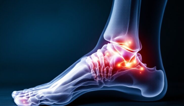
What is Osteoid Osteoma?
Osteoid osteoma is a type of benign (non-cancerous) bone tumor first identified by a researcher named Jaffe in 1935. It makes up approximately 10% of all benign bone tumors. However, it doesn’t have the potential to turn into a cancerous tumor and is not locally aggressive, which means it doesn’t readily spread to other areas of the body. This type of bone tumor often occurs in the long bones of the thigh (femur) and lower leg (tibia).
In this discussion, we’ll focus on osteoid osteomas affecting the foot and ankle, an area which is less commonly affected, representing 2-10% of cases. Among foot cases, the talus bone, located within the ankle, is most frequently involved.
An osteoid osteoma is recognized by a core area known as a ‘nidus’ of vascular osteoid (immature bone), surrounded by harder, denser bone. These tumors are usually pretty small, not exceeding a diameter of 2 cm. They can be categorized into three types – cortical (found in the outer layer of bones), cancellous (found within the inner, softer part of bones), and subperiosteal (found under the outer layer of bones).
When osteoid osteomas occur in long bones, they are primarily found in the cortex (the hard outer layer of the bone). In contrast, osteoid osteomas appearing in the foot show minimal reaction on the periosteum (outer fibrous layer of bone) and are usually of the cancellous or subperiosteal types.
We differentiate osteoid osteomas from another form of benign tumor, known as an osteoblastoma, based on the size of the nidus. Osteoblastomas are usually larger than 2 cm.
What Causes Osteoid Osteoma?
The cause of osteoid osteomas, a type of bone tumor, is still unclear. Some experts think it’s a non-cancerous growth, while others believe it could be due to injury or inflammation.
Risk Factors and Frequency for Osteoid Osteoma
Osteoid osteomas are a type of benign (non-cancerous) bone tumor, making up 10% of these cases. They are typically found in people between the ages of 5 and 25, and it’s more likely to occur in males, who are three times as likely to be affected.
Signs and Symptoms of Osteoid Osteoma
Osteoid osteoma is a medical condition characterized by several recognizable symptoms. These symptoms usually begin with periodic, localized pain that tends to get worse at night but is relieved by aspirin or other anti-inflammatory drugs. This condition is often associated with swelling, thought to be caused by increased blood flow to the tumor.
- Periodic, localized pain that tends to get worse at night but is relieved by aspirin or nonsteroidal anti-inflammatory drugs
- Swelling due to the increased blood flow to the tumor
- Bone deformity
- Muscle atrophy
- Walking disturbances
- If the tumor is within or near a joint, there might be inflammation of the joint, fluid accumulation, arthritis-like changes, and muscle contractions
- In some cases, osteoid osteomas may cause unequal leg lengths, especially when it affects the femur and tibia.
In one study involving four patients, it was found that the affected leg became longer than the unaffected leg. This phenomenon is believed to be due to the increased blood supply to a lesion near an open growth plate.
Testing for Osteoid Osteoma
When checking for a bone tumor such as an osteoid osteoma, the first examination is usually through a straightforward X-ray. On the X-ray, an osteoid osteoma generally appears as a small, round shadow with thickened bone around it. This shadow might show areas of hardened tissue or calcification.
If the X-ray results are unclear, a three-phase bone scan might be performed. A specific indicator of an osteoid osteoma in this type of scan is known as the “double density sign”. This reveals a central area of intense activity surrounded by a less active zone.
A CT scan, which uses X-rays and a computer to create detailed images of the body, is considered the preferred way to examine the condition. CT scans are especially good at identifying exactly where this shadow, or nidus, is located. The nidus usually appears as a target-shaped area on a CT scan.
On the other hand, MRI scans, which use magnetic fields and radio waves to create body images, may not be as useful as CT scans for checking osteoid osteomas. This is because the typical features of this type of tumor can be hidden by changes in the bone marrow detected by an MRI. However, MRIs are generally more accurate than CT scans when it comes to detecting lesions in the soft, spongy bone tissue, also known as cancellous bone. A study that compared the diagnostic accuracy of MRI and CT scans in children showed that correct diagnosis of osteoid osteoma was possible only 3% of the time with MRI images, compared to 67% of the time with CT images.
Treatment Options for Osteoid Osteoma
Using over-the-counter pain relievers, also known as Nonsteroidal Anti-Inflammatory Drugs (NSAIDs), can be a reasonable method for treating osteoid osteomas – a type of benign bone tumor. While NSAIDs can ease symptoms, complete symptom relief might take up to 33 months. But, it’s important to note that long-term use of these medications is not recommended due to their potential side effects. Also, there’s no definitive evidence about whether the tumor might reappear once the NSAID use is stopped.
Traditionally, complete surgical removal of the osteoma, usually including the tumor’s central core or ‘nidus’, has been the favored treatment. However, there are potential complications, especially if the tumor is located in a weight-bearing part of the body. These complications can include a lengthy period of restricted physical activity, potential bone fractures, and the need for a bone graft and internal fixation.
According to current medical practices, a less invasive surgical procedure called CT-guided percutaneous radiofrequency ablation is the preferred option. In this procedure, a radiofrequency electrode is inserted into the ‘nidus’ of the tumor under the guidance of a CT scan. Then, the nidus is destroyed using heat generated by radiofrequency waves. This method has a high success rate, being effective in up to 90% of cases.
What else can Osteoid Osteoma be?
When it comes to identifying osteoid osteoma, a type of bone tumor, doctors have to rule out other conditions that could be causing similar symptoms. These may include:
- Chondroblastoma (a rare, non-cancerous bone tumor)
- Bone infarction (death of bone tissue due to lack of blood supply)
- Brodie’s abscess (a type of bone infection)
- Stress fracture (a small crack in a bone due to overuse)
- Chronic osteomyelitis (a long-lasting bone infection)
- Focal cortical bone abscess (a localized infection in the bone)
- Glomus tumor (rare, benign tumor of the skin)
- Sclerosing osteitis (bone inflammation that leads to hardening of the bone)
- Solitary enostosis (a benign, solitary bone lesion)
- Early stage of Ewing’s sarcoma (a rare, aggressive bone cancer)
It’s crucial for healthcare professionals to consider these potential conditions and make use of necessary tests for an accurate diagnosis.
What to expect with Osteoid Osteoma
An osteoid osteoma has a good outlook because it is a benign or non-cancerous condition that does not have the risk of becoming malignant or cancerous. If other treatments are not effective, completely removing it through surgery usually cures it.
Possible Complications When Diagnosed with Osteoid Osteoma
When an osteoid osteoma, a type of benign bone tumor, affects the end of a bone, it can cause the limb to grow longer, resulting in uneven limb lengths. If a treatment called radiofrequency ablation is used to treat the tumor, there are some possible complications, but these are rare. Complications can include a skin infection called cellulitis, inflammation of a vein caused by a blood clot, nerve damage, skin burns, and a pain disorder called reflex sympathetic dystrophy.
In order to lessen the chance of issues with nerves or blood vessels, or damage to the skin, it is usually recommended to only use radiofrequency ablation for tumors that are larger than 1.5 cm from any nerves or blood vessels, and further than 1.0 cm from the skin surface.
Possible Complications of Treatment:
- Skin infection (cellulitis)
- Inflammation of a vein due to a blood clot (thrombophlebitis)
- Nerve damage
- Skin burns
- Pain disorder (reflex sympathetic dystrophy)
Recovery from Osteoid Osteoma
In the past, a surgery called ‘wide en bloc excision’ was used to treat bone tumors called ‘osteoid osteomas’. This involved removing a lot of the surrounding healthy bone, which often resulted in a weakened bone structure and forced the patient to remain immobile for extended periods.
However, thanks to modern technology and new surgical techniques, treatment and recovery have significantly improved. Procedures such as ‘percutaneous techniques’, like radiofrequency ablation, cause minimal damage to the nearby healthy tissue. This means patients can bear weight on the affected area immediately after surgery, enabling them to return to their everyday activities much quicker.
Preventing Osteoid Osteoma
An osteoid osteoma is a non-cancerous tumor that forms in the bones. It never turns into cancer. It often triggers serious pain during the night, which can be managed using anti-inflammatory medications. Sometimes, it can disappear by itself without any treatment. If the pain persists, the first line of treatment typically involves using medications that reduce inflammation. If this approach doesn’t ease the symptoms, the tumor can be surgically removed.


