What is Pars Interarticularis Defect (Spondylolysis)?
A Pars Interarticularis Defect, also known as Spondylolysis, is a common reason for back pain in teenagers, especially those who play sports regularly. The Pars Interarticularis is a part of each vertebra in your spinal column, situated between the upper and lower joint processes. You can think of it as the region between two connecting points on the vertebra – one above and one below. The term ‘Pars Interarticularis Defect’ refers to a fracture in this region due to overuse or repeated stress. The fracture usually happens in the lower part of the spine, most often at a level called L5.
In some cases, the Pars Interarticularis defect can lead to a condition called Spondylolisthesis, where one vertebra slips out of line with the one above or below it. This slippage is graded based on how much one vertebra has moved in relation to the other.
Commonly, a Pars Interarticularis defect is discovered in one of two ways. It might be found incidentally when a teenager or adult has an imaging test for some other reason. Or, it might be suspected in a young athlete who has back pain that starts suddenly or gradually and gets worse when they stress the lower back. It should be noted that these symptoms could be from other conditions as well. So, it’s important to confirm the diagnosis of a Pars Interarticularis defect with an imaging test. Depending on when the problem is discovered and how severe it is, most cases can be treated successfully with rest and other non-surgical treatments.
What Causes Pars Interarticularis Defect (Spondylolysis)?
The exact reason for certain conditions isn’t always crystal clear, and this is the case for some problems related to the pars interarticularis – a part of the vertebrae, or the bones in your spine. Right now, it’s believed that repeated stress – specifically from continually bending and twisting your lower back – can cause overuse or tiny fractures to this part of the spine.
Studies back this theory because they’ve found no instances of damage to the pars interarticularis in newborn babies or in patients who can’t walk – which suggests this damage happens over time due to repeated strain.
In fact, findings show that over time, damage that first only affects one side can progress to affect both sides – again, suggesting that ongoing strain leads to both the initial injury and worsening over time. However, it’s also possible that these injuries can occur due to a single, sudden overload of stress.
The pars interarticularis is particularly vulnerable to ongoing strain as it’s a weak spot in the vertebrae that takes on the most stress when you’re bending or straightening your back. This weakness is due to a few factors including a genetic component and a physical one. The pars interarticularis is physically narrower compared to other parts of the vertebrae and in the lower part of the spine, it has patchy thickness. These inherent structure issues, combined with the high stress on the lower back, make this part of the spine susceptible to stress fractures.
There’s also a strong link reported with spina bifida occulta, which is a condition where the spine doesn’t form properly.
Risk Factors and Frequency for Pars Interarticularis Defect (Spondylolysis)
The disease in question is more common in certain groups and stages of life, and multiple studies have been conducted to understand this better. Here’s a summary of the key findings:
- Its overall occurrence rate ranges from 4% to 6% in teenagers and adults.
- White people are 2 to 3 times more likely to have this issue than African Americans.
- Males are 2 to 3 times more likely to have it than females.
- A study examining 4200 cadavers found an incidence rate of 4.2%.
- Between 8% to 15% of athletes in their teens who don’t show any symptoms have the condition.
- Almost half (47%) of young athletes with low back pain also suffer from this.
- In another study of 185 teenagers under 19 with the condition, 180 were actively participating in sports.
In connection with certain body parts, the L5 level was found to be affected from 85% to 95% of the time, according to a study in 2000, while the L4 level was affected only 5% to 15% of the time. A newborn study found that none of the 500 newborns had this condition from their X-rays. Furthermore, a study with 143 patients mostly suffering from cerebral palsy found this condition was present. Looking at the incidence in sports, 11% of 100 female gymnasts had this condition according to a 1976 study. Finally, it was found that this condition becomes visible on X-ray about 25% of the time when associated with a shift in the spine, and can also be associated with a mild form of spina bifida.
Signs and Symptoms of Pars Interarticularis Defect (Spondylolysis)
Pars defects are usually discovered in two common scenarios. The first involves an accidental discovery when a child or teenager undergoes a scan for another reason, and the scan reveals a pars defect. There may be no physical signs of this defect because the person doesn’t experience any symptoms. This kind of discovery often happens during abdominal CT scans or MRI imaging.
The second common situation involves adolescent athletes who participate in sports that put continuous strain on the lower back. They may have sudden or gradual onset of lower back pain that gets worse with strenuous activities and improves with rest. In fact, about half of the young athletes complaining of lower back pain have a pars defect, suggesting that special attention should be paid to their diagnosis.
When a patient has symptoms, they usually include lower back pain that increases with heavy activity or overstretching, and gets better with rest. The pain mainly stays in the lower back, sometimes radiating to the buttock or upper leg. Numbness or tingling in the lower limbs is rare.
- Sports involving regular lower back strain
- Pain onset is sudden or gradual over weeks
- Low back pain worsens with heavy activity or overstretching
- Pain improves with rest
- Pain mainly in the low back, sometimes radiating to buttock or upper leg
- Numbness/tingling in the lower limbs is uncommon
The physical exam for pars defect includes a single-leg hyperextension, also known as the Stork test. The patient is asked to stand on one leg while overstretching the lower back. If this test recreates the pain, and if it is worse when standing on the affected side, then it is deemed positive. This test is essentially the only potentially definitive physical sign of a pars defect. Sometimes, other physical signs like an excessive inward curve in the lower back and tight hamstrings may also be noticed.
Testing for Pars Interarticularis Defect (Spondylolysis)
If your doctor suspects you have a pars defect – which is a common type of spinal injury, they might recommend additional tests. This condition is especially prevalent in children and adolescents. That’s why it’s essential to keep measures in place to limit exposure to radiation during testing.
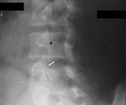
The first step is usually to take X-rays of the back from different angles, including front-to-back (AP), side (lateral), and angled (oblique) views. The AP/lateral X-ray can spot most pars defects, but about 20% may require the oblique view for a clear image. Sometimes, your doctor might request dynamic X-rays if they’re worried about instability caused by a condition called ‘spondylolisthesis’, where one bone in your back slides forward over the bone below it.
When examining an oblique plain X-ray, doctors often describe a characteristic fracture to the pars interarticularis – a specific part of a spinal vertebra, as the ‘collar’ on the ‘Scottie dog’. This is due to the image resembling the shape of a Scottish terrier.
However, X-rays don’t always reveal all pars defects, especially in the early stages. If that’s the case, the doctor may suggest other tests such as a CT scan, MRI, or bone scan. A bone scan is highly sensitive and excellent for detecting pars defects early. It can be particularly helpful if the X-rays aren’t conclusive, and the doctor strongly suspects a pars defect based on your symptoms.
After X-rays, the doctor might suggest either an MRI or CT scan without contrast. A CT scan gives the best picture of the fracture’s size and extent and is the best option when tracking how the injury is healing over time. However, CT scans come with additional radiation exposure, a significant concern for younger patients.
MRI, on the other hand, has no radiation exposure and can spot early-stage injuries by showing bone marrow swelling on T2 weighted sequences. However, it may not always clearly show incomplete fractures.
Treatment Options for Pars Interarticularis Defect (Spondylolysis)
The best approach to manage a problems with the pars interarticularis, part of the bone in your spine, isn’t completely clear. However, here’s a generally preferred treatment plan:
Firstly, for patients not experiencing any symptoms with their spondylolysis (a stress fracture in the pars interarticularis) or low-grade spondylolisthesis (when one vertebra slides forward over the one below it), doctors might just observe the condition without limiting the patient’s activities. These patients can even participate in contact sports.
If you’re experiencing symptoms with the spondylolysis or low-grade spondylolisthesis, though, the doctor might recommend physical therapy along with restricting some of your activities. This usually involves a six-month physical therapy program where you’ll do exercises like hamstring stretches and core strengthening.
In some cases, the doctor may recommend using a TLSO (Thoraco-Lumbo-Sacral-Orthosis) brace for six to 12 weeks. This kind of medical brace is usually recommended when you have an acute pars stress reaction (early signs of a stress fracture before it actually happens), if physical therapy didn’t help your spondylolysis or low-grade spondylolisthesis, or if brace immobilization is expected to give better results.
Finally, if these treatments don’t work or if your condition is more severe, surgery might be necessary. For instance, if the non-surgical treatments failed, if there are multiple fractures in the pars interarticularis, or if the spondylolisthesis is either getting worse or likely to progress, the doctor might suggest surgery. There are different surgical approaches that can be used depending on your specific situation.
Also, the doctor may check your vitamin D levels. If those levels are low, you might need to supplement your diet with more vitamin D.
What else can Pars Interarticularis Defect (Spondylolysis) be?
Lower back pain can be caused by various conditions, including:
- Myofascial pain in the lower back and hip area
- Pain originating from the sacroiliac joint, the joint between the sacrum and the ilium bones of the pelvis
- Pain caused by issues with the facet joints in the spine
- Pain caused by intervertebral disc problems, also known as Discogenic mediated pain
- Pars stress reaction, which is a condition that precedes a type of spine disorder called spondylolysis
- Spondylolisthesis, a spine condition where one vertebra slips forward over the one below it
What to expect with Pars Interarticularis Defect (Spondylolysis)
Defects in the pars interarticularis, a part of the spine, usually have a good prognosis or likely outcome. In fact, a whopping 80% of cases may not show any symptoms at all. Among these symptom-free cases, most will not worsen over time.
For teenagers experiencing symptoms relating to this spine defect, around 75% to 95% will see improvements with appropriate non-surgical treatments, such as physical therapy or wearing a back brace. However, around 9% to 15% of cases showing symptoms might eventually need surgery.
One study showed that between the ages of 20 and 80, there’s no increased occurrence of such spine defects. This suggests that the risk is highest during the teenage years, after which the bones have fully hardened and the risk levels off.
Interestingly, about 25% of the time, people whose X-rays show spondylolysis, a type of spinal condition, are also likely to have spondylolisthesis, a condition where a bone in the spine slips out of place.
In terms of this slagging effect, research shows different rates. One study found that in a follow-up of around 16 years, 23% of their 272 child and teenage patients showed a greater than 10% slip, whereas another study found that, in a follow-up of about four years, 3% of their 311 patients showed a greater than 20% slip. It is thought that patients are most likely to experience this slip during growth spurts.
Possible Complications When Diagnosed with Pars Interarticularis Defect (Spondylolysis)
Common Issues:
- Neurological problems
- False joint formation
- Progressive slippage of the bones
- Implanted medical device malfunction
- Chronic pain
Recovery from Pars Interarticularis Defect (Spondylolysis)
If we’ve talked in the past about the need for physical therapy, it’s usually recommended that the physical therapy program should last for about six months. The main goal of this program should be to concentrate on stretching the hamstring muscles, to bear additional stretching forces, making your core muscles stronger, and doing pelvic tilt exercises. Along with these, the physical therapy program should initially focus more on bending-based exercises. Over time, as these exercises stop causing discomfort, gradually, more stretching exercises can be slowly introduced.
Preventing Pars Interarticularis Defect (Spondylolysis)
There are certain sports that can increase your risk for injuries. These sports might include gymnastics, football (particularly for those playing on the line), weightlifting, wrestling, dancing, swimming styles like diving, volleyball, and soccer. It doesn’t mean that you can’t engage in these activities, but those who do are potentially at a higher risk of getting hurt than their counterparts in less physical sports.


