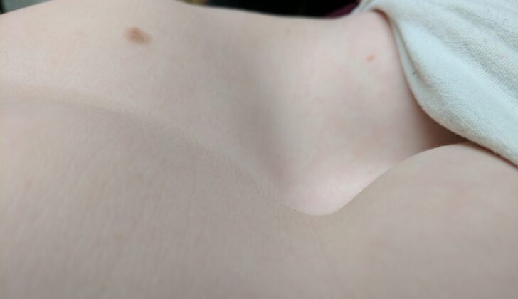
What is Pectus Excavatum?
Around 95% of birth defects related to the chest wall are due to pectus deformities, with pectus excavatum being the most common type. This condition leads to a depression or dip in the front of the chest, which resembles a “funnel chest”. This defect typically affects the third to seventh ribs, and is most evident near the lower part of the breastbone, known as the xiphisternum. The deformity may be even on both sides, but it is usually uneven and can affect other parts of the chest as well. A child with a pectus deformity may have it from birth, or it could develop later during childhood.
What Causes Pectus Excavatum?
There are several theories about what causes pectus excavatum, a condition where the chest appears sunken or caved in, resembling a funnel. Some believe it may be due to a weak or overly flexible sternum (the long flat bone at the front of your chest), an overgrowth of the ribs, or problems with the development of the chest area. Regardless of the cause, the result is a pushed in and shifted sternum and nearby ribs or cartilage, to varying degrees. While no specific genetic issue has been found, genetics may play a role, as over 40% of people with the condition have a family history of it.
Risk Factors and Frequency for Pectus Excavatum
Pectus excavatum, also known as “funnel chest,” is a chest wall deformity that affects around 1 in 300 to 1,000 newborns. The condition is more common in males with a ratio of 5 males for every 1 female. This deformity accounts for 90% of all chest wall defects.
- Usually, the defect is noticed within the first year after birth, and severe forms can be seen right at birth.
- The condition often becomes more visible during the growth spurt in puberty.
- Pectus excavatum can exist by itself or along with other birth defects.
- Among these conditions, connective tissue disorders are rarely associated with pectus excavatum, the chances being less than 1%.
Signs and Symptoms of Pectus Excavatum
Patients with certain conditions often have a common appearance – typically they are tall, thin males who seem to be slouching. They might also have thoracic scoliosis, which is a type of abnormal curve in the spine. These patients could have heart-related symptoms due to a indented breastbone that affects the heart’s position. This can lead to issues like having difficulty doing physical activities. This could also cause a heart murmur, a sound that can be heard when listening to the heart, which might be due to problems with the heart’s mitral valve, such as its falling out of place or leaking. It’s not entirely clear why, but struggling with physical activities is often one of the first symptoms these patients experience. This condition can also impact their psychological health, particularly in their teenage years.
Testing for Pectus Excavatum
When it comes to Pectus Excavatum, a condition which causes a sunken or caved-in appearance of the chest, a side view chest X-ray can clearly show the indentation in the sternum (central part of the ribcage). Additional scans might show displaced or shifted spinal bones and varying degrees of scoliosis, which is a condition that causes the spine to curve to one side.
Some people with mild Pectus Excavatum may not exhibit any symptoms. However, it’s crucial to perform heart and lung examinations every one to two years to establish normal functioning levels and check for any progression of the chest deformity. These examinations can involve tests like a chest X-ray, pulmonary function testing (assessing lung function), an EKG (electrocardiogram to check for heart conditions), and an echocardiogram (an ultrasound of the heart to check for secondary or associated problems).
Pulmonary function testing can identify possible cases of lung disease in older patients and air trapping, which leads to increased residual volume, meaning air remains in the lungs after a complete exhale. This could happen due to issues affecting the muscles used for breathing. The presence of unusual patterns in the EKG could suggest a shift of the heart towards the left side of the chest. Arrhythmias (irregular heartbeats) such as first-degree heart block, right bundle branch block, and Wolff-Parkinson-White syndrome can occur in 16% of these patients. Heart and lungs stress testing might reveal limitations that aren’t otherwise apparent when the body is at rest. An echocardiogram can assess for heart compression, heart valve defects, and heart muscle function. Some patients may have a heart shifted to the left and other conduction defects.
The severity of Pectus Excavatum is quantified with a measurement called the Haller Index (HI). It’s a ratio derived from the width and front-to-back diameter of the chest. These measurements are obtained from a CT scan; a normal value is 2.5 or less. Measurements above 3.2 are considered severe. Patients with a HI above 7 are four times more likely to have a restrictive breathing pattern in their pulmonary function testing (their lungs cannot fully expand).
MRI scans, which offer detailed images of the body, have been used to assess chest deformities before surgery. Breath-hold MRI, specifically,can provide a more accurate picture of chest shape in Pectus Excavatum. Additional techniques can be used to study how the lungs in these patients work. For example, ‘oculo-electronic plethysmography’ (OEP) can show that the sunken portion of the chest and the adjacent chest walls do not move as they should during breathing, which causes a reduction in lung volume.
Treatment Options for Pectus Excavatum
Early surgical correction of pectus excavatum, a condition where the chest appears sunken due to a deformity in the chest wall, was initially based on a rigorous method involving resection (removal) and reconstruction of the chest wall. However, the modern approach is to delay the surgical repair until after puberty and to perform a more limited cartilage resection. The primary reason for surgical correction is to improve the function of the heart and lungs, not to enhance physical appearance. It’s recommended that the surgery is performed after the child’s growth spurt during puberty.
The first known surgical repair was performed by Ravitch in 1949. Influenced by a rigorous chest wall resection method used to manage asphyxiating thoracic dystrophy (a fatal genetic disorder), Ravitch performed a subperichondrial resection (removal of tissue under the lining of the cartilage) of all deformed rib cartilages and the xiphoid process (the lower part of the sternum), as well as a transverse sternotomy (a surgical procedure where the sternum is divided). More modifications took place over the years, including the implantation of various metallic bars to stabilize the sternum. Patients have these bars removed after one year. Nowadays, the Ravitch procedure uses orthopedic metal plates and screws, which are specifically molded to the patient’s defect and do not require a second surgery for removal.
The Nuss procedure, a less invasive technique, involves the insertion of a special metal bar under the sternum in the most depressed area and along the most outward curving lines on both sides of the chest. The bar is placed through small incisions along the middle of the chest with the help of small cameras. This bar is rotated so the indented side is directed backwards, pushing the sternum forwards. The bar is secured in place with stabilizers or wires to prevent migration or movement. The bar is usually left in place for around three years.
There have been several modifications to both the Ravitch and Nuss procedures. Some of these involve excision of the lower costal cartilage, leaving the tissue around the cartilage in place, stabilizing the sternum after an osteotomy (a surgical procedure where bone is cut), or restraining the sternum anteriorly (in a forward direction) using magnetic forces.
Another non-surgical option is the use of negative pressure on the thorax. This involves applying a vacuum bell to the chest wall defect and then the patient uses a hand pump to apply negative pressure. This procedure may be suitable for less severe defects or younger symptomatic patients where surgery before puberty is not desired. Long term results are still to be determined.
What else can Pectus Excavatum be?
Here are some conditions to consider:
- Ehlers-Danlos syndrome
- Marfan syndrome
- Noonan syndrome
- Scoliosis
Possible Complications When Diagnosed with Pectus Excavatum
Minor pectus excavatum defects, or small indents in the chest, often don’t cause symptoms or complications. However, more severe indents can affect the functioning of the heart and lungs, causing symptoms such as:
- Chest pain
- Fatigue
- Shortness of breath during exertion
- Recurring respiratory tract infections
- Asthma
- Heart palpitations
- Heart murmurs (mitral regurgitation)
- Irregular heart rhythms (arrhythmias)
- Fainting (syncope)
Complications are more likely to occur in patients who have surgery to correct the defect, and can include:
- The sternal bar moving out of place
- Infections
- Pneumothorax, or collapsed lung
- Bleeding
- Cuts or tears in the heart (cardiac laceration)
- Chronic pain
- The chest defect coming back (recurrence)
A recent comparison of two surgical procedures for pectus excavatum, the Ravitch and Nuss procedures, found they had similar impacts on patients’ overall health and hospital stays. However, patients undergoing the Nuss procedure had higher rates of complications such as infections in the chest, collapsed lung and having to return to the operating room. The analysis compared expenses, medication use and complications and found the Nuss group had more of each. However, when looking at quality-of-life outcomes, the Nuss and Ravitch procedures were comparable.


