What is Pelvic Trauma (Hip and Lower Abdomen Injury)?
Traumatic injuries can vary from minor cuts to serious ones that result in shock and organ failure. The primary cause of death among individuals aged 15 to 24 is trauma. It also accounts for about 30% of all intensive care unit (ICU) admissions every year. Particularly concerning is pelvic trauma, due to the great force usually needed to cause such injury. It’s often linked to further injuries, the need for blood transfusions, and long-term rehabilitation.
The pelvis is a ring-like bone structure. It is made up of the sacrum, coccyx, and the innominate bones: the pubis, ischium, and ilium. These innominate bones join to form the acetabulum and meet at the front at the pubic symphysis. This area houses blood vessels, nerves, urogenital organs, and the rectum.
The pelvis is closely linked with many blood vessels. The aorta splits into the common iliac arteries at about the L4 level, which then further divide into the internal and external branches at the sacroiliac joint. The most frequently injured vessel in pelvic trauma is the superior gluteal artery, which branches from the internal iliac artery and exits the pelvis at the sciatic notch. Other arteries prone to injury are the inferior gluteal artery, rectal arteries, obturator artery, and the vesical artery. Veins, situated near the arteries, are also susceptible to injury. The close proximity of these arteries and veins explains the high occurrence of combined injury. Pelvic fractures are serious due to their association with other injuries and potential for massive bleeding and shock.
In comparison to vascular injuries, nerve damage is less common with pelvic trauma. Most nerve injuries are associated with damage to the lumbosacral plexus, which is near the sacroiliac joint and the acetabulum, common sites of pelvic injury. Severe pelvic trauma may cause root avulsion, a serious nerve injury. The lumbar plexus can also be injured but this is less common. These injuries typically result from stretching or compression due to internal bleeding.
What Causes Pelvic Trauma (Hip and Lower Abdomen Injury)?
The pelvic ring is a strong structure in the body and it takes a lot of force to damage or break it. As a result, pelvic fractures don’t happen very often. Most of the time, these fractures are caused by things like car crashes, falls from high places, or being hit by a vehicle while walking or cycling. However, it’s important to note that people who have a pelvic fracture often have other injuries as well. Between 12% and 62% of patients with a pelvic fracture also have injuries in other parts of their body.
Risk Factors and Frequency for Pelvic Trauma (Hip and Lower Abdomen Injury)
Pelvic fractures are a type of injury that account for around 10% of all blunt trauma fractures. It’s important to note that over 16% of people with pelvic fractures will also have another associated injury. The organs most likely to be injured along with the pelvis are the liver, spleen, and kidneys. Around 40% of these related injuries affect the genitourinary system.
It’s estimated that roughly 24% of patients with pelvic trauma will have urethral injury and 20% will have bladder laceration. Urethral injuries in males are typically found at the bulbomembranous junction.
Rectal injuries due to pelvic fractures are relatively rare, representing only 1-2% of cases. Vaginal lacerations are slightly more common, occurring in 2-4% of pelvic fractures. The American Association for the Surgery of Trauma categorizes vaginal injuries into three degrees, from I to III.
- First-degree injuries comprise bruises, blood clots (hematomas), and surface cuts that only involve the mucosal layer of the vagina.
- Second-degree injuries are deeper cuts that reach into the fat or muscle tissue.
- Third-degree injuries are the most severe and can extend into the cervix or peritoneum (lining of the abdomen), or even damage adjacent organs.
Pelvic nerve and blood vessel injuries are also reasonably common with pelvic fracture. The most frequently injured arteries are the anterior branches of the internal iliac artery: the superior gluteal artery, lateral sacral artery, and pedestal and obturator arteries. Veins often injured include the presacral plexus and prevesical veins. Additionally, blood loss can occur from the fractured bones themselves.
Signs and Symptoms of Pelvic Trauma (Hip and Lower Abdomen Injury)
The Advanced Trauma Life Support (ATLS) course gives a well-planned approach for managing a trauma patient. The primary goal is to identify and manage life-threatening injuries quickly. At the start, a complete history of the patient isn’t necessary. The Emergency Medical Services (EMS) team will give a brief summary of what happened when they bring the patient to the emergency department. After the patient is stable, a detailed history can be collected. It’s important to know factors like what caused the injury, if the patient could move at the scene, any lack of bowel or bladder control, and any feeling of numbness or weakness. In conscious patients, the patient’s history and physical exam can be very reliable in identifying pelvic trauma.
Testing for Pelvic Trauma (Hip and Lower Abdomen Injury)
The first step in evaluating a patient in a traumatic situation involves a primary survey. This survey consists of five key parts, summarized by the acronym ABCDE:
A stands for Airway and cervical spine protection. A simple test to check if the airway is open is to ask the patient to speak.
B stands for Breathing -The medical provider will listen to the patient’s breathing and monitor their chest movements during breathing.
C stands for Circulation, which helps to determine whether the patient might be in shock.
D represents Disability, evaluated using the Glasgow coma scale (GCS).
E refers to Exposure and environmental control, where the patient is undressed for a full-body injury assessment.
Following the primary survey is the secondary survey, which includes a complete body examination. A rectal exam is also part of this survey. There can be several indicators of injury such as blood in certain areas or suspicion of a pelvic bone injury.
Lab tests can be helpful in trauma situations. Certain markers can give clues about conditions like bleeding, for example, serum lactate level and base deficit. Other useful markers might be coagulation panels, which can flag problems with blood clotting – a risk factor that can increase fatality rates in trauma patients. Techniques like Thromboelastography (TEG) or rotational thromboelastometry (ROTEM) can assist in managing patient care by providing targeted resupply of blood products.
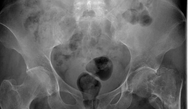
Images can play a key role in assessing pelvic trauma. For instance, ultrasounds can help identify fluid build up in certain areas, though they can’t always tell the difference between blood and bowel contents. X-rays can be helpful to quickly identify serious injuries in patients who are not stable. They can also help spot hip issues, such as a broken bone or dislocation that needs quick repair. However, X-rays can’t provide the same level of detailed information about fractures that a computed tomography (CT) scan can. A CT scan is the most accurate method available for identifying fractures, and can provide three-dimensional images which can be useful for surgical planning. It also allows for the use of contrast, which can help highlight injuries and better visualize urologic structures.
In case of suspected injury to the urethra, a urethrogram can be performed which involves imaging the structure with the help of a special dye. However, since urethral injuries are not immediately life-threatening, urgent treatment should first be given to any vascular or visceral injuries present.
Treatment Options for Pelvic Trauma (Hip and Lower Abdomen Injury)
Pelvic fractures, regardless of the severity, have the potential to be deadly. Treatment should focus first on ensuring the patient can breathe and has a clear airway and stable circulation. It’s crucial for the patient to be resuscitated well. Two large intravenous lines are generally put in when these patients arrive at the hospital. Any low blood pressure should be addressed with substantial fluid replacement, and then blood products, if needed.
If there are signs of disruption in the pelvic ring, a pelvic binder should be used, whether the patient is stable or not. The binder helps reduce bleeding in two ways. It compresses the bleed from the bones and reduces space in the pelvis – this helps form a blood clot to stop the bleeding. However, this is a temporary solution until a more definitive repair can be performed. The location of the binder is essential; it needs to be positioned around the larger outer thigh bone and the bone connecting the hip bones at the front to correctly reduce the pelvic space. It should not be used on a sideways fracture as it can increase bleeding.
A stable pelvic fracture can be identified by a bleeding artery visible on a CT scan, and the patient should be directed for a procedure called angioembolization. This procedure is usually performed by an interventional radiologist and works effectively in 85% to 100% of cases. It involves blocking the bleeding vessel(s) by inserting a substance that promotes clotting. After the embolization, an angiogram – an X-ray of the blood vessels – is used to confirm whether the bleeding has stopped.
Patients with stable pelvic fractures should stay in the hospital for observation.
For unstable pelvic fractures, immediate surgery might be required if the patient’s condition gets worse. However, some argue that angioembolization should be performed first since there’s a strong link between survival rates and the time to embolization.
An alternative to angioembolization is ‘pre-peritoneal packing’ (PPP), which involves putting packing against the outer lining of the abdomen to help form a blood clot. This quicker procedure can be done if interventional radiology is unavailable, or as an initial step towards more time-consuming procedures like angioembolization.
A technique called ‘Resuscitative endovascular balloon occlusion of the aorta’ (REBOA) can also be used, where a balloon catheter stops the bleeding by occluding the aorta.
For unstable pelvic fractures, surgery is needed for fixation. Early fixation can help control bleeding, improve pain, provide a better fracture reduction, and permit earlier movement. A few surgical options can provide initial stabilization in unstable patients or those with pelvic contamination.
The best time for definitive repair, if required, depends on the patient’s condition. It’s usually carried out once the patient has been adequately resuscitated and stabilized.
Various injuries might be associated with pelvic fractures, like bladder injuries, urethral injuries, and intestinal injuries. The treatment would depend on the type and location of these injuries. Vaginal lacerations associated with pelvic fractures might also require treatment based on their severity.
What else can Pelvic Trauma (Hip and Lower Abdomen Injury) be?
- Fracture in the hip socket (Acetabular pelvic fracture)
- Damage to the bladder
- Broken hip
- Dislocated hip
- Cut or tear in the urethra (Urethral transection)
- Fracture that splits the pelvis into two halves (Open book pelvic fracture)
- Injury to the rectum
- Damage to the sigmoid colon
- Cut or tear in the urethra, again (Urethral transection)
- Broken pelvis due to an upward force (Vertical shear pelvic fracture)
What to expect with Pelvic Trauma (Hip and Lower Abdomen Injury)
Unstable pelvic fractures are serious injuries with a mortality rate of around 8%. If a patient with such fracture also shows signs of hemorrhagic shock – a severe amount of blood loss – then the mortality rate increases. Open pelvic fractures, a type where the broken bone penetrates the skin, are even more dangerous with a risk of death going up to 45%.
What makes pelvic fractures particularly severe are the additional injuries that often accompany them. However, if a pelvic fracture happens in isolation, without any other injuries, the risk of death is relatively low, ranging between 0.4% to 0.8%.
Possible Complications When Diagnosed with Pelvic Trauma (Hip and Lower Abdomen Injury)
The injuries that come along with pelvic fractures can make it hard to anticipate the patient’s progress or outcome. Research indicates that over 60% of patients with traumatic pelvic fractures end up experiencing chronic pelvic pain. This constant pain is linked to mental health problems like depression and anxiety. An external fixation method (stabilising the fracture using a metal frame) can sometimes lead to infections at the pin site, but these can usually be treated with oral or intravenous antibiotics and possibly cleaning out the infected site. There is also a risk of damage to the lateral femoral cutaneous nerve due to external fixation.
Alongside this, pelvic fractures can lead to urogenital injuries which may cause sexual dysfunction including painful intercourse, erectile dysfunction, and limited movement. There may also be instances of urinary and fecal incontinence. Within patients undergoing complete urethral transection (cutting of the urethra), urethral strictures (abnormal narrowing of the urethra) have been noted in about 31% to 69% of cases. Doctors usually choose to treat these initially through simple dilation. If this does not work, a surgical procedure called posterior urethroplasty, specifically bulbomembranous anastomosis (BMA), may be performed. Fortunately, BMA has a high success rate of over 90%.
Urinary incontinence is first attempted to be treated non-surgically. Techniques that can strengthen the pelvic floor and biofeedback are commonly used. The use of Duloxetine, a medication that increases serotonin and norepinephrine in the brain, along with physiotherapy, often results in improvement for many patients. Alternately, a device that stimulates the sacral nerve can be implanted within the upper buttock area, which has also been successful in treating incontinence. If everything else fails, doctors may resort to the implantation of an artificial urinary sphincter.
For fecal incontinence, a similar non-surgical approach is initially used. Adjusting diet and adding fiber supplements can enhance stool consistency and reduce urgency. Various medications can be used to slow down bowel movements. If necessary, surgical options like sphincteroplasty (repairing the anal sphincter), implanting artificial or magnetic anal sphincters, and using sacral nerve stimulators are available.
One point to note is that even a common treatment tool like a pelvic binder – a compression belt to stabilise the pelvic area – can have its complications. It should not be used for more than 24 hours as it can cause skin cell death and pressure sores in as little as 2 to 3 hours. Other risks include deep vein blood clots due to the decreased mobility and bone fractures associated with all trauma patients. Hence, until the patients are able to move around on their own, they should be treated with devices that aid in blood flow and preventative medicines.
Complications from angioembolization (a procedure that blocks blood flow to specific areas) occur in up to 5% of cases. Problems can arise at the insertion site that include blood pooling, formation of false aneurysm, dissection, or clot formation. Life-threatening tissue death of the pelvic region is a severe complication that requires surgical exploration and repair. There can also be reactions to the contrast dye used in this procedure or contrast-induced kidney damage and acute kidney injury.
The use of REBOA (Resuscitative Endovascular Balloon Occlusion of the Aorta), a technique used to control bleeding, has several potential complications mainly related to vessel injuries. These can include an arterial rupture, perforation, or dissection. The catheter itself can create lower extremity lack of blood flow and subsequent blood flow restoration injury and compartment syndrome (a painful and dangerous condition caused by pressure buildup). The balloon can also lead to lack of blood supply below the insertion point. Prolonged lack of blood supply leads to organ damage, which can be irreversible. Restoration of the blood supply can lead to multiorgan dysfunction or failure, including acute kidney injury, liver failure, intestinal lack of blood supply, spinal cord infarction, and death. Access site problems can be treated with patch repairs, arterial reconstructions with native or artificial tissue, or bypasses. If these measures fail, then amputation of the limb may be required. Aortic injuries from REBOA are generally life or limb-threatening. The multiorgan dysfunction is generally treated medically.
Preventing Pelvic Trauma (Hip and Lower Abdomen Injury)
Using your cell phone while driving leads to roughly 1.6 million car accidents every year. In fact, texting behind the wheel is a factor in one out of every four crashes. All drivers should stick to the speed limit, never drive impaired, and stay focused on the road. Pedestrians, too, need to stay alert and should never assume that a car will see them or let them go first.
It’s crucial to prepare patients for what they’ll face after a severe pelvic fracture. Rehabilitation and physical therapy will be a long-term commitment. Patients must also understand that they may experience ongoing pain. They should learn about ways to manage this pain, as well as how to cope with the potential depression and anxiety that may arise. For patients needing a colostomy due to a colon injury, training on how to manage their ostomy is crucial. Conversations about the potential for sexual dysfunction should also be initiated, with specialist referrals to gynecologists or urologists provided as needed.


